Observational Study
. 2024 Oct 4;14(1):23071.
doi: 10.1038/s41598-024-73681-5.

Patterns of intersectional tumor volumes in T2-weighted MRI and [18F]FET PET in adult glioma: a prospective, observational study

Affiliations
Observational Study

Patterns of intersectional tumor volumes in T2-weighted MRI and [18F]FET PET in adult glioma: a prospective, observational study

Jonathan Weller et al. Sci Rep. .

Abstract

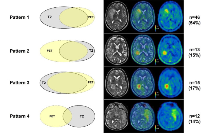
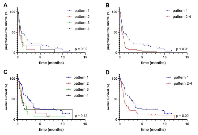
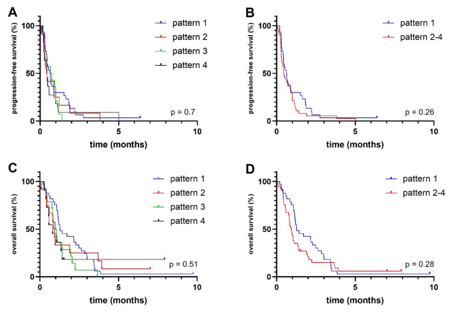
Brain tumor volumes as assessed by magnetic resonance imaging (MRI) do not always spatially overlap with biological tumor volumes (BTV) measured by [18F]Fluoroethyltyrosine positron emission tomography ([18F]FET PET). We prospectively investigated volumetric patterns based on the extent of tumor volume overlap between the two modalities. Eighty-six patients with newly diagnosed glioma who had undergone MRI and [18F]FET PET between 2007 and 2009 were included in this prospective study and (re-)classified according to CNS WHO 2021 (Classification of Tumors of the Central Nervous System by the World Health Organization). Four different patterns of volume overlap were defined mathematically according to the extent of overlap between MRI-based T2 tumor volume (non-enhancing tumor volume, nCEV) and BTVs. Progression-free (PFS) and overall survival (OS) were determined. Seventy patients were diagnosed with isocitrate dehydrogenase wildtype (IDHwt) glioblastoma and 16 with IDH-mutant glioma, respectively. The most common pattern was characterized by a larger non-contrast-enhancing tumor volume (nCEV) that enclosed all or most of the BTV and was observed in 46 patients (54%) (pattern 1). This pattern was more frequent in IDH-mutant gliomas than in IDH-wildtype glioblastoma (81% versus 47%, p = 0.02). In multivariate analyses, pattern 1 was associated with prolonged PFS (HR 0.59; 95 CI 0.34-1.0; p = 0.05), but not OS (HR 0.66; 95 CI 0.4-1.08; p = 0.1). For OS, presence of an IDH mutation (p = 0.05) and lower age (p = 0.03) were associated with prolonged OS. The spatial relation between nCEV and BTV varies within and between glioma entities. Most frequently, a larger nCEV encases the BTV. Some patients show spatially dissociated nCEVs and BTVs. Not accounting for this phenomenon in surgery or radiotherapy planning might lead to undertreatment.

Keywords: Glioblastoma; Glioma; Imaging; Positron emission tomography; Radiotherapy.


Conflict of interest statement

The authors declare no competing interests.

Figures

Fig. 1
Fig. 1
Schematic illustration and examples of different volume patterns in juxtaposition. Different patterns were defined based on the extent of overlap between non-enhancing tumor volume and biological tumor volume as determined by volumetric analysis of [18F]FET PET imaging. The number of patients in each group and percentages are given. T2 = non-enhancing tumor volume in T2-weighted MRI-scan; PET = biological tumor volume in [18F]FET PET.
Fig. 2
Fig. 2
Progression-free survival (PFS) (A,B) and overall survival (OS) (C, D) of different intersectional tumor volume patterns. Pattern 1 was associated with significantly longer PFS than other patterns individually and combined ((A,B); p = 0.02 and p < 0.01). Pattern 1 was associated with longer OS than other patterns combined ((D), p = 0.02). Pattern 1: non-enhancing tumor volume (nCEV) is larger than the biological tumor volume (BTV) and encloses all or most of it. Pattern 2: BTV is larger than nCEV and encloses all or most of it. Pattern 3: nCEV and BTV are similar in size and there are minimal exclusive tumor volumes. Pattern 4: nCEV and BTV are similar in size, the exclusive tumor volumes are larger than the intersectional tumor volume.
Fig. 3
Fig. 3
Progression-free survival (PFS) (A,B) and overall survival (OS) (C,D) of different intersectional tumor volume patterns among patients with newly diagnosed glioblastoma. There were no significant differences between the patterns in the glioblastoma cohort. Pattern 1: non-enhancing tumor volume (nCEV) is larger than the biological tumor volume (BTV) and encloses all or most of it. Pattern 2: BTV is larger than nCEV and encloses all or most of it. Pattern 3: nCEV and BTV are similar in size and there are minimal exclusive tumor volumes. Pattern 4: nCEV and BTV are similar in size, the exclusive tumor volumes are larger than the intersectional tumor volume.

Similar articles

Cited by

References

    1. Brat, D. J. et al. cIMPACT-NOW update 5: recommended grading criteria and terminologies for IDH-mutant astrocytomas. Acta Neuropathol.139 (3), 603–608 (2020). - PMC - PubMed
    1. Brat, D. J. et al. cIMPACT-NOW update 3: recommended diagnostic criteria for diffuse astrocytic glioma, IDH-wildtype, with molecular features of glioblastoma, WHO grade IV. Acta Neuropathol.136 (5), 805–810 (2018). - PMC - PubMed
    1. Louis, D. N. et al. The 2021 WHO classification of tumors of the Central Nervous System: a summary. Neuro Oncology23 (8), 1231–1251 (2021). - PMC - PubMed
    1. Ostrom, Q. T. et al. CBTRUS Statistical Report: primary brain and other Central Nervous System tumors diagnosed in the United States in 2013–2017. Neuro Oncology22 (12 Suppl 2), iv1–iv96 (2020). - PMC - PubMed
    1. Buckner, J. C., Chakravarti, A. & Curran, W. J. Radiation plus chemotherapy in low-grade glioma. N. Engl. J. Med.375 (5), 490–491 (2016). - PubMed

Publication types