

High androgen level during controlled ovarian stimulation cycle impairs endometrial receptivity in PCOS patients
- PMID: 39367050
- PMCID: PMC11452613
- DOI: 10.1038/s41598-024-74295-7
High androgen level during controlled ovarian stimulation cycle impairs endometrial receptivity in PCOS patients
Abstract
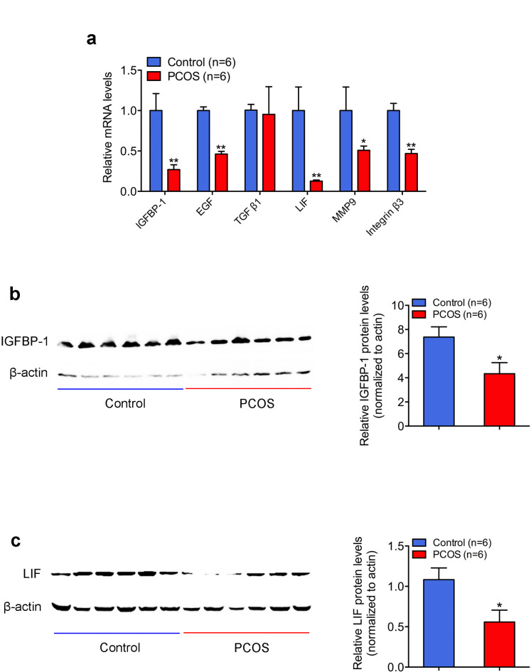
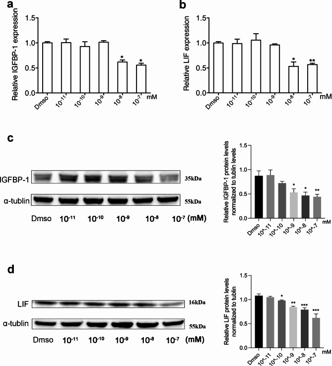
PCOS is one of the most common endocrine disorders among women of reproductive age. While the mechanism involved is not yet fully characterized. Our study aims to examine the pregnancy outcomes of embryo transfers in women with PCOS after pretreatment, and to explore the possible effect of high androgen levels on endometrial receptivity. Retrospective cohort study was conducted to analyze pregnancy outcomes among 2714 infertile women with tubal factor and 452 PCOS women. Endometrium samples were collected from 6 controls and 6 PCOS patients to detect the expression of endometrial receptivity marks. The implantation rate, clinical and ongoing pregnancy rates and live birth rate in women with PCOS followed fresh embryo transfers were obviously decreased even after the pretreatment. Similar pregnancy outcomes were found in frozen-thawed embryo transfer cycles between women with or without PCOS. Strikingly, serum total testosterone (TT) levels on trigger day were significantly higher in PCOS women. Women with high TT levels presented significantly lower clinical and ongoing pregnancy rates, and the expression of insulin-like growth factor binding protein 1 (IGFBP-1), and leukemia inhibitory factor (LIF) in the endometria decreased significantly as well. High doses of testosterone significantly down-regulated the expression of IGFBP-1 and LIF in Ishikawa cells. Although endocrine abnormalities had been improved before the controlled ovarian stimulation (COS) cycle started, higher serum TT levels were detected on the trigger day of the COS cycle in PCOS patients, which may contribute to the decreased fresh embryo implantation by impairing endometrial receptivity.
Keywords: Androgen; Embryo transfer; Endometrial receptivity; PCOS.
© 2024. The Author(s).
Conflict of interest statement
The authors declare no competing interests.
Figures
Similar articles
-
Follicular development and endometrial receptivity of different androgen phenotypes in women with polycystic ovary syndrome.Front Endocrinol (Lausanne). 2024 Dec 12;15:1400880. doi: 10.3389/fendo.2024.1400880. eCollection 2024. Front Endocrinol (Lausanne). 2024. PMID: 39726841 Free PMC article.
-
Endometrial fluid visualized through ultrasonography during ovarian stimulation in IVF cycles impairs the outcome in tubal factor, but not PCOS, patients.Hum Reprod. 2005 Apr;20(4):906-9. doi: 10.1093/humrep/deh737. Epub 2005 Jan 7. Hum Reprod. 2005. PMID: 15640251
-
The depot GnRH agonist protocol improves the live birth rate per fresh embryo transfer cycle, but not the cumulative live birth rate in normal responders: a randomized controlled trial and molecular mechanism study.Hum Reprod. 2020 Jun 1;35(6):1306-1318. doi: 10.1093/humrep/deaa086. Hum Reprod. 2020. PMID: 32478400
-
An Update on the Progress of Endometrial Receptivity in Women with Polycystic Ovary Syndrome.Reprod Sci. 2022 Aug;29(8):2136-2144. doi: 10.1007/s43032-021-00641-z. Epub 2021 Jun 2. Reprod Sci. 2022. PMID: 34076874 Review.
-
Endometrium in PCOS: Implantation and predisposition to endocrine CA.Best Pract Res Clin Endocrinol Metab. 2006 Jun;20(2):235-44. doi: 10.1016/j.beem.2006.03.005. Best Pract Res Clin Endocrinol Metab. 2006. PMID: 16772154 Review.
Cited by
-
The emerging roles of N6-methyladenosine (m6A) deregulation in polycystic ovary syndrome.J Ovarian Res. 2025 May 23;18(1):107. doi: 10.1186/s13048-025-01690-7. J Ovarian Res. 2025. PMID: 40410881 Free PMC article. Review.
-
Genetically predicted serum metabolites mediate the association between inflammatory proteins and polycystic ovary syndrome: a Mendelian randomization study.Front Med (Lausanne). 2024 Dec 3;11:1433612. doi: 10.3389/fmed.2024.1433612. eCollection 2024. Front Med (Lausanne). 2024. PMID: 39691364 Free PMC article.
-
Dysregulation of Leukaemia Inhibitory Factor (LIF) Signalling Pathway by Supraphysiological Dose of Testosterone in Female Sprague Dawley Rats During Development of Endometrial Receptivity.Biomedicines. 2025 Jan 24;13(2):289. doi: 10.3390/biomedicines13020289. Biomedicines. 2025. PMID: 40002703 Free PMC article.
References
-
- Bozdag, G., Mumusoglu, S., Zengin, D., Karabulut, E. & Yildiz, B. O. The prevalence and phenotypic features of polycystic ovary syndrome: a systematic review and meta-analysis. Hum. Reprod. 31(12), 2841–2855 (2016). - PubMed
-
- Li, R. et al. Prevalence of polycystic ovary syndrome in women in China: a large community-based study. Hum. Reprod. 28(9), 2562–2569 (2013). - PubMed
-
- Lizneva, D. et al. Criteria, prevalence, and phenotypes of polycystic ovary syndrome. Fertil. Steril. 106(1), 6–15 (2016). - PubMed
MeSH terms
Substances
Grants and funding
- IPMCH2022CR1-03/Shanghai Frontiers Science Research Base of Reproduction and Development and the Clinical Research Plan of IPMCH
- 2018YFC1005001/National Key Research and Development Program of China
- 82071730/National Natural Science Foundation of China
- SHDC12019X17/Research Grant from Shanghai Hospital Development Center
LinkOut - more resources
Full Text Sources
Medical
Research Materials
