

Comparison of human pluripotent stem cell differentiation protocols to generate neuroblastoma tumors
- PMID: 39367051
- PMCID: PMC11452544
- DOI: 10.1038/s41598-024-73947-y
Comparison of human pluripotent stem cell differentiation protocols to generate neuroblastoma tumors
Abstract
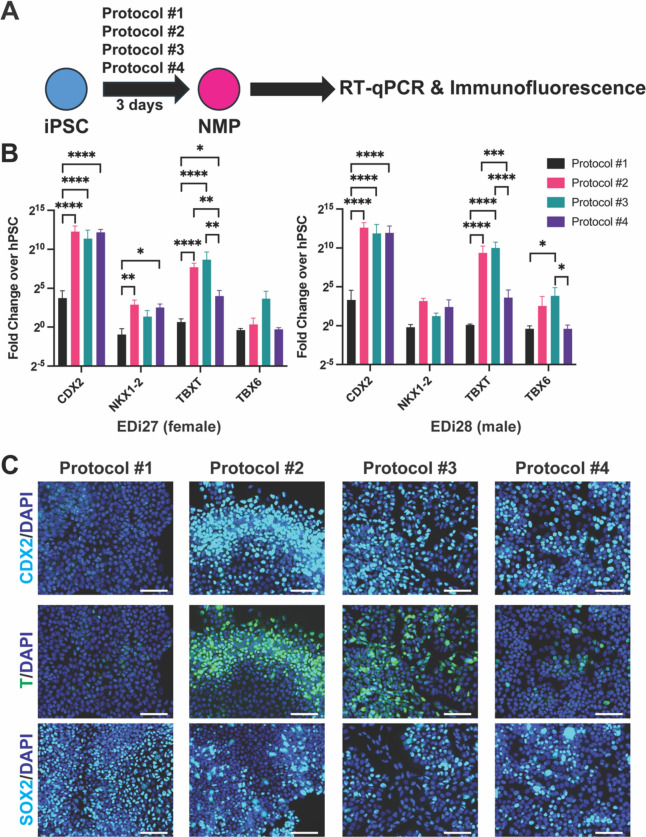
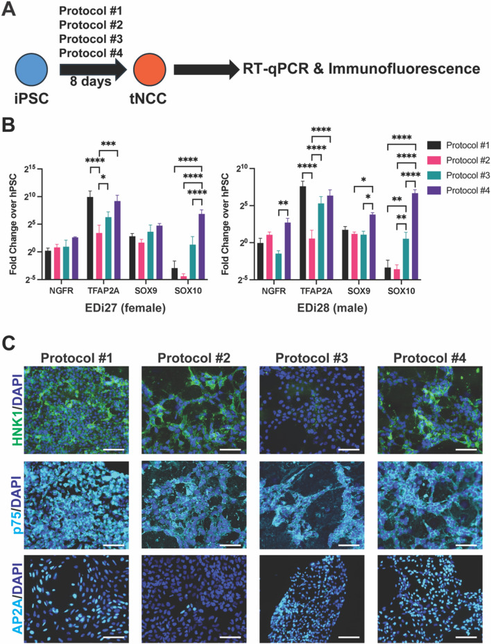
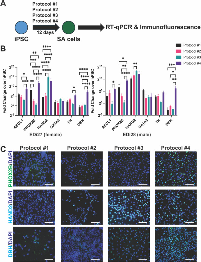
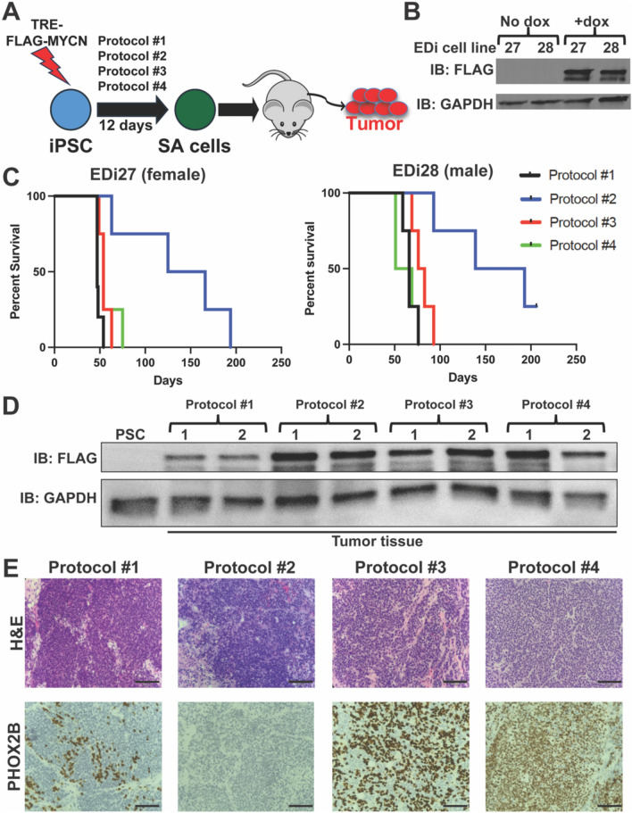
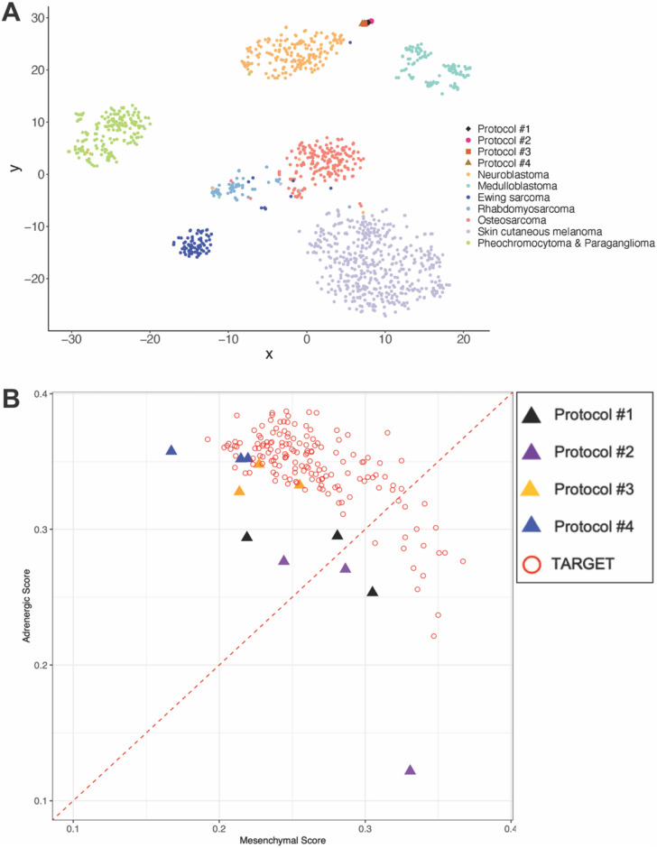
Neuroblastoma is the most common pediatric extracranial solid tumor and is derived from trunk neural crest cells (tNCC) and its progenitor sympathoadrenal (SA) cells. While human pluripotent stem cell (PSC) models of neuroblastoma have been described, the PSC were differentiated using protocols that made neural crest cells, but not specifically the trunk subtype. Here, we compared four recent protocols to differentiate pluripotent stem cells (PSC) toward SA cells and examined their efficiency at generating SA cells along with earlier cell states (neuromesodermal progenitors [NMP], tNCC), as well as generating MYCN-driven tumors. Interestingly, the protocols that created cells with the highest level of NMP markers did not produce cells with the highest tNCC or SA cell markers. We identified a protocol that consistently produced cells with the highest level of SA markers using two PSC lines of different genders. This protocol also generated tumors with the highest level of PHOX2B, a marker of neuroblastoma. Transcriptionally, however, each protocol generates tumors that resemble neuroblastoma. Two of the protocols repeatedly produced adrenergic neuroblastoma whereas the other two protocols were ambiguous. Thus, we identified a protocol that reliably generates adrenergic neuroblastoma.
Keywords: Human pluripotent stem cells; MYCN; Neuroblastoma; Sympathoadrenal cell.
© 2024. The Author(s).
Conflict of interest statement
The authors declare no competing interests.
Figures
Similar articles
-
Alternative NHEJ pathway proteins as components of MYCN oncogenic activity in human neural crest stem cell differentiation: implications for neuroblastoma initiation.Cell Death Dis. 2017 Dec 13;8(12):3208. doi: 10.1038/s41419-017-0004-9. Cell Death Dis. 2017. PMID: 29238067 Free PMC article.
-
METTL3/MYCN cooperation drives neural crest differentiation and provides therapeutic vulnerability in neuroblastoma.EMBO J. 2024 Dec;43(24):6310-6335. doi: 10.1038/s44318-024-00299-8. Epub 2024 Nov 11. EMBO J. 2024. PMID: 39528654 Free PMC article.
-
Generating trunk neural crest from human pluripotent stem cells.Sci Rep. 2016 Jan 27;6:19727. doi: 10.1038/srep19727. Sci Rep. 2016. PMID: 26812940 Free PMC article.
-
The MYCN oncogene and differentiation in neuroblastoma.Semin Cancer Biol. 2011 Oct;21(4):256-66. doi: 10.1016/j.semcancer.2011.08.001. Epub 2011 Aug 9. Semin Cancer Biol. 2011. PMID: 21849159 Review.
-
Neuroblastoma-derived v-myc avian myelocytomatosis viral related oncogene or MYCN gene.J Clin Pathol. 2023 Aug;76(8):518-523. doi: 10.1136/jcp-2022-208476. Epub 2023 May 23. J Clin Pathol. 2023. PMID: 37221048 Review.
Cited by
-
A systematic review and embryological perspective of pluripotent stem cell-derived autonomic postganglionic neuron differentiation for human disease modeling.Elife. 2025 Mar 12;14:e103728. doi: 10.7554/eLife.103728. Elife. 2025. PMID: 40071727 Free PMC article.
References
-
- Huber, K. Segregation of neuronal and neuroendocrine differentiation in the sympathoadrenal lineage. Cell Tissue Res 359(1), 333–341 (2015). - PubMed
Publication types
MeSH terms
Substances
Grants and funding
LinkOut - more resources
Full Text Sources
Medical
Molecular Biology Databases
Miscellaneous
