. 2024 Oct 4;7(1):1215.
doi: 10.1038/s42003-024-06878-z.

Preselection of potential target spaces based on partial information by the supplementary eye field

Affiliations

Preselection of potential target spaces based on partial information by the supplementary eye field

Osamu Yokoyama et al. Commun Biol. .

Abstract

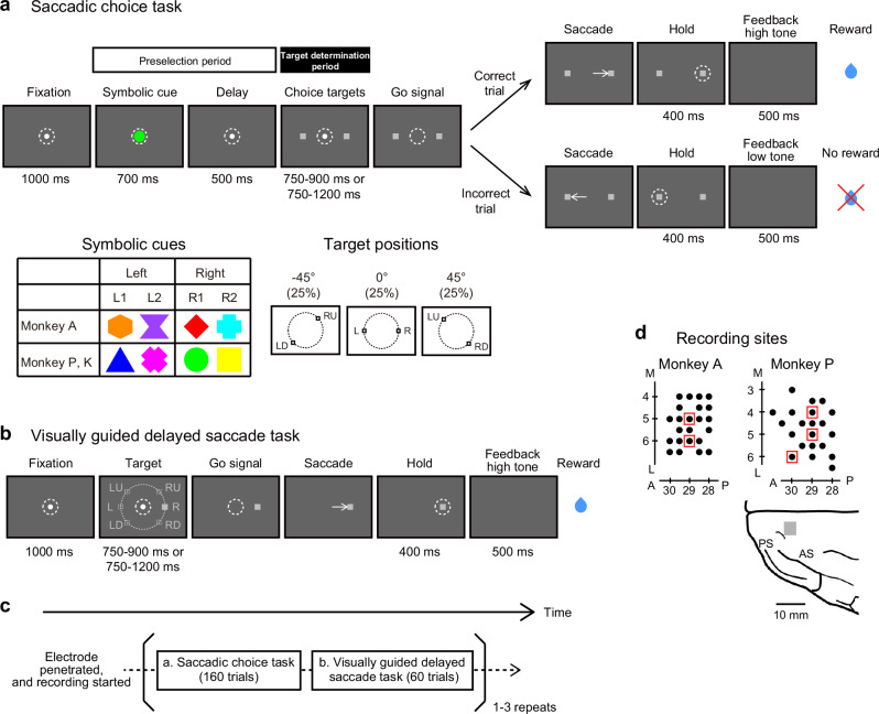
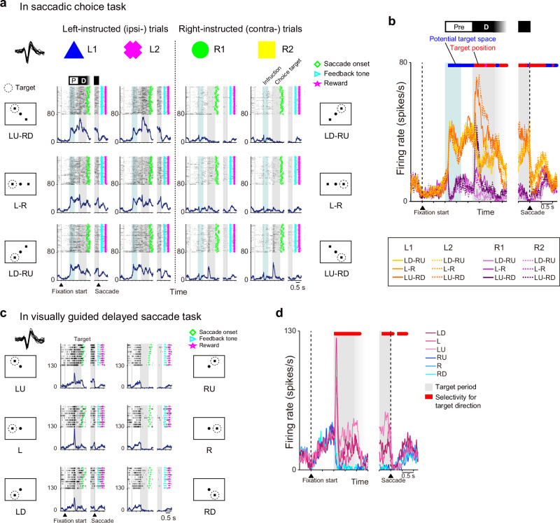
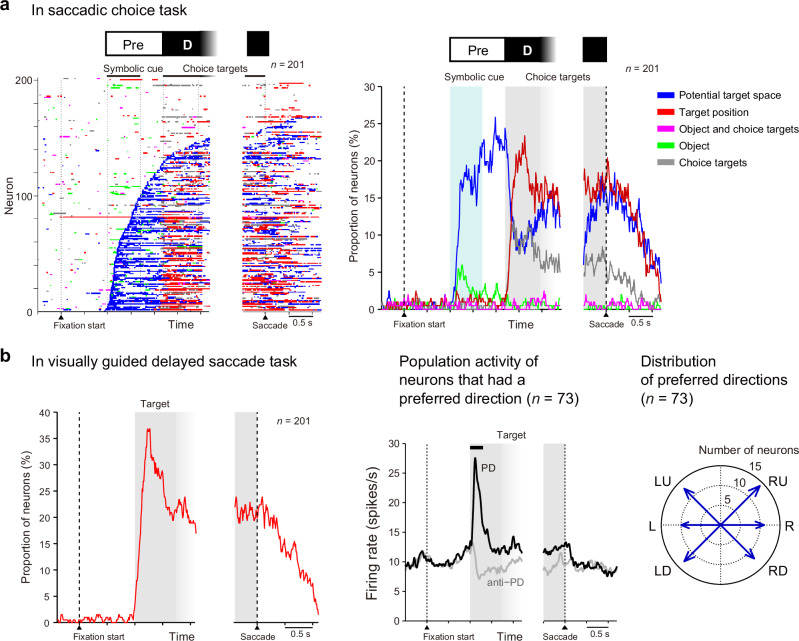
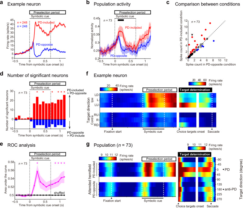
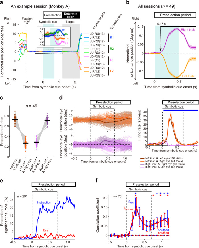
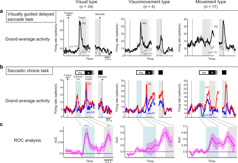
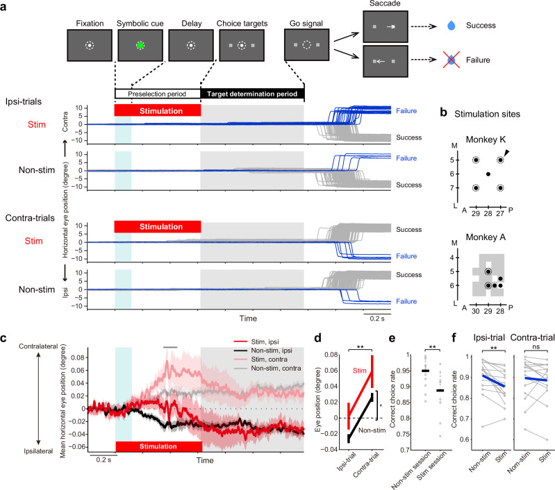
Before selecting a saccadic target, we often acquire partial information about the location of the forthcoming target and preselect a region of visual space even before the target becomes visible. To determine whether the supplementary eye field (SEF) represents information signifying the potential target space, we examined neuronal activity in the SEF of monkeys performing a behavioral task designed to isolate the process of visuospatial preselection under uncertainty from the process of selecting a specified location. Our data showed that the activity of SEF neurons represented information about the potential target space instructed by symbolic cues. Increased activity of visuospatially selective SEF neurons encoded the potential target space, which could be a mechanism facilitating subsequent selection of an appropriate target. Furthermore, electrical stimulation of the SEF during the preselection period disrupted subsequent target selection. These results demonstrate that the SEF contributes to the preselection of potential target spaces based on partial information.


Conflict of interest statement

The authors declare no competing interests.

Figures

Fig. 1
Fig. 1. Behavioral tasks and recording sites in the SEF.
a Temporal sequence of behavioral events in the saccadic choice task. The appearance of visual cues (symbolic cues) that instructed the monkey to select a target in the L or R visual hemifield and the main configurations of the choice targets are shown below the task sequence. b Temporal sequence of behavioral events in the visually guided delayed saccade task. c Task order in each recording session. The position of the inserted electrode was fixed throughout the session once the recording began. d Recording sites in the SEF. Neuronal activity was recorded from the SEF in the left hemisphere of two monkeys. (Left and right) Filled black circles indicate the recording sites. Red squares indicate the sites where intracortical microstimulation evoked saccades. (bottom) Top view of the frontal cortex in the left hemisphere of monkey P. The gray rectangle indicates the recorded area. A anterior, L lateral, M medial, P posterior, AS arcuate sulcus, PS principal sulcus.
Fig. 2
Fig. 2. Activity of an example neuron recorded from the SEF.
a Raster plots and mean spike density functions of an example neuron in the saccadic choice task. The ordinate represents the instantaneous firing rate (spikes s−1). The plots are arranged such that trials with a certain symbolic cue are grouped in each column, and trials with a chosen target in a certain location are shown in the corresponding location of the panel. Neuronal activity is aligned with symbolic cue onset and saccade initiation (left and right gray vertical lines, respectively). Light blue shading indicates the period during which a symbolic cue was presented. Gray shading indicates the period from the choice target onset to the go signal (750–1200 ms). The spike waveforms are shown in the upper-left corner. b Merged spike density functions of the neuron shown in (a). The color of each line plot indicates the trial type, as shown below the plot. The color of the horizontal bar above the plot indicates the factors (blue: potential target space; red: target position) represented by the activity as statistically determined. c Raster plots and mean spike density functions of the same neuron are shown in (a) and (b) in the visually guided delayed saccade task. Gray shading indicates the period from the target onset to the go signal (750–1200 ms). Gray vertical bars indicate the fixation initiation and saccade onset times. The spike waveforms are shown in the upper-left corner. d Merged spike density functions of the neuron shown in (c). The color of each line plot indicates the trial type, as shown to the right of the plot. The red horizontal bar above the plot indicates the period during which the neuron exhibited directional selectivity.
Fig. 3
Fig. 3. SEF neurons represent the potential target space and target position.
a Left, time courses of the representation of task-related factors by individual SEF neurons in the saccadic choice task. Different colors indicate selectivity for potential target space (blue), target position (red), object (green), choice targets (gray), and object and choice targets (magenta). Each row represents a single neuron. Neurons were ordered according to the onset time of selectivity for the potential target space. Right, the proportion of neurons representing task-related factors over time. The color code is the same as in the left panel. b Left, the proportion of neurons that exhibited direction selectivity over time in the visually guided delayed saccade task. Center, mean activity of neurons that had direction selectivity. The PD of each neuron was determined based on the spike counts during the 0–300 ms period after target onset (horizontal black bar above the plot). Population-averaged activity was calculated separately for the PD (black) and anti-PD (gray) trials. Right, distribution of the PDs of the neurons.
Fig. 4
Fig. 4. SEF neurons encode the instructed visual hemifield if it includes their PDs.
a Activity of an example neuron in the PD-included and PD-opposite conditions (mean ± SEM) of the saccadic choice task. b Normalized activity for the population of neurons (mean ± SEM) for which a PD could be determined. c Comparison of spike counts at 0.1–0.3 s (black line in [b]) after symbolic cue onset. Red/blue-filled circles: neurons exhibiting greater/lesser activity in the PD-included than PD-opposite condition (two-sample t-test, α = 0.05), respectively. Black circles: neurons exhibiting no significance. Arrowhead: the example neuron in (a). d The number of neurons exhibiting different activity (two-sample t-test, p ≤ 0.0484, α = 0.05) between the PD-included and PD-opposite conditions. Asterisks indicate that the number of neurons showing significantly greater activity in the PD-included condition (red bar) was larger (binomial test, p ≤ 0.015, α = 0.05, FDR-corrected) than that showing greater activity in the PD-opposite condition (blue bar). e Time course of AUC values (mean ± SEM) for the PD-included vs PD-opposite condition across the neurons. Asterisks indicate that the AUC value of the experimental data (magenta) was significantly greater (Wilcoxon signed-rank test, p ≤ 0.010, α = 0.05, FDR-corrected) than that of shuffled data (black). f Changes in spatial encoding by an example neuron. The mean firing rate is color-coded as a function of time (abscissa) and saccade target direction (ordinate). The color ranges are scaled separately for the periods before and after the choice target onset for illustrative purposes. g Changes in spatial encoding by a subpopulation of neurons for which a PD could be determined. The mean firing rate across the neurons is color-coded. For the period before choice target onset, the trial types (ordinate) were ordered so that the hemifield that included the PD and the hemifield that did not were aligned (shown on the left y-axis) across the neurons. For the period after the choice target onset, the trial types (ordinate) were ordered so that the PDs were aligned (to 0°, shown on the right y-axis) across the neurons. The color ranges are scaled separately for the periods before and after the choice target onset for illustrative purposes (shown by color scales above the plot).
Fig. 5
Fig. 5. Eye position bias and its effects on SEF activity.
a Horizontal eye position in the saccadic choice task of an example session. Each line indicates the mean for each trial type (i.e., a combination of instruction and configurations of choice targets). The color and number of trials for each trial type are shown on the right. The inset shows an enlarged view of the period marked by the dotted rectangle (from 0.5 s before the cue appearance until the choice target appearance) for trials grouped according to symbolic cue. b Normalized horizontal eye position (mean ± SEM across sessions and monkeys) during the preselection period. The black bar above the plot indicates the period during which eye position was different between the right- and left-instructed trials (paired t-test, α = 0.01 for ≥20 ms); the arrow indicates the onset time. c The proportion of the four types of trials classified by instructed visual hemifield (left/right) and the direction of eye position shift (left/right) across sessions. The black line indicates the mean. d Effects of instruction and eye position shift on neuronal activity. Left, eye position traces in individual trials (thin lines) and their mean (thick lines) for the four trial types classified by the instructed hemifield (left/right) and the direction of the eye position shift (left/right) of an example session. Right, the activity of an example neuron in the four trial types (mean ± SEM). The blue line above the plot indicates the period in which the effect of the instructions was significant (two-way ANOVA, p < 0.01 in consecutive ≥3 bins). e The proportion of neurons that were modulated according to the instructed hemifield (blue) and that of neurons for which activity depended on the direction of eye position shift (red). f Effect of eye position on neuronal activity. The blue line shows the mean correlation coefficient (βhemi) obtained when the spike counts were regressed by the instructed hemifield across neurons (mean ± SEM). The red line shows the mean partial correlation coefficient (βhemi,eye) of the instructed hemifield when spike counts were regressed by the instructed hemifield and horizontal eye position across neurons. Asterisks indicate that the βhemi (blue) and βhemi,eye (red) values were greater than those of shuffled data (Wilcoxon signed-rank sum test, p ≤ 0.013, α = 0.05, FDR-corrected).
Fig. 6
Fig. 6. Visual-type neurons encode potential target space.
ac Left, visual type; center, visuomovement type; right, movement type. a Mean activity of the neurons in the PD (black) and anti-PD (gray) trials in the visually guided delayed saccade task. b Mean activity of the neurons in the PD (red) and anti-PD (blue) trials in the saccadic choice task. c Time course of the AUC values (mean ± SEM) for activity across the neurons in the PD vs anti-PD conditions. Red dots above the line plots indicate windows in which the AUC value of the experimental data (solid line) was significantly greater than that of shuffled data (dotted line) (Wilcoxon signed-rank test, α = 0.05, FDR-corrected).
Fig. 7
Fig. 7. Electrical stimulation of the SEF affects choice behavior based on spatial selection under uncertainty.
a Experimental procedure and horizontal eye position in an example session. Gray and blue lines indicate eye traces in the success and failure trials, respectively. Positive and negative values indicate the contralateral (right) and ipsilateral (left) sides, respectively. Ipsi- and Contra-trials indicate trials with the correct target in the visual field ipsilateral and contralateral to the stimulated (left) hemisphere, respectively. Stim, stimulation trials; non-stim, nonstimulation trials. b Stimulation sites are indicated with black-filled circles and the sites where intracortical microstimulation evoked saccades during free viewing outside of the task are encircled with a dotted line. Gray shading indicates the sites where neuronal activity was recorded. The arrow indicates the stimulation site in the example session shown in (a). c Horizontal eye position in two monkeys (mean ± SEM, n = 16). The scale of the ordinate was magnified from (a). d Horizontal eye position at 0.35–0.45 s (gray bar in [c]) after symbolic cue onset (and stimulation onset) in the two monkeys (mean ± SEM, n = 16; monkey A, n = 9 sessions; monkey K, n = 7 sessions). The horizontal eye position was more contralateral in trials with SEF stimulation than in those without SEF stimulation (two-way ANOVA, *p = 0.032) and more contralateral in Contra-trials than in Ipsi-trials (**p = 0.00026). e Effect of stimulation on correct choice rate (nonstimulation and stimulation sessions, n = 16, each; monkey A, n = 9 [circles]; monkey K, n = 7 [Xs]; two-sample t-test, **p = 0.0022). The black bar indicates the mean values; the light gray mark indicates individual sessions. f Correct choice rate in stimulation sessions (n = 16; nonkey A, n = 9 [circles]; nonkey K, n = 7 [Xs]). Blue indicates mean values; light gray indicates individual sessions. For the Ipsi-trials (left), the correct choice rate was higher in the Stim trials than in the non-stim trials (paired t-test, **p = 0.0024), whereas no difference was observed between the Stim and non-stim trials for the Contra-trials (right, p = 0.4625).

Similar articles

References

    1. Lee, Y.-C., Lee, J. D. & Ng Boyle, L. The interaction of cognitive load and attention-directing cues in driving. Hum. Factors51, 271–280 (2009). - PubMed
    1. Theeuwes, J. Exogenous and endogenous control of attention: the effect of visual onsets and offsets. Percept. Psychophys.49, 83–90 (1991). - PubMed
    1. Stuphorn, V., Taylor, T. L. & Schall, J. D. Performance monitoring by the supplementary eye field. Nature408, 857–860 (2000). - PubMed
    1. Huerta, M. F. & Kaas, J. H. Supplementary eye field as defined by intracortical microstimulation: connections in macaques. J. Comp. Neurol.293, 299–330 (1990). - PubMed
    1. Tanji, J. & Hoshi, E. Role of the lateral prefrontal cortex in executive behavioral control. Physiol. Rev.88, 37–57 (2008). - PubMed

LinkOut - more resources