. 2024 Oct 4;38(1):3.
doi: 10.1007/s10548-024-01078-8.

Distinct Longitudinal Changes in EEG Measures Reflecting Functional Network Disruption in ALS Cognitive Phenotypes

Affiliations

Distinct Longitudinal Changes in EEG Measures Reflecting Functional Network Disruption in ALS Cognitive Phenotypes

Marjorie Metzger et al. Brain Topogr. .

Abstract

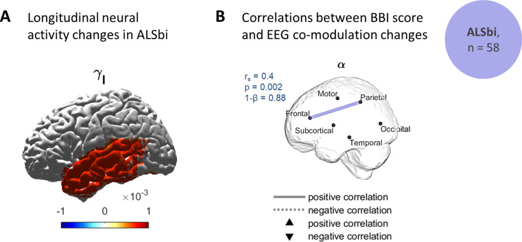
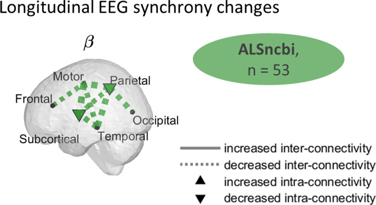
Amyotrophic lateral sclerosis (ALS) is characterised primarily by motor system degeneration, with clinical evidence of cognitive and behavioural change in up to 50% of cases. We have shown previously that resting-state EEG captures dysfunction in motor and cognitive networks in ALS. However, the longitudinal development of these dysfunctional patterns, especially in networks linked with cognitive-behavioural functions, remains unclear. Longitudinal studies on non-motor changes in ALS are essential to further develop our understanding of disease progression, improve care and enhance the evaluation of new treatments. To address this gap, we examined 124 ALS individuals with 128-channel resting-state EEG recordings, categorised by cognitive impairment (ALSci, n = 25), behavioural impairment (ALSbi, n = 58), or non-impaired (ALSncbi, n = 53), with 12 participants meeting the criteria for both ALSci and ALSbi. Using linear mixed-effects models, we characterised the general and phenotype-specific longitudinal changes in brain network, and their association with cognitive performance, behaviour changes, fine motor symptoms, and survival. Our findings revealed a significant decline in [Formula: see text]-band spectral power over time in the temporal region along with increased [Formula: see text]-band power in the fronto-temporal region in the ALS group. ALSncbi participants showed widespread β-band synchrony decrease, while ALSci participants exhibited increased co-modulation correlated with verbal fluency decline. Longitudinal network-level changes were specific of ALS subgroups and correlated with motor, cognitive, and behavioural decline, as well as with survival. Spectral EEG measures can longitudinally track abnormal network patterns, serving as a candidate stratification tool for clinical trials and personalised treatments in ALS.

Keywords: Cognitive-behavioural impairments; Functional connectivity; Motor neuron disease; Neurodegeneration; Source localisation; Spectral resting-state EEG.


Conflict of interest statement

The authors declare no competing interests.

Figures

Fig. 1
Fig. 1
Changes in neural activity include a decrease in slow oscillations and increase in faster oscillations. Longitudinal changes of EEG spectral power in ALS were measured in term of significant longitudinal spectral power variations, based on the time fixed-effect and the time ROI-specific random-effects (Bootstrapping, q < 0.1). Longitudinal changes were mapped to get a spatial visualisation. The neural activity showed a significant decrease in formula image-band and an increase in formula image-band
Fig. 2
Fig. 2
Widespread increased EEG co-modulation in ALSci, and associations with cognitive decline. (A) Regions of longitudinal changes of EEG co-modulation and synchrony in ALSci group. The significant longitudinal connectivity changes were mapped to get a spatial visualisation of their magnitudes. The longitudinal variations represent the combined estimated slope (significance by bootstrapping, q < 0.1). A widespread increase in formula image- and formula image-band co-modulation was observed in ALSci. The dashed lines represent a decrease while the solid lines represent an increase in connectivity. A filled node represents significant intra-region connectivity. (B-C) Regions with significant correlations between participant/ROI-specific co-modulation progressions and cognitive decline, in ALSci. For each significant correlation, the correlation coefficient, rs, the p-value, p, and the statistical power,1-β are given. An adaptive FDR was applied to Spearman’s correlation coefficients. (B) Correlations between EEG co-modulation and ECAS total score progressions. (C) Correlations between EEG co-modulation and ECAS verbal fluency changes. ALSci: individuals with ALS and impaired cognition; MM: intra-motor connectivity; MP: connectivity between motor and parietal regions; PP: intra-parietal connectivity; FO: connectivity between frontal and occipital regions; ST: connectivity between subcortical and temporal regions; SO: connectivity between subcortical and occipital regions
Fig. 3
Fig. 3
Increased EEG neural activity in the temporal lobe in ALSbi and associations with behavioural impairment. (A) Longitudinal changes of EEG spectral power in ALSbi. The significant temporal spectral power variations, in terms of the time fixed-effect and the time ROI-specific random-effects (Bootstrapping, q < 0.1), were mapped to get a spatial visualisation. An increase in formula image-band co-modulation was observed in the temporal lobe for the ALSbi group. (B) Regions with significant correlations between participant/ROI-specific co-modulation progressions and cognitive decline, in the ALSbi group. A higher rate of change in co-modulation between the frontal and parietal lobes was correlated with an increased rate of change in BBI scores. For each significant correlation, the correlation coefficient, rs, the p-value, p, and the statistical power,1-β, are given. An adaptive FDR was applied to Spearman’s correlations. ALSbi: individuals with ALS and impaired behaviour
Fig. 4
Fig. 4
Widespread decreased formula image-band EEG synchrony in the ALSncbi group. Regions of longitudinal changes of EEG synchrony in the ALSncbi group. The significant longitudinal connectivity changes were mapped to get a spatial visualisation of their magnitudes. A widespread decrease in formula image-band synchrony was observed in the ALSncbi group. The longitudinal variations represent the combined estimated slope (significance by bootstrapping, q < 0.1). The dashed lines represent a decrease while the solid lines represent an increase in connectivity. A filled node represents significant intra-region connectivity. ALSncbi: individuals with ALS with normal cognition and behaviour
Fig. 5
Fig. 5
Survival and EEG functional connectivity in ALSci, ALSbi and ALSncbi subgroups. Regions with significant correlations between survival and participant/ROI-specific connectivity (AEC and iCoh) progressions. Solid lines (or upper triangles) depict positive correlations, indicating that higher rates of functional connectivity change are associated with a better prognosis. In contrast, dashed lines (or lower triangles) represent negative correlations, signifying that higher rates of functional connectivity change are linked to a worse prognosis. In ALSci, ALSbi, and ALSncbi subgroups, the correlation coefficient, formula image, the p-value, p, and the statistical power,1-β, are given for each significant correlation. An adaptive FDR was applied to Spearman’s correlations. ALSci: individuals with ALS and impaired cognition; ALSbi: individuals with ALS and impaired behaviour; ALSncbi: individuals with ALS with normal cognition and behaviour
Fig. 6
Fig. 6
Summary of the main findings. Distinct changes in functional networks characterise cognitive phenotypes in ALS and are clinically relevant. Positive correlations, indicates that higher rates of functional connectivity change are associated with a better prognosis/increased rate of change in cognition or behaviour. In contrast, negative correlations, signify that higher rates of functional connectivity change are linked to a worse prognosis/lower rate of change in cognition or behaviour

References

    1. Abrahams S, Leigh PN, Harvey A, Vythelingum GN, Grisé D, Goldstein LH (2000) Verbal fluency and executive dysfunction in amyotrophic lateral sclerosis (ALS). Neuropsychologia 38(6):734–747. 10.1016/S0028-3932(99)00146-3 - PubMed
    1. Abrahams S, Newton J, Niven E, Foley J, Bak TH (2014) Screening for cognition and behaviour changes in ALS. Amyotroph Lateral Scler Frontotemporal Degeneration 15(1–2):9–14. 10.3109/21678421.2013.805784 - PubMed
    1. Baldo JV, Schwartz S, Wilkins D, Dronkers NF (2006) Role of frontal versus temporal cortex in verbal fluency as revealed by Voxel-based lesion symptom mapping. J Int Neuropsychol Soc 12(06). 10.1017/S1355617706061078 - PubMed
    1. Balendra R, Jones A, Jivraj N, Knights C, Ellis CM, Burman R, Turner MR, Leigh PN, Shaw CE, Al-Chalabi A (2014) Estimating clinical stage of amyotrophic lateral sclerosis from the ALS Functional Rating Scale. Amyotroph Lateral Scler Frontotemporal Degeneration 15(3/4):279–284. 10.3109/21678421.2014.897357 - PubMed
    1. Beasley TM, Erickson S, Allison DB (2009) Rank-based inverse normal transformations are increasingly used, but are they Merited? Behav Genet 39(5):580–595. 10.1007/s10519-009-9281-0 - PMC - PubMed

LinkOut - more resources