. 2024 Oct;8(10):1308-1321.
doi: 10.1038/s41551-024-01260-0. Epub 2024 Oct 4.

Immunometabolic cues recompose and reprogram the microenvironment around implanted biomaterials

Affiliations

Immunometabolic cues recompose and reprogram the microenvironment around implanted biomaterials

Chima V Maduka et al. Nat Biomed Eng. 2024 Oct.

Abstract

Circulating monocytes infiltrate and coordinate immune responses in tissues surrounding implanted biomaterials and in other inflamed tissues. Here we show that immunometabolic cues in the biomaterial microenvironment govern the trafficking of immune cells, including neutrophils and monocytes, in a manner dependent on the chemokine receptor 2 (CCR2) and the C-X3-C motif chemokine receptor 1 (CX3CR1). This affects the composition and activation states of macrophage and dendritic cell populations, ultimately orchestrating the relative composition of pro-inflammatory, transitory and anti-inflammatory CCR2+, CX3CR1+ and CCR2+ CX3CR1+ immune cell populations. In amorphous polylactide implants, modifying immunometabolism by glycolytic inhibition drives a pro-regenerative microenvironment principally by myeloid cells. In crystalline polylactide implants, together with arginase-1-expressing myeloid cells, T helper 2 cells and γδ+ T cells producing interleukin-4 substantially contribute to shaping the metabolically reprogrammed pro-regenerative microenvironment. Our findings inform the premise that local metabolic states regulate inflammatory processes in the biomaterial microenvironment.


Conflict of interest statement

Competing interests

C.V.M. and C.H.C. are inventors on a pending patent application (PCT/US23/11733) filed by Michigan State University on metabolic reprogramming to biodegradable polymers. The other authors declare no competing interests.

Figures

Extended Data Fig. 1|
Extended Data Fig. 1|. Deficiency of CCR2 and CX3CR1 differentially affects the proportion and activation states of cells in the amorphous polylactide biomaterial microenvironment.
a, Ccr2RFP/RFPCx3cr1GFP/GFP (CCR2- and CX3CR1-deficient) mice were surgically incised (sham group) or implanted with reprocessed amorphous polylactide (aPLA). Afterwards, intravital microscopy and flow cytometric analysis of tissues around incision sites (sham group) or implants were undertaken. b, Representative intravital microscopy images at different time points around incision (sham group) or implants (scale bars, 50 μm). c-e, Flow cytometry quantification of CCR2+(c), CX3CR1+(d) and CCR2+CX3CR1+(e) cells. f-h, Quantification of proinflammatory (CD86+CD206) cells among CCR2+(f), CX3CR1+(g) and CCR2+CX3CR1+(h) populations. i-k, Quantification of anti-inflammatory (CD206+) cells among CCR2+(i), CX3CR1+(j) and CCR2+CX3CR1+(k) populations. l-n, Quantification of transition (CD86+CD206+) cells among CCR2+(l), CX3CR1+(m) and CCR2+CX3CR1+(n) populations. o, Nucleated hematopoietic (CD45+) cells. p-q, Fold change of proinflammatory (H1; CD86+CD206) CD45+ cells with respect to transition (T; CD86+CD206+) CD45+ cells (p) or anti-inflammatory (H2; CD206+) CD45+ cells (q). r-s, Fold change of T (r) or H2 (s) CD45+ cells with respect to H1 CD45+ cells. t, Neutrophils (CD45+Ly6G+ cells). u, Monocytes (CD45+CD11b+ cells). v, Macrophages (CD45+F4/80+ cells). w, Dendritic cells (CD45+CD11c+ cells). x, Dendritic cells expressing MHCII (CD45+CD11c+MHCII+ cells). Unpaired t-test (two-tailed), n = 3, mean (SD). Extended Data Fig. 1a was created with BioRender.com.
Extended Data Fig. 2|
Extended Data Fig. 2|. CD86 and CD206 proportions in myeloid populations are not modulated by locally targeting immunometabolism at 11 weeks post-implantation of crystalline polylactide biomaterials
a-b, Fold change of proinflammatory (H1; CD86+CD206) cells with respect to transition (T; CD86+CD206+) cells (a) or anti-inflammatory (H2; CD206+) cells (b), gated for nucleated hematopoietic (CD45+) populations. c-d, Fold change of T (c) or H2 (d) cells with respect to H1 cells. e-f, Fold change of proinflammatory (M1; CD86+CD206) monocytes (CD45+CD11b+) with respect to transition (T; CD86+CD206+) monocytes (e) or anti-inflammatory (M2; CD206+) monocytes (f). g-h, Fold change of T (g) or M2 (h) monocytes with respect to M1 monocytes. i-j, Fold change of proinflammatory (M1; CD86+CD206) macrophages (CD45+F4/80+) with respect to transition (T; CD86+CD206+) macrophages (i) or anti-inflammatory (M2; CD206+) macrophages (j). k-l, Fold change of T (k) or M2 (l) macrophages with respect to M1 macrophages. m-n, Fold change of proinflammatory (D1; CD86+CD206) dendritic (CD45+CD11c+) cells with respect to transition (T; CD86+CD206+) dendritic cells (m) or anti-inflammatory (D2; CD206+) dendritic cells (n). o-p, Fold change of T (o) or D2 (p) dendritic cells with respect to D1 dendritic cells. q-r, Fold change of proinflammatory (D1; CD86+CD206) MHCII+ dendritic cells with respect to transition (T; CD86+CD206+) MHCII+ dendritic cells (q) or anti-inflammatory (D2; CD206+) MHCII+ dendritic cells (r). s-t, Fold change of T (s) or D2 (t) MHCII+ dendritic cells with respect to D1 MHCII+ dendritic cells. One-way ANOVA followed by Tukey’s multiple comparison test, n = 3, mean (SD); reprocessed crystalline polylactide, cPLA; aminooxyacetic acid, a.a.; 3-(3-pyridinyl)-1-(4-pyridinyl)-2-propen-1-one, 3PO.
Fig. 1|
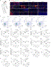
Fig. 1|. Locally rewiring immune cell metabolism in the aPLA biomaterial environment affects CCR2- and CX3CR1-dependent trafficking
a, B6 albino mice were crossed to Ccr2RFP/RFPCx3cr1GFP/GFP mice to generate Ccr2RFP/+Cx3cr1GFP/+ mice, which were surgically incised (sham group) or implanted with biomaterials. Intravital microscopy preceded flow cytometric analysis of tissues around incision sites (sham group) or biomaterials. b, Representative intravital microscopy images around incision sites (sham group), reprocessed aPLA, aPLA incorporating a.a. or 2DG; scale bars, 50 μm. c, Representative CCR2 and CX3CR1 flow cytometry plots gated on live cells. df, Flow cytometry quantification of CCR2+ (d), CX3CR1+ (e) and CCR2+CX3CR1+ (f) cells. gi, Quantification of pro-inflammatory (CD86+CD206) cells among CCR2+ (g), CX3CR1+ (h) and CCR2+CX3CR1+ (i) populations. jl, Quantification of anti-inflammatory (CD206+) cells among CCR2+ (j), CX3CR1+ (k) and CCR2+CX3CR1+ (l) populations. mo, Quantification of transition (CD86+CD206+) cells among CCR2+ (m), CX3CR1+ (n) and CCR2+CX3CR1+ (o) populations. p, Nucleated hematopoietic (CD45+) cells. q,r, Fold change of pro-inflammatory (H1; CD86+CD206) CD45+ cells with respect to transition (T; CD86+CD206+) CD45+ cells (q) or anti-inflammatory (H2; CD206+) CD45+ cells (r). s,t, Fold change of T (s) or H2 (t) cells with respect to H1 CD45+ cells. u, Neutrophils (CD45+Ly6G+ cells). One-way ANOVA followed by Tukey’s or Newman–Keuls’ multiple comparison test, n = 3, mean (s.d.). Panel a created with BioRender.com.
Fig. 2|
Fig. 2|. Polarization states of myeloid cells are regulated by targeting immunometabolism in the aPLA biomaterial microenvironment.
a, Representative flow cytometry plots gated on CD45. b, Monocytes (CD45+CD11b+ cells). c,d, Fold change of pro-inflammatory (M1; CD86+CD206) monocytes with respect to transition (T; CD86+CD206+) monocytes (c) or anti-inflammatory (M2; CD206+) monocytes (d). e,f, Fold change of T (e) or M2 (f) monocytes with respect to M1 monocytes. g, Macrophages (CD45+F4/80+ cells). h,i, Fold change of pro-inflammatory (M1; CD86+CD206) macrophages with respect to transition (T; CD86+CD206+) macrophages (h) or anti-inflammatory (M2; CD206+) macrophages (i). j,k, Fold change of T (j) or M2 (k) macrophages with respect to M1 macrophages. l, Dendritic (CD45+CD11c+) cells. m,n, Fold change of pro-inflammatory (D1; CD86+CD206) dendritic cells with respect to transition (T; CD86+CD206+) dendritic cells (m) or anti-inflammatory (D2; CD206+) dendritic cells (n). o,p, Fold change of T (o) or D2 (p) dendritic cells with respect to D1 dendritic cells. q, Dendritic cells expressing MHCII molecules (CD45+CD11c+MHCII+ cells). r,s, Fold change of pro-inflammatory (D1; CD86+CD206) MHCII+ dendritic cells with respect to transition (T; CD86+CD206+) MHCII+ dendritic cells (r) or anti-inflammatory (D2; CD206+) MHCII+ dendritic cells (s). t,u, Fold change of T (t) or D2 (u) MHCII+ dendritic cells with respect to D1 MHCII+ dendritic cells. One-way ANOVA followed by Tukey’s or Newman–Keuls’ multiple comparison test, n = 3, mean (s.d.); reprocessed aPLA; a.a.; 2DG.
Fig. 3|
Fig. 3|. Using an acid more favourably modulates activation states of immune cells around aPLA biomaterials compared with traditional neutralization techniques.
a, Wild-type B6 mice were surgically incised (sham group) or implanted with reprocessed aPLA, aPLA incorporating a.a., 2DG or HA. Afterwards, flow cytometric analysis of tissues around incision sites (sham group) or biomaterials was undertaken. b, Neutrophils (CD45+Ly6G+ cells). c,d, Fold change of pro-inflammatory (H1; CD86+CD206) cells with respect to transition (T; CD86+CD206+) cells (c) or anti-inflammatory (H2; CD206+) cells (d), gated for nucleated hematopoietic (CD45+) populations. e,f, Fold change of T (e) or H2 (f) cells with respect to H1 cells. g,h, Fold change of pro-inflammatory (M1; CD86+CD206) monocytes (CD45+CD11b+) with respect to transition (T; CD86+CD206+) monocytes (g) or anti-inflammatory (M2; CD206+) monocytes (h). i,j, Fold change of T (i) or M2 (j) monocytes with respect to M1 monocytes. k,l, Fold change of pro-inflammatory (M1; CD86+CD206) macrophages (CD45+F4/80+) with respect to transition (T; CD86+CD206+) macrophages (k) or anti-inflammatory (M2; CD206+) macrophages (l). m,n, Fold change of T (m) or M2 (n) macrophages with respect to M1 macrophages. o, Dendritic (CD45+CD11c+) cells. p,q, Fold change of pro-inflammatory (D1; CD86+CD206) dendritic cells with respect to transition (T; CD86+CD206+) dendritic cells (p) or anti-inflammatory (D2; CD206+) dendritic cells (q). r,s, Fold change of T (r) or D2 (s) dendritic cells with respect to D1 dendritic cells. t, Dendritic cells expressing MHCII molecules (CD45+CD11c+MHCII+ cells). u,v, Fold change of pro-inflammatory (D1; CD86+CD206) MHCII+ dendritic cells with respect to transition (T; CD86+CD206+) MHCII+ dendritic cells (u) or anti-inflammatory (D2; CD206+) MHCII+ dendritic cells (v). w,x, Fold change of T (w) or D2 (x) MHCII+ dendritic cells with respect to D1 MHCII+ dendritic cells. One-way ANOVA followed by Tukey’s multiple comparison test, n = 3, mean (s.d.). Panel a created with BioRender.com.
Fig. 4|
Fig. 4|. Locally targeting immunometabolism in the cPLA environment elevates IL-4-expressing T-cell subsets with differential effects on myeloid populations.
a, Wild-type B6 mice were surgically incised (sham group) or implanted with reprocessed cPLA, cPLA incorporating a.a. or 3PO. Afterwards, flow cytometric analysis of tissues around incision sites (sham group) or biomaterials was undertaken. b, Nucleated hematopoietic (CD45+) cells. c, Neutrophils (CD45+Ly6G+ cells). d, Monocytes (CD45+CD11b+ cells). e, Macrophages (CD45+F4/80+ cells). f, Dendritic (CD45+CD11c+) cells. g, Dendritic cells expressing MHCII molecules (CD45+CD11c+MHCII+ cells). hl, Nucleated hematopoietic cells (h), monocytes (i), macrophages (j), dendritic cells (k) and MHCII+ dendritic cells (l) expressing arginase 1 (Arg1+). m, Cytotoxic T lymphocytes (CD45+CD3+CD8+ cells). n, T helper lymphocytes (CD45+CD3+CD4+ cells). o, T helper 1 cells expressing IFNγ. p, T helper 2 cells expressing IL-4. q, T helper 17 cells expressing interleukin-17A (IL-17A). r, Gamma delta (γδ) T (CD45+CD3+γδ+) cells. su, γδ+ T cells producing IFNγ (s), IL-4 (t) and IL-17A (u). v, Innate lymphoid cells (CD45+CD3+Thy1.2+) producing IL-17A. w, B cells (CD45+CD11bCD19+). One-way ANOVA followed by Tukey’s multiple comparison test, n = 3, mean (s.d.). Panel a created with BioRender.com.

References

    1. Sadtler K et al. Design, clinical translation and immunological response of biomaterials in regenerative medicine. Nat. Rev. Mater. 1, 1–17 (2016).
    1. Whitaker R, Hernaez-Estrada B, Hernandez RM, Santos-Vizcaino E & Spiller KL. Immunomodulatory biomaterials for tissue repair. Chem. Rev. 121, 11305–11335 (2021). - PubMed
    1. Li C et al. Design of biodegradable, implantable devices towards clinical translation. Nat. Rev. Mater. 5, 61–81 (2020).
    1. Narayanan G, Vernekar VN, Kuyinu EL & Laurencin CT. Poly (lactic acid)-based biomaterials for orthopaedic regenerative engineering. Adv. Drug Deliv. Rev. 107, 247–276 (2016). - PMC - PubMed
    1. Serbina NV & Pamer EG. Monocyte emigration from bone marrow during bacterial infection requires signals mediated by chemokine receptor CCR2. Nat. Immunol. 7, 311–317 (2006). - PubMed

LinkOut - more resources