. 2024 Dec;30(5-6):513-523.
doi: 10.1007/s13365-024-01229-4. Epub 2024 Oct 4.

Regulation of voltage-gated sodium channels by TNF-α during herpes simplex virus latency establishment

Affiliations

Regulation of voltage-gated sodium channels by TNF-α during herpes simplex virus latency establishment

Qiaojuan Zhang et al. J Neurovirol. 2024 Dec.

Abstract

During lytic or latent infection of sensory neurons with herpes simplex virus type 1 (HSV-1) there are significant changes in the expression of voltage-gated Na+ channels, which may disrupt the transmission of pain information. HSV-1 infection can also evoke the secretion of various pro-inflammatory cytokines, including TNF-α and IL-6. In this work, we hypothesized that TNF-α regulates the expression of Na+ channels during HSV-1 latency establishment in ND7/23 sensory-like neurons. Latency establishment was mimicked by culturing HSV-1 infected ND7/23 cells in the presence of acyclovir (ACV) for 3 days. Changes in the functional expression of voltage-gated Na+ channels were assessed by whole-cell recordings. Our results demonstrate that infection of ND7/23 cells with the HSV-1 strain McKrae with GFP expression (M-GFP) causes a significant decrease in sodium currents during latency establishment. Exposure of ND7/23 cells to TNF-α during latency establishment reverses the effect of HSV-1, resulting in a significant increase in sodium current density. However, Na+ currents were not restored by 3 day-treatment with IL-6. There were no changes in the pharmacological and biophysical properties of sodium currents promoted by TNF-α, including sensitivity to tetrodotoxin and the current-voltage relationship. TNF-α stimulation of ND7/23 cells increases p38 signaling. Inhibition of p38 signaling with SB203580 or SB202190 eliminates the stimulatory effect of TNF-α on sodium currents. These results indicate that TNF-α signaling in sensory neurons during latency establishment upregulates the expression of voltage-gated Na+ channels in order to maintain the transmission of pain information.

Keywords: Electrical excitability; Pain; Sensory neuron; Sodium channel; Tumor necrosis factor-alpha.


Conflict of interest statement

Declarations. Conflicts of interest: The authors declare no potential conflicts of interest regarding the research, authorship, and/or publication of this article.

Figures

Fig. 1
Fig. 1
Morphology of differentiated ND7/23 cells following M-GFP infection. (A) Differentiated ND7/23 cells present long dendritic processes. (B, a-b) Culture of differentiated ND7/23 cells with acyclovir (ACV, 100 μM) for up to 4 d does not affect cell morphology or GFP expression. (C, a-b) Lytic infection of differentiated ND7/23 with M-GFP causes significant cell death and a reduction in dendritic outgrowth and GFP expression. (D-E, a-b) ACV-induced HSV-1 latency establishment in the presence or absence of TNF-α had minimal effect on dendritic outgrowth while showing GFP labeling of infected neurons. The arrows in Ca-b, Da-b, and Ea-b identify GFP-labeled cells
Fig. 2
Fig. 2
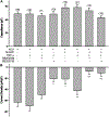
Effect of M-GFP infection on Na+ currents in differentiated ND7/23 cells. (A) Typical family of Na+ currents generated in a differentiated ND7/23 following a series of voltage steps from a holding potential of −100 mV. Recordings from control (non-treated) and treated cells were performed at the end of the 3 d period following latency establishment. In this and subsequent figures, the voltage step protocol is shown below the current trace. (B) Comparison of cell capacitance in differentiated ND7/23 cells following M-GFP infection and treatment with ACV, TNF-α, or IL-6. Note that infection of differentiated ND7/23 with M-GFP had no significant effect on cell capacitance even in the presence of ACV or TNF-α. However, following latency establishment and treatment with IL-6 there is a significant increase in cell capacitance (** denotes p ≤ 0.05 vs. ACV pre-treated cells). The number of cells recorded under each condition is presented in parenthesis from at least 3 different cell cultures. (C) Mean Na+ current densities generated in differentiated ND7/23 cells following M-GFP infection (with or without ACV) and treatment with TNF-α or IL-6. Na+ current densities were calculated from the peak current amplitude generated by depolarizing voltage steps from a holding potential of −100 mV (* denotes p ≤ 0.05 vs. control (non-treated) cells; ** denotes p ≤ 0.05 vs. ACV pre-treated cells; *** denotes p ≤ 0.05 vs. M-GFP infected cells pre-treated with ACV; ns denotes no significant difference). (D-E) Density plot of Na+ current densities generated in differentiated ND7/23 cells during latency establishment with or without TNF-α. Treatment of ND7/23 cells with TNF-α during latency establishment caused an increase in current densities in the range of 1 to < 100 or > 100 pA/pF. (F) Current-voltage relationship in ACV pre-treated cells and following latency establishment and treatment with TNF-α. There is no significant shift in the voltage dependence of Na+currents following treatment with TNF-α compared to ACV pre-treated cells
Fig. 3
Fig. 3
Effect of TTX on the Na+ currents generated in differentiated ND7/23 cells following latency establishment. (A) Examples of whole-cell Na+ currents generated in a differentiated ND7/23 cell following latency establishment and treatment with TNF-α in the presence of TTX. Note that the inward Na+ current generated in differentiated ND7/23 cells following latency establishment and treatment with TNF-α was eliminated by TTX (250 nM, arrow). (B) Comparison of cell capacitance in differentiated ND7/23 cells following latency establishment and treatment with TTX. (C) Overall effect of TTX on Na+ current densities in control (ACV-treated) cells and cells latently infected cells treated with TNF-α. TTX evokes a significant reduction in the current densities under all conditions tested. (* denotes p < 0.05 vs. ACV-treated cells; ** denotes p < 0.05 vs. ACV + M-GFP + TNF-α treated cells)
Fig. 4
Fig. 4
Effect of latency establishment and TNF-α treatment on the expression of different sodium channel Nav transcripts as assessed by real-time PCR analysis. (A-F) Effect of ACV, ACV + TNF-α, latency establishment, and latency establishment with TNF-α treatment on the transcript expression of the Nav1.1 (A), Nav1.2 (B), Nav1.3 (C), Nav1.6 (D), and Nav1.7 (E) channel subunits. In ACV-treated cells, exposure of differentiated ND7/23 cells to TNF-α evoked a significant reduction in the expression of Nav1.3 and Nav1.7 channel transcripts (* denotes p < 0.05 vs. ACV-treated cells). Latency establishment (following ACV pre-treatment and M-GFP infection) resulted in a reduction in Nav1.7 transcripts compared to ACV-treated cells. Latency establishment followed by TNF-α treatment evokes a significant increase in the expression of the Nav1.1, Nav1.2, and Nav1.6 mRNA compared with latently infected cells. (* denotes p < 0.05 vs. M-GFP + ACV treated cells)
Fig. 5
Fig. 5
Effect of latency establishment and TNF-α treatment on viral replication and p38 signaling. (A) M-GFP infection of differentiated ND7/23 cells generates an increase in the expression of TK viral transcripts, which is reversed by ACV treatment. TNF-α does not have any effect on TK expression following latency establishment (* denotes p ≤ 0.05 vs. M-GFP-infected cells; n = 4). (B) TNF-α treatment of differentiated ND7/23 cells evokes a significant increase in p38 signaling, without altering the level of sodium channel expression as detected with a pan-Na channel antibody. (C) Quantification of changes in p38 activation following latency establishment and TNF-α treatment (* denotes p ≤ 0.05 vs. ACV-treated cells; n = 4)
Fig. 6
Fig. 6
Effect of TNF-α signaling on the functional expression of Na+ channels in differentiated ND7/23 cells following latency establishment. (A) TNF-α stimulation of differentiated ND7/23 cells following ACV treatment or latency establishment does not affect cell capacitance. The p38 inhibitors SB203580 (10 μM) and SB202190 (10 μM) evoked no significant changes in cell capacitance under any treatment conditions. (B) Latency establishment (ACV + M-GFP) causes a significant reduction in the Na+ current densities compared to ACV-treated cells (* denotes p < 0.05 vs. ACV-treated cells). TNF-α stimulation of differentiated ND7/23 cells following latency establishment (ACV + M-GFP) evoked a significant increase in Na+ current densities compared to ACV + M-GFP-treated cells (** denotes p < 0.05 vs. ACV + M-GFP-treated cells). However, the stimulatory effect of TNF-α on Na+ channels during latency establishment was eliminated following treatment with SB203580 or SB202190 (*** denotes p < 0.05 vs. ACV + M-GFP + TNF-α treated cells)

Similar articles

References

    1. Andoh T, Shiraki K, Kurokawa M (1995) Paresthesia induced by cutaneous infection with herpes simplex virus in rats. Neurosci Lett 190(2):101–104 - PubMed
    1. Azher T, Yin X, Stuart P (2017) Understanding the role of chemokines and cytokines in experimental models of herpes simplex keratitis. J. Immunol. Res 2017:7261980. - PMC - PubMed
    1. Chen X, Pang RP, Shen KF, Zimmermann M, Xin WJ, Li YY, Liu XG (2011) TNF-α enhances the currents of voltage gated sodium channels in uninjured dorsal root ganglion neurons following motor nerve injury. Exp Neurol 227(2):279–286 - PubMed
    1. Czeschik JC, Hagenacker T, Schäfers M, Büsselberg D (2008) TNF-alpha differentially modulates ion channels of nociceptive neurons. Neurosci Lett 434(3):293–298 - PubMed
    1. David AT, Saied A, Charles A, Subramanian R, Chouljenko VN, Kousoulas KG (2012) A herpes simplex virus 1 (McKrae) mutant lacking the glycoprotein K gene is unable to infect via neuronal axons and egress from neuronal cell bodies. MBio 3(4):e00144–e00112 - PMC - PubMed

Publication types

MeSH terms

LinkOut - more resources