. 2024 Oct 4;24(1):535.
doi: 10.1186/s12872-024-04192-4.

Quercetin regulates pulmonary vascular remodeling in pulmonary hypertension by downregulating TGF-β1-Smad2/3 pathway

Affiliations

Quercetin regulates pulmonary vascular remodeling in pulmonary hypertension by downregulating TGF-β1-Smad2/3 pathway

Rui-Juan Gao et al. BMC Cardiovasc Disord. .

Abstract

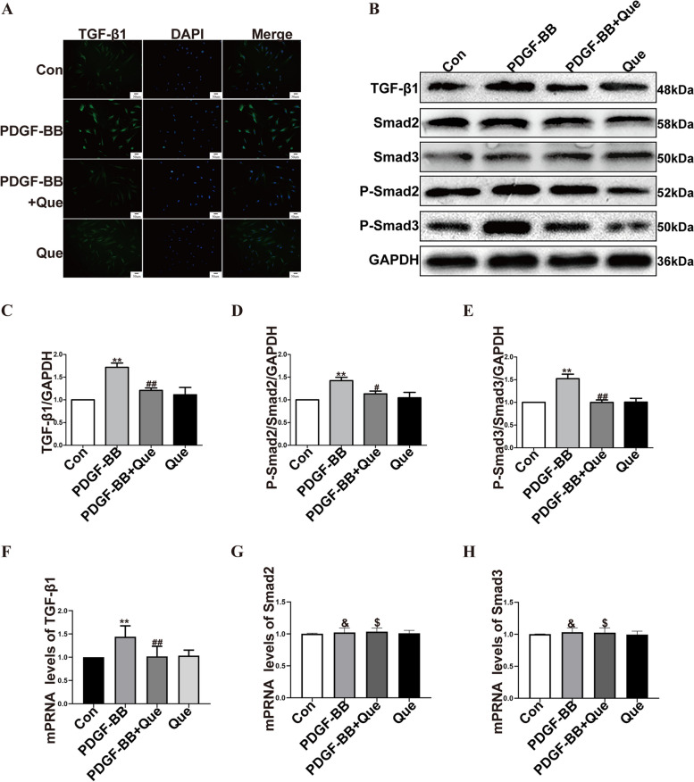
Background: Pulmonary arterial hypertension (PAH) is a worldwide challenging disease characterized by progressive elevation of pulmonary artery pressure. The proliferation, migration and phenotypic transformation of pulmonary smooth muscle cells are the key steps of pulmonary vascular remodeling. Quercetin (3,3', 4', 5, 6-pentahydroxyflavone, Que) is a natural flavonol compound that has antioxidant, anti-inflammatory, anti-tumor and other biological activities. Studies have shown that Que has therapeutic effects on PAH. However, the effect of quercetin on pulmonary vascular remodeling in PAH and its mechanism remain unclear.

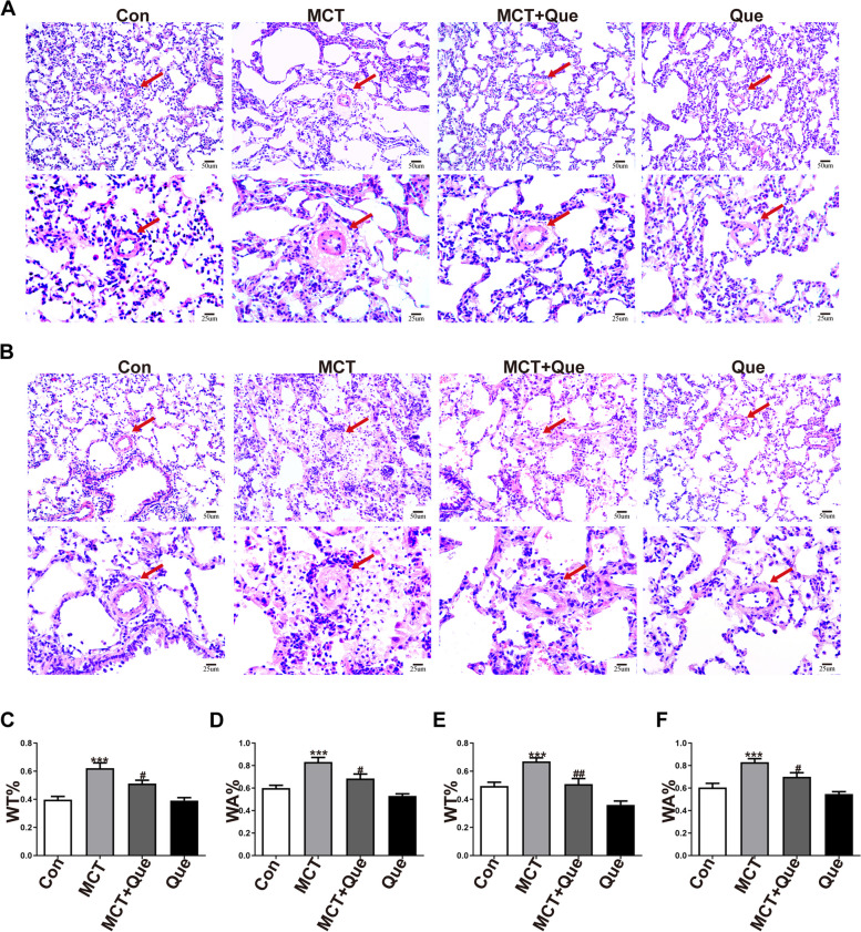
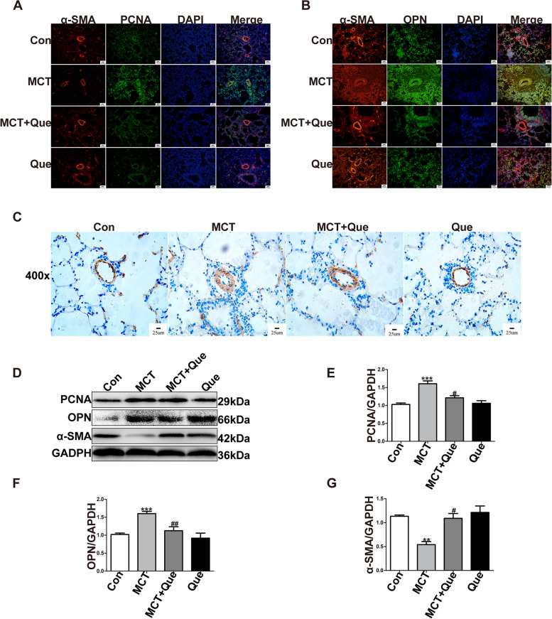
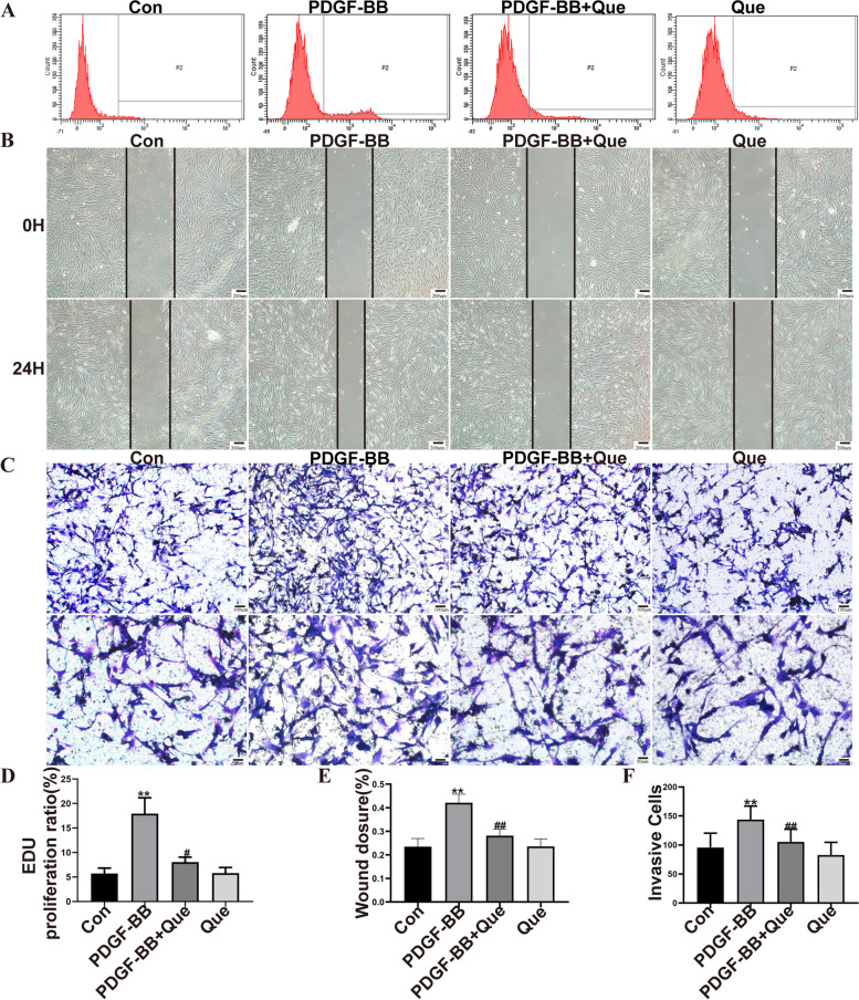
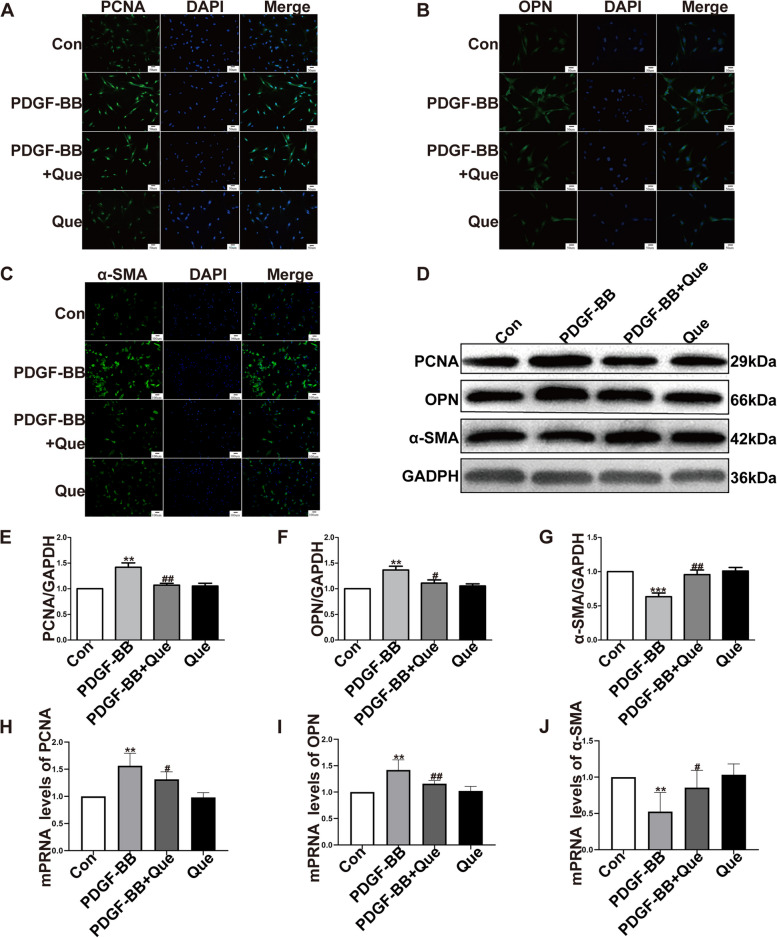
Methods and results: In vivo, PAH rats were constructed by intraperitoneal injection of monocrotaline (MCT) at 60 mg/kg. Human pulmonary artery smooth muscle cells (HPASMCs) were treated with platelet-derived growth factor BB (PDGF-BB) 20 ng/mL to construct PAH cell model in vitro. The results showed that in vivo studies, MCT could induce right ventricular wall hyperplasia, narrow the small and medium pulmonary artery cavity, up-regulate the expression of proliferating and migration-related proteins proliferating cell nuclear antigen (PCNA) and osteopontin (OPN), and down-regulate the expression of alpha-smooth muscle actin (α-SMA). Que reversed the MCT-induced results. This process works by down-regulating the transforming growth factor-β1 (TGF-β1)/ Smad2/3 signaling pathway. In vitro studies, Que had the same effect on PDGF-BB-induced proliferation and migration cell models.

Conclusions: Que inhibits the proliferation, migration and phenotypic transformation of HPASMCs by down-regulating TGF-β1/Smad2/Smad3 pathway, thereby reducing right ventricular hyperplasia (RVH) and pulmonary vascular remodeling, providing potential pharmacological and molecular explanations for the treatment of PAH.

Keywords: Migration; PAH; Proliferation; Que; TGF-β1.


Conflict of interest statement

The authors declare no competing interests.

Figures

Fig. 1
Fig. 1
Que improves hemodynamics and right ventricular remodeling in PAH rats. A The pulmonary haemodynamic spectrum; B Cardiac cross-sectional HE results; C The comparison of PAAT; D The comparison of mPAP; E The comparison of RVHI%. MCT vs CON, ** P < 0.01; MCT + Que vs MCT, # P < 0.05, ## P < 0.01. (n = 6)
Fig. 2
Fig. 2
Que improves pulmonary artery remodeling in PAH rats. A HE staining of lung tissue and pulmonary small artery at × 200 and × 400; B He staining of lung tissue and medium pulmonary artery at × 200 and × 400; C The statistical analysis of the small pulmonary artery’s WT%; D The statistical analysis of the small pulmonary artery’s WA%; E The statistical analysis of the medium pulmonary artery’s WT%; F The statistical analysis of the medium pulmonary artery’s WA%. The arrow points to the pulmonary artery. MCT vs CON, *** P < 0.001; MCT + Que vs MCT, # P < 0.05, ## P < 0.01. (n = 6)
Fig. 3
Fig. 3
Effects of Que on the distribution and expression of PCNA, OPN α- SMA in pulmonary blood vessels of MCT-induced PAH rats. A Immunofluorescence of α-SMA and PCNA at × 200; B Immunofluorescence of α-SMA and OPN at × 200; C Immunohistochemical expression of α- SMA at × 400; D Western blotting of PCNA, OPN, α-SMA and GAPDH; E Analysis of relative expression of PCNA by Western blot; F Analysis of relative expression of OPN by Western blot;  G Analysis of relative expression of α- SMA by Western blot. MCT vs CON, ** P < 0.01, *** P < 0.001; MCT + Que vs MCT, # P < 0.05, ## P < 0.01. (n = 3 to 6)
Fig. 4
Fig. 4
Effects of Que on the distribution and expression of TGF-β1-Smad2/3 singal patnway in pulmonary blood vessels of MCT-induced PAH rats. A Immunohistochemical expression of TGF-β1 at × 400; B Western blotting of TGF-β1, Smad2/3, p-Smad2/3 and GAPDH; C Analysis of relative expression of TGF-β1 by Western blot; D Analysis of relative expression of p-Smad2/ Smad2 by Western blot; E Analysis of relative expression of p-Smad3/ Smad3 by Western blot. MCT vs CON, * P < 0.05, ** P < 0.01; MCT + Que vs MCT, # P < 0.05. (n = 3 to 6)
Fig. 5
Fig. 5
Effect of Que on proliferation, migration and phenotypic transformation of HPASMCs induced by PDGF-BB in vitro. A HPASMCs proliferation was detected by flow cytometry (EDU); B The migration ability of HPASMCs was tested by scratch test at × 50; C Transwell ™ cell crystal violet staining was used to detect the migration of HPASMCs at × 100 and × 200; D HPASMCs proliferation rate was analyzed by flow cytometry (EDU); E The migration ratio of scratch was quantitatively analyzed; F Cell invasion relative quantitative analysis of migration ratio. PDGF-BB vs CON, ** P < 0.01; PDGF-BB + Que vs PDGF-BB, # P < 0.05, ## P < 0.01. (n = 3 to 6)
Fig. 6
Fig. 6
Effects of Que on PCNA, OPN and α-SMA distribution and expression in PDGF-BB induced HPASMCs. A Immunofluorescence of PCNA of HPASMCs at × 200; B Immunofluorescence of OPN of HPASMCs × 200; C Immunofluorescence of α-SMA of HPASMCs at × 200; D Western blotting of PCNA, OPN, α-SMA and GAPDH of HPASMCs; E Analysis of relative expression of PCNA by Western blot; F Analysis of relative expression of OPN by Western blot; G Analysis of relative expression of α-SMA by Western blot; H Analysis of relative expression of mRNA levels of PCNA; I Analysis of relative expression of mRNA levels of OPN; J Analysis of relative expression of mRNA levels of α-SMA. PDGF-BB vs CON. ** P < 0.01, *** P < 0.001; PDGF-BB + Que vs PDGF-BB, # P < 0.05, ## P < 0.01. (n = 3 to 6)
Fig. 7
Fig. 7
Effects of Que on TGF-β1-Smad2/3 signal pathway distribution and expression in PDGF-BB induced HPASMCs. A Immunofluorescence of TGF-β1 of HPASMCs at × 200; B Western blotting of TGF-β1, Smad2/3, p-Smad2/3 and GAPDH of HPASMCs; C Analysis of relative expression of TGF-β1 by Western blot; D Analysis of relative expression of p-smad3/Smad2 by Western blot; E Analysis of relative expression of p-smad3/Smad3 by Western blot; F Analysis of relative expression of mRNA levels of TGF-β1; G Analysis of relative expression of mRNA levels of Smad2; H Analysis of relative expression of mRNA levels of Smad3. PDGF-BB vs CON, ** P < 0.01,& P> 0.05; PDGF-BB + Que vs PDGF-BB, # P < 0.05, ## P < 0.01, $ P> 0.05. (n = 3 to 6)

Similar articles

Cited by

References

    1. Jose A, Eckman MH, Elwing JM. Anticoagulation in pulmonary arterial hypertension: a decision analysis. Pulm Circ. 2019;9:2045894019895451. - PMC - PubMed
    1. Lei S, Peng F, Li ML, et al. LncRNA-SMILR modulates RhoA/ROCKsignaling by targeting miR-141 to regulate vascular remodeling in pulmonaryarterial hypertension. Am J Physiol Heart Circ Physiol. 2020;319:H377–91. - PubMed
    1. Bisserier M, Mathiyalagan P, Zhang S, et al. Regulation ofthe methylation and expression levels of the BMPR2 Gene by SIN3a as a Novel Therapeutic Mechanism in Pulmonary Arterial Hypertension. Circulation. 2021;144:52–73. - PMC - PubMed
    1. Courboulin A, Barrier M, Perreault T, et al. Plumbaginreverses proliferation and resistance to apoptosis in experimental PAH. EurRespir J. 2012;40:618–29. - PubMed
    1. Wang SC. PCNA: a silent housekeeper or a potential therapeutic target. Trends Pharmacol Sci. 2014;35:178–86. - PubMed

MeSH terms

LinkOut - more resources