Review
. 2024 Oct 4;25(1):932.
doi: 10.1186/s12864-024-10847-5.

A decade of dinoflagellate genomics illuminating an enigmatic eukaryote cell

Affiliations
Review

A decade of dinoflagellate genomics illuminating an enigmatic eukaryote cell

Senjie Lin. BMC Genomics. .

Abstract

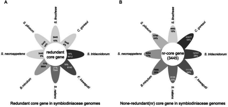
Dinoflagellates are a remarkable group of protists, not only for their association with harmful algal blooms and coral reefs but also for their numerous characteristics deviating from the rules of eukaryotic biology. Genome research on dinoflagellates has lagged due to their immense genome sizes in most species (~ 1-250 Gbp). Nevertheless, the last decade marked a fruitful era of dinoflagellate genomics, with 27 genomes sequenced and many insights attained. This review aims to synthesize information from these genomes, along with other omic data, to reflect on where we are now in understanding dinoflagellates and where we are heading in the future. The most notable insights from the decade-long genomics work include: (1) dinoflagellate genomes have been expanded in multiple times independently, probably by a combination of rampant retroposition, accumulation of repetitive DNA, and genome duplication; (2) Symbiodiniacean genomes are highly divergent, but share about 3,445 core unigenes concentrated in 219 KEGG pathways; (3) Most dinoflagellate genes are encoded unidirectionally and are not intron-poor; (4) The dinoflagellate nucleus has undergone extreme evolutionary changes, including complete or nearly complete loss of nucleosome and histone H1, and acquisition of dinoflagellate viral nuclear protein (DVNP); (5) Major basic nuclear protein (MBNP), histone-like protein (HLP), and bacterial HU-like protein (HCc) belong to the same protein family, and MBNP can be the unifying name; (6) Dinoflagellate gene expression is regulated by poorly understood mechanisms, but microRNA and other epigenetic mechanisms are likely important; (7) Over 50% of dinoflagellate genes are "dark" and their functions remain to be deciphered using functional genetics; (8) Initial insights into the genomic basis of parasitism and mutualism have emerged. The review then highlights functionally unique and interesting genes. Future research needs to obtain a finished genome, tackle large genomes, characterize the unknown genes, and develop a quantitative molecular ecological model for addressing ecological questions.

Keywords: Dinoflagellate; Gene regulation; Genome expansion; Histone; MicroRNA (miRNA); Nuclear protein; Omics; Parasitic; Retroposition; Symbiotic.


Conflict of interest statement

The authors declare no competing interests.

Figures

Fig. 1
Fig. 1
Light micrographs of dinoflagellates and a bioluminescence scene (A-G) and phylogenetic relationship of dinoflagellates, apicomplexa, and other alveolates (H). A. Tripos platycorne. B. Tripos muelleri. C. Erythropsidinium agile. The arrow points at the ocelloid. D. Gymnodinium catenatum. E. Gessnerium (=Alexandrium) monilatum. F. Prorocentrum micans. Scale bars = 20 µm. G. Bioluminescence by Noctiluca scintillans at Zhejiang, China. Images A-F courtesy of Dr. Fernando Gómez; all in apex facing up view except E and F being lateral view. Image G courtesy of Dr. Hao Luo. In H), core dinoflagellates (a) include naked dinoflagellate with two flagella (longitudinal and transverse) and thecate dinoflagellate with two flagella (longitudinal and transverse) and the conspicuous thecal plates. Non-core dinoflagellates include Marine Alveolate Group I (MAGI, euduboscquellids), Syndiniales (also known as MAGII), and the basal lineages Oxyrrhis and Noctiluca. Modified from Lin 2011 with up-to-date information
Fig. 2
Fig. 2
Distribution pattern of telomeres in a tertiary endosymbiosis dinoflagellate and schematic of TADs in dinoflagellate chromosomes and associated gene distribution and expression machinery. A Chromosomes of Karenia brevis stained with fluorescent in situ hybridization of telomere probe. B DNA DAPI stain (Blue) and telomere FISH (red) in F. kawagutii. C TAD organization highlighting gene distribution and gene expression machinery. Reproduced with permission from Cuadrado et al. 2019 [37] (A), Lin et al. 2015 [18] (B), and Lin et al. 2021 [33] (C)
Fig. 3
Fig. 3
Evolutionary dynamics of dinoflagellate genome size showing no phylogenetic trend (A) and double peak in 21-mer profile of Polarella glacialis showing evidence of genome duplication (B). A Vertical arrow indicates phylogenetic trend from the dinoflagellate sister group perkinsids, basal dinoflagellate Oxyrrhis, to later diverging taxa. Size of oval in the middle illustrates genome size (not to scale) as depicted by numbers shown on the right. B Reproduced from Stephans et al. 2020 [21] under the terms of the Creative Commons CC BY license
Fig. 4
Fig. 4
Phylogenetic tree of major basic nuclear protein (MBNP), histone-like protein (HLP), and bacterial DNA binding protein (HU)-like protein (Hcc). The tree is rooted with bacterial DNA binding protein (HU). The lack of monophyletic separation indicates that these three proteins belong to the same protein family under different names. Species name abbreviations: Abor, Alcanivorax borkumensis; Acar, Amphidinium carterae; Bmin, Breviolum minutum; Cbur, Coxiella burnetiid; Ccoh, Crypthecodinium cohnii; Cgor, Cladocopium goreaui; Fkaw, Fugacium kawagutii; Gcat, Gymnodinium catenatum; Kven, Karlodinium veneficum; Nsci, Noctiluca scintillans; Pgla, Polarella glacialis; Pmin, Prorocentrum minimum; Ppis, Pfiesteria piscicida; Pshi, Prorocentrum shikokuence; Rbal, Rhodopirellula baltica; Sacu, Scrippsiella acuminata; Smic, Symbiodinium microadriacticum; Snat, Symbiodinium natans; Stri, Symbiodinium tridacnidorum
Fig. 5
Fig. 5
Shared and unique genes in Symbiodiniaceae genomes. A Core genes (3,445 after de-redundancy) in proportion of total genes in each species. B Unique genes in each species. Darker colors indicate a larger number of genes
Fig. 6
Fig. 6
Organization of cold shock domain (CSD) in various proteins in dinoflagellates. Amphidinium, A. massarti, data source MMETSP0689; CSD2, domain PS51857; CSD8, domain SM00357; CmR, Calcium regulated mRNA-binding domain (IPR052069); Gymnodinium, G. catenatum, data source MMETSP0784-20121206; Smic, Symbiodinium microadriaticum, data source CAE7767176.1 dbp2, Helicase = helicase superfamily ATP-binding domain; Sym_CCMP2592, Symbiodinium strain CCMP2592; Fkaw38550, Fugacium kawagutii, BEST = bestrophin
Fig. 7
Fig. 7
Evidence that Symbiodiniaceae miRNA may interact with coral host to modulate host gene expression. A Phylogenetic tree of double-stranded RNA channel (SID-1). The close relationship between the homologs in Symbiodinium and the cnidarians suggests horizontal gene transfer between the two lineages. B Protein interaction network and metabolic pathways predicted to be impacted by miRNA. C GO categorization of predicted miRNA target genes in both F. kawagutii and coral
Fig. 8
Fig. 8
Correlation between ribosomal rRNA and ribosome-bound mRNA abundances in Lingulodinium polyedra. Data from Bowazolo et al. (2022) [129]
Fig. 9
Fig. 9
Comparison of dinoflagellate photosystem (PS) components with counterparts in other algal lineages. A PSI. Reproduced from Lin et al. 2024 [149]. B PSII. Information source: Fromme et al. 2001 [150], Pi et al. 2018 [151], Qin et al. 2019 [152], Gisriel et al. 2022 [153], Su et al. 2019, 2022 [154, 155], Zhao et al. 2023 [156], Li et al. 2024 [82]
Fig. 10
Fig. 10
Two episodes of retroposition discovered in two Symbiodiniacea genomes coincident with two major periods of symbiosis evolution. A) Fugacium kawagutii. B) Breviolum minutum. a, first episode led to expansion of sugar and fatty acid metabolism, oxidative stress response, and transport. b, second episode that promoted photosynthesis and adhesion. Based on Song et al. 2017 [49]
Fig. 11
Fig. 11
Macrophage migration inhibitory factors (MIF) detected in dinoflagellates. A Schematic of MIF structure. B Cell surface localization of MIF in L. polyedra (green fluorescence pointed by arrows). From Jaouannet et al. 2020 [205]. C Phylogenetic tree showing clustering of dinoflagellate MIF with homologs from prasinophytes and other lineages. D Alignment of dinoflagellate MIFs with humans MIF showing high similarity. Homsa, Homo sapiens; Alemo, Alexandrium monilatum; Fugka, Fugacium kawagutii; Linpo, Lingulodinium polyedra; SymbB, Breviolum sp.; Symmi, Symbiodinium microadriaticum

Similar articles

Cited by

References

    1. Hackett JD, Anderson DM, Erdner DL, Bhattacharya D. Dinoflagellates: a remarkable evolutionary experiment. Am J Bot. 2004;91(10):1523–34. - PubMed
    1. Lin S. Genomic understanding of dinoflagellates. Res Microbiol. 2011;162(6):551–69. - PubMed
    1. Fensome RA. A classification of living and fossil dinoflagellates. Micropaleontology Spec Publ. 1993;7:351.
    1. Gómez F. A checklist and classification of living dinoflagellates (Dinoflagellata, Alveolata). Cicimar Oceánides. 2012;27(1):65–140.
    1. Logares R, Shalchian-Tabrizi K, Boltovskoy A, Rengefors K. Extensive dinoflagellate phylogenies indicate infrequent marine–freshwater transitions. Mol Phylogenet Evol. 2007;45(3):887–903. - PubMed

LinkOut - more resources