. 2024 Oct 4;22(1):474.
doi: 10.1186/s12964-024-01836-y.

GZMA suppressed GPX4-mediated ferroptosis to improve intestinal mucosal barrier function in inflammatory bowel disease

Affiliations

GZMA suppressed GPX4-mediated ferroptosis to improve intestinal mucosal barrier function in inflammatory bowel disease

Rongwei Niu et al. Cell Commun Signal. .

Abstract

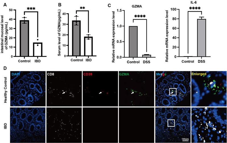
Background: Our previous study has demonstrated a decreased colonic CD8+CD39+ T cells, enrichment of granzyme A (GZMA), was found in pediatric-onset colitis and inflammatory bowel disease (IBD) characterized by impaired intestinal barrier function. However, the influence of GZMA on intestinal barrier function remains unknown.

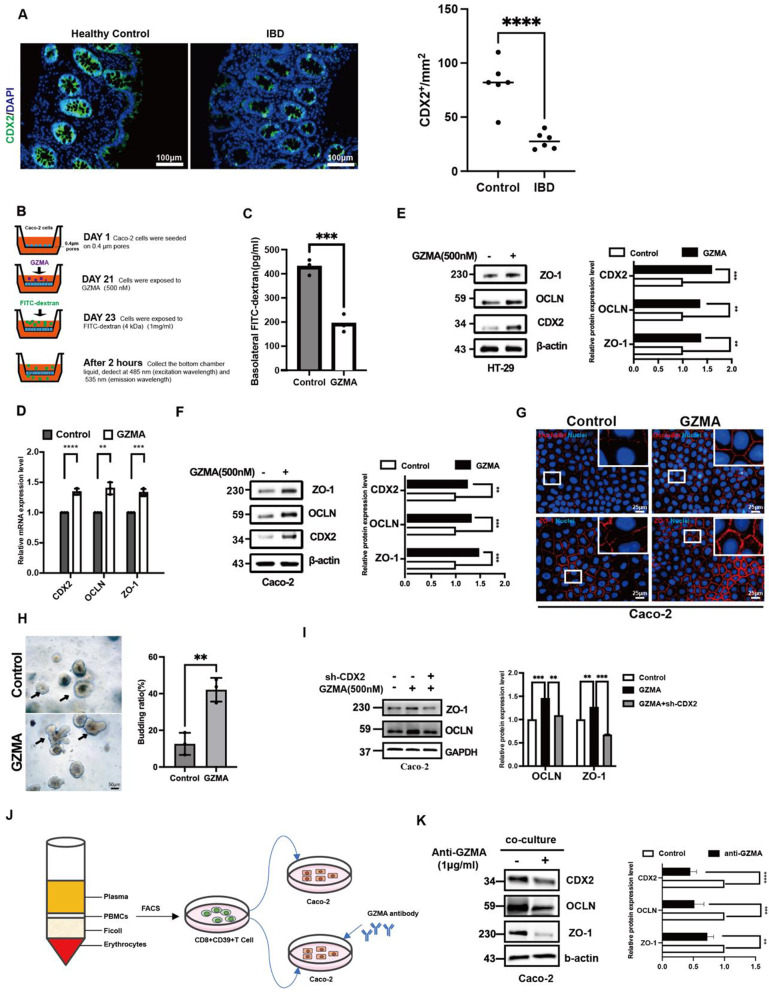
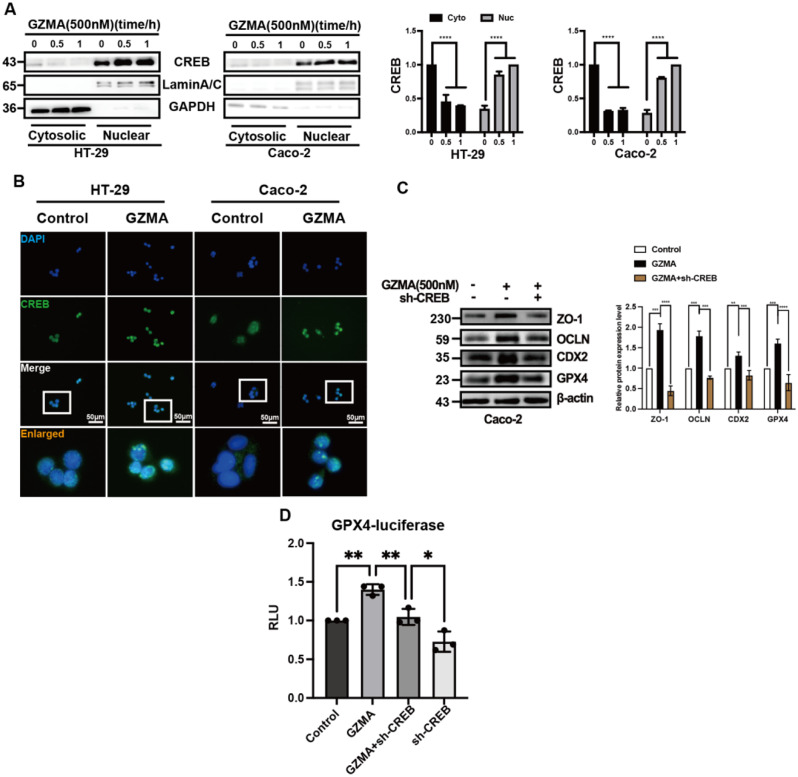
Methods: Western blotting(WB), real-time PCR (qPCR), immunofluorescence (IF) and in vitro permeability assay combined with intestinal organoid culture were used to detect the effect of GZMA on intestinal epithelial barrier function in vivo and in vitro. Luciferase, immunoprecipitation (IP) and subcellular fractionation isolation were performed to identify the mechanism through which GZMA modulated intestinal epithelial barrier function.

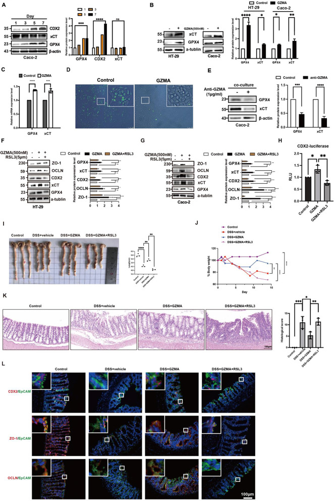
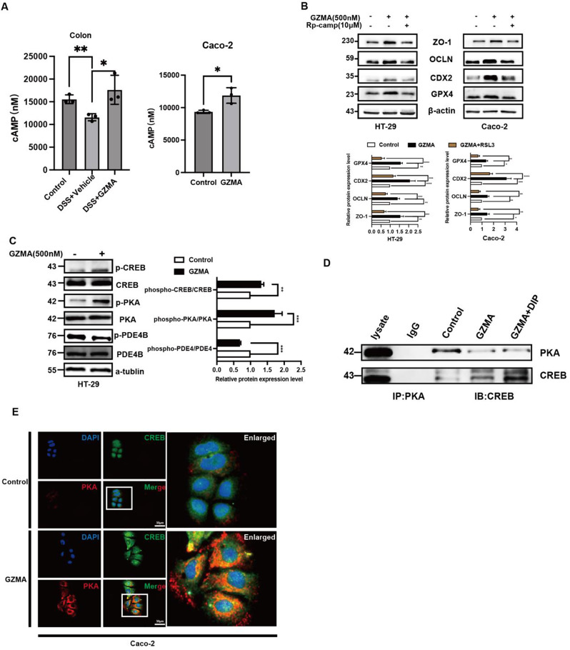
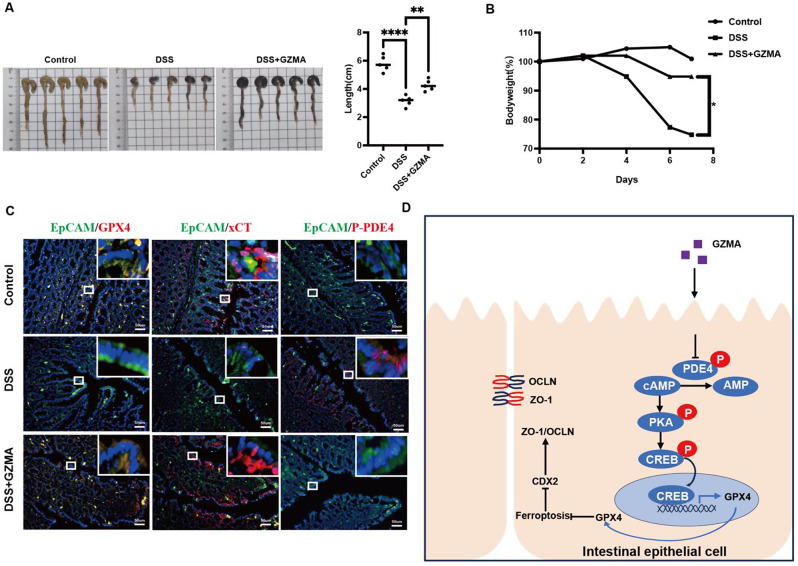
Results: Herein, we, for the first time, demonstrated that CD8+CD39+ T cells promoted intestinal epithelial barrier function through GZMA, leading to induce Occludin(OCLN) and Zonula Occludens-1(ZO-1) expression, which was attributed to enhanced CDX2-mediated cell differentiation caused by increased glutathione peroxidase 4(GPX4)-induced ferroptosis inhibition in vivo and in vitro. Mechanically, GZMA inhibited intestinal epithelial cellular PDE4B activation to trigger cAMP/PKA/CREB cascade signaling to increase CREB nuclear translocation, initiating GPX4 transactivity. In addition, endogenous PKA interacted with CREB, and this interaction was enhanced in response to GZMA. Most importantly, administration of GZMA could alleviate DSS-induced colitis in vivo.

Conclusion: These findings extended the novel insight of GZMA contributed to intestinal epithelial cell differentiation to improve barrier function, and enhacement of GZMA could be a promising strategy to patients with IBD.

Keywords: CDX2; Ferroptosis; GZMA; IBD; Intestinal barrier function.


Conflict of interest statement

The authors declare no competing interests.

Figures

Fig. 1
Fig. 1
Decreased GZMA in patients with IBD and DSS-induced colitis. (A-B) The level of GZMA in serum and colonic mucosa were measured by ELISA according to the instruction. Data was displayed as the means ± s.d. of three independent experiments and analyzed by two-sample t test for significance, ***p < 0.001, **p < 0.01. (C) Real-time PCR was employed to assess colonic GZMA and IL-6 mRNA level in indicated group. Data was exhibited as the means ± s.d. of three independent experiments and analyzed by one-sample t-test for significance, ****p < 0.0001. (D) Immunofluorescence assay was performed to detect CD8, CD39, and GZMA expression in indicated group (Scale bar: 100 μm)
Fig. 2
Fig. 2
GZMA promoted intestinal epithelial cell differentiation. (A) Immunofluorescence assay was performed to detect CDX2 expression of colonic mucosa from clinical sample (Scale bar: 100 μm), quantitation was performed by Image J, and analyzed by two-sample t test, ∗∗∗p < 0:001. (B-C) Caco-2 cells were seeded onto transwell polycarbonate membranes (0.4 μm pores). Upon confluence (21 days after seeding), the cells were treated with GZMA (500 nM) for up to 48 h. The permeability of the monolayer to FITC-dextran (4 kDa) was assessed by measuring the fluorescence intensity in the bottom chamber at Ex/Em = 485/535 nm. Data was displayed as means ± s.d. of three independent experiments and analyzed by two-sample t test for significance, ***p < 0.001.(D) Real-time PCR and (E-F) western blotting as well as (G) immunofluorescence staining were conducted to analyze the indicated gene expression at mRNA and protein level in HT-29 and Caco-2 cells treated with or without GZMA (500 nM) for 48 h (Scale bar: 25 μm), Data was showed as the means ± s.d. of three independent experiments and quantified by one-sample t test for significance, ***p < 0.001, **p < 0.01, *p < 0.05. (H) intestinal crypt isolated from mice was used to explore the effect of GZMA (500 nM) on intestinal organoid generation, microscopic examination of organoids was employed to calculate the proportion of budding organoids among every average 100 organoids. Data was exhibited as means ± s.d. of three independent experiments and analyzed by two sample t test, **p < 0.01 (Scale bar: 50 μm). (I) western blotting was conducted to analyze the indicated proteins in Caco-2 cells after transferred with sh-CDX2 plasmid, followed by stimulation with GZMA (500 nM) for 48 h, with β-actin serving as the internal control. (J-K) CD8+CD39+ T cell subsets isolated from peripheral blood of healthy donors were cultured in medium for 24 h was collected to co-culture with Caco-2 cells for 48 h combined with or without GZMA antibody supplementation. The total lysate was harvested to detect indicated proteins, Data was displayed as mean ± s.d. of three independent experiments and analyzed by one-sample t test for significance, ***p < 0.001, **p < 0.01
Fig. 3
Fig. 3
GZMA promoted intestinal epithelial cell differentiation through inhibition of ferroptosis. (A) Lysate was extracted from Caco-2 at day 1/3/5/7 post-confluence, and the expression of target proteins were analyzed by western blotting. Data was represented the mean ± s.d. of three independent experiments and determined by one-way ANOVA for significance, ****p < 0.0001, ***p < 0.001. HT-29 and Caco-2 cells were treated with GZMA (500 nM) for 48 h, WB (B) and qPCR (C) were employed to determine indicated gene at mRNA and protein level. The control was normalized as 1. The band intensity and relative mRNA expression was represented as mean ± s.d. of three independent experiments and analyzed by one-sample t test for significance, ***p < 0.001, **p < 0.01, *p < 0.05. (D) After serum starvation for 24 h, Caco-2 cells were treated with GZMA (500 nM) for 48 h and the ROS level was detected. (E) immunoblotiting was performed to detect indicated protein in coculture model established by CD39+CD8+ T cell and Caco-2 cells combined with or without anti-GZMA addition, Data was displayed as the mean ± s.d. of three independent experiments and analyzed using one sample t test for significance, ****p < 0.0001, ***p < 0.001. The control was normalized to 1. (F-G) HT-29 and Caco-2 cells were treated with GZMA (500 nM) combined with or without RSL3 (5 µM) for 48 h, WB was conducted to analyze the indicated protein. Data was presented as the means ± s.d. of three independent experiments and analyzed by one-way ANOVA and Dunnett’s multiple comparison test, ****p < 0.0001, ***p < 0.001, **p < 0.01, *p < 0.05. (H) After co-transfection with CDX2-luc plasmid and a control renilla luciferase vector for 12 h, 293T cells were treated with GZMA (500 nM) or GZMA (500 nM) combined with RSL3 (5 µM) for 48 h, the relative luciferase unit (RLU) was measured. Data was presented as the means ± s.d. of three independent experiments and analyzed by one-way ANOVA, *p < 0.05,**p < 0.01. (I) Image of colon length and (J) body weight change in indicated group were exhibited. the percentage of initial body weight at the start of the experiments as 100%. Statistical difference was determined by one way ANOVA analysis, ****p < 0.0001, **p < 0.01. (K) HE staining of representative colon mucosa and scored to determine difference using one way ANOVA in indicated groups, *p < 0.05,**p < 0.01,***p < 0.001. (L) Immunofluorescence was performed to detect CDX2, ZO-1, and OCLN expression in indicated group
Fig. 4
Fig. 4
GZMA modulated CREB nuclear translocation to promote intestinal epithelial integirty (A) HT-29 and Caco-2 cells were serum-starved for 24 h, followed by stimulation with GZMA (500 nM) for indicated time. Cell cytoplasmic and nuclear proteins were extracted to detect CREB. Data represent the mean ± s.d. of three independent experiments and were analyzed by one-way ANOVA with multiple comparisons, followed by Dunnett post hoc test for significance versus Control, ****p < 0.0001. the control was normalized as 1. (B) Immunofluorescence of CREB localization in HT-29 and Caco-2 cells treated with or without GZMA for 1 h after serum starved for 24 h. (C) western blotting was conducted to analyze the expression of the specific proteins in Caco-2 cells after transferd with sh-CREB plasmid, followed by stimulation with GZMA (500 nM) for 48 h. Data represent the mean ± s.d. of three independent experiments and were analyzed by one-way ANOVA with multiple comparisons, followed by Dunnett post hoc test for significance versus Control, **p < 0.01, ***p < 0.001, ****p < 0.0001. The control was normalized to 1. (D) After co-transfected with indicated plasmids combined with GPX4-Luc plasmid, sh-CREB plasmids and a control Renilla luciferase expression vector for 48 h, 293T cells were treated with or without GZMA(500 nM) for 48 h, the relative luciferase unit (RLU) was presented as the fold activation relative to Renilla luciferase activity. Data represent the mean ± s.d. of three independent experiments and were analyzed by two-way ANOVA, followed by Dunnett post hoc test for significance versus Control. *p < 0.05, ***p < 0.01
Fig. 5
Fig. 5
GZMA modulated PDE4/PKA/CREB cascade signaling. (A) The level of cAMP was measured by Elisa according to the instruction. Data presented as the means ± s.d. of three independent experiments and were analyzed by one-way ANOVA (left panel) and two sample t test (right panel), *p < 0.05. (B) HT-29 and Caco-2 cells were serum-starved for 24 h, followed by stimulation with GZMA (500 nM) for 1 h followed by addition of with Rp-cAMPS (10 µM) for 48 h, WB was conducted to analyze the indicated protein. The band was quantified and analyzed by one-sample t test for significance, the control was normalized as 1, data represent the mean ± s.d. ***p < 0.001, **p < 0.01, ****p < 0.0001. (C) after starvation overnight, HT-29 cells were treated with GZMA for 1 h, and the total protein was collected to detect indicated protein, Data was presented as the mean ± s.d. of three independent experiments and were analyzed by one-way ANOVA (left panel) and two sample t test (right panel), **p < 0.01, ***p < 0.001. (D) Caco-2 cells were serum starved for 24 h after 80% confluence, then stimulated as indicated for 1 h. Immunoprecipitated (IP) was employed to analyze the interaction between PKA and CREB. (E) Immunofluorescence of co-localization between PKA and CREB in Caco-2 cells treated with or without GZMA for 1 h after serum starved for 24 h. scale bar = 50 μm
Fig. 6
Fig. 6
GZMA improved DSS-induced colitis in vivo (A) Representative colon length and (B) body weight changes in indicated group were measured and analyzed by one way ANOVA, The body weight changes were expressed as the percentage of initial body weight at the start of the experiments as 100%, *** p < 0.001, ** p < 0.01. (C) Immunofluorescence was performed to detect GPX4, xCT and phosphorylation of PDE4 expression in indicated group. (D) Schematic model of GZMA regulating IEC differentiation through ferroptosis

References

    1. Peterson LW, Artis D. Intestinal epithelial cells: regulators of barrier function and immune homeostasis. Nat Rev Immunol. 2014;14(3):141–53. - PubMed
    1. Zeisel MB, Dhawan P, Baumert TF. Tight junction proteins in gastrointestinal and liver disease. Gut. 2019;68(3):547–61. - PMC - PubMed
    1. Horowitz A, et al. Paracellular permeability and tight junction regulation in gut health and disease. Nat Rev Gastroenterol Hepatol. 2023;20(7):417–32. - PMC - PubMed
    1. Fanning AS, et al. The tight junction protein ZO-1 establishes a link between the transmembrane protein occludin and the actin cytoskeleton. J Biol Chem. 1998;273(45):29745–53. - PubMed
    1. Pan W et al. Vitronectin Destroyed Intestinal Epithelial Cell Differentiation through Activation of PDE4-Mediated Ferroptosis in Inflammatory Bowel Disease. Mediators of Inflammation, 2023. 2023: p. 6623329. - PMC - PubMed

MeSH terms

Substances

LinkOut - more resources