Review
. 2024 Oct 4;22(1):898.
doi: 10.1186/s12967-024-05441-y.

Insights to Ang/Tie signaling pathway: another rosy dawn for treating retinal and choroidal vascular diseases

Affiliations
Review

Insights to Ang/Tie signaling pathway: another rosy dawn for treating retinal and choroidal vascular diseases

Lulu Sha et al. J Transl Med. .

Abstract

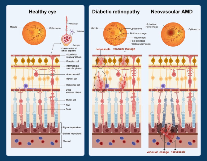
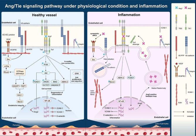
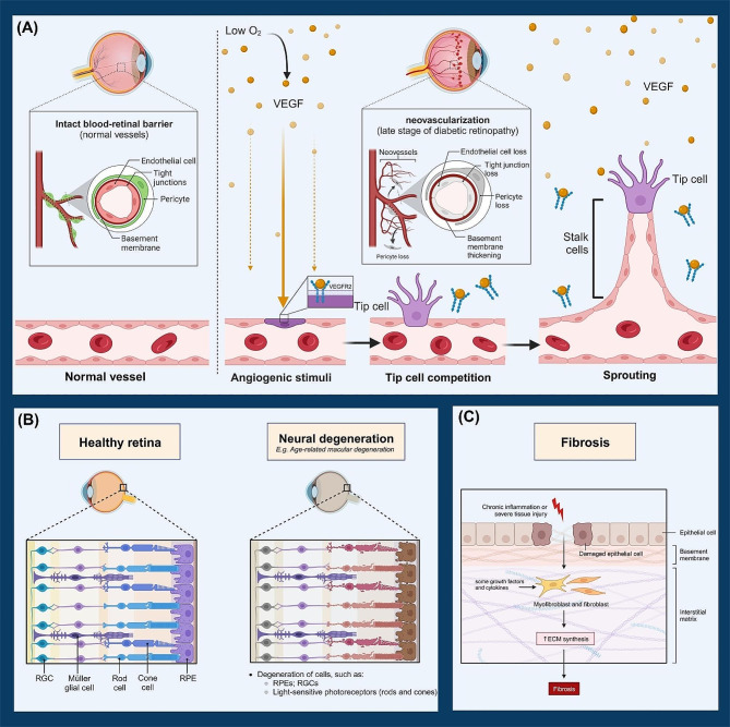
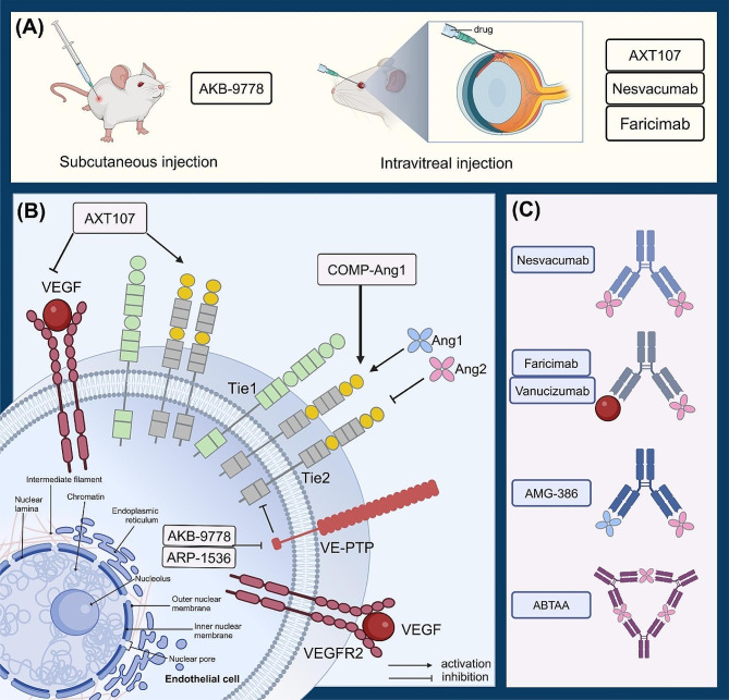
Retinal neurovascular unit (NVU) is a multi-cellular structure that consists of the functional coupling between neural tissue and vascular system. Disrupted NVU will result in the occurrence of retinal and choroidal vascular diseases, which are characterized by the development of neovascularization, increased vascular permeability, and inflammation. This pathological entity mainly includes neovascular age-related macular degeneration (neovascular-AMD), diabetic retinopathy (DR) retinal vein occlusion (RVO), and retinopathy of prematurity (ROP). Emerging evidences suggest that the angopoietin/tyrosine kinase with immunoglobulin and epidermal growth factor homology domains (Ang/Tie) signaling pathway is essential for the development of retinal and choroidal vascular. Tie receptors and their downstream pathways play a key role in modulating the vascular development, vascular stability, remodeling and angiogenesis. Angiopoietin 1 (Ang1) is a natural agonist of Tie2 receptor, which can promote vascular stability. On the other hand, angiopoietin 2 (Ang2) is an antagonist of Tie2 receptor that causes vascular instability. Currently, agents targeting the Ang/Tie signaling pathway have been used to inhibit neovascularization and vascular leakage in neovascular-AMD and DR animal models. Particularly, the AKB-9778 and Faricimab have shown promising efficacy in improving visual acuity in patients with neovascular-AMD and DR. These experimental and clinical evidences suggest that activation of Ang/Tie signaling pathway can inhibit the vascular permeability, neovascularization, thereby maintaining the normal function and structure of NVU. This review seeks to introduce the versatile functions and elucidate the modulatory mechanisms of Ang/Tie signaling pathway. Recent pharmacologic therapies targeting this pathway are also elaborated and summarized. Further translation of these findings may afford a new therapeutic strategy from bench to bedside.

Keywords: Angiopoietin; Clinical translation; Diabetic retinopathy; Neovascular age-related macular degeneration; Vascular endothelial growth factor.


Conflict of interest statement

There are no competing interests.

Figures

Fig. 1
Fig. 1
Retinal and choroidal vasculatures in the healthy eye (left), the eye with diabetic retinopathy (middle) and the eye with neovascular AMD (right). In diabetic retinopathy, the formation of retinal neovessels is accompanied by vascular leakage, which disrupts the nerve layer structure. Choroidal neovessels grow through Bruch’s membrane into the retinal pigment epithelium or subretinal space, causing destruction of the retinal pigment epithelial cells and photoreceptors. Created with BioRender.com
Fig. 2
Fig. 2
Modulating mechanisms of the Ang/Tie signaling pathway. In a healthy vessel (left), Ang1 binds the extracellular domain of Tie2 and activates Tie2 intracellular signaling pathway at EC-EC junctions. The activated PI3K/Akt signaling pathway mediates the phosphorylation of FOXO1 and transcription of ang2. Akt can also maintain cell survival by inducing eNOS and survivin, and exert anti-apoptotic effects by inhibiting caspase-9 and BAD. Activated Tie2 can inhibit the activation of NF-κB by interacting with ABIN-2, thereby inhibiting the expression of inflammatory genes. Ang1/Tie2 signaling can stabilize vessels by activating the GTPase signaling pathways. RAC1 dephosphorylates MLC2 to stabilize the actin cytoskeleton. Src, the downstream molecule of VEGF/VEGFR2, is sequestered by mDia, thereby prevent phosphorylation and internalization of VEC. In motile/non-contact ECs, Tie2 is localised at the EC-ECM and preferentially activates ERK to conduct cell migration and proliferation. Under inflammatory conditions (middle), cleavage of the Tie1 extracellular domain correlates with the switch of Ang2 from an agonist to an antagonist of Tie2. Suppression of Tie2 signaling pathway leads to cytoskeleton instability, inflammation, pericyte detachment and Ang2 positive feedback formation. At this point, VE-PTP inhibits the intracellular domain of Tie2. The meaning of the symbols involved in the figure (right). Created with BioRender.com
Fig. 3
Fig. 3
Pathological conditions of ocular vascular diseases. (A): At the late stage of diabetic retinopathy, changes occur to the vasculature including basement membrane thickening, loss of endothelial tight junctions, loss of pericytes and abnormal neovascularization. Angiogenesis occurs in regions of low O2, which induces VEGF production. VEGF binds to VEGFR2, triggering competition between neighboring cells as they differentiate. Therefore, these adjacent vascular endothelial cells become stalk cells and form the body of the sprouting vessel. (B): Retinal structure is intact in a healthy eye. Nevertheless, in an eye with neural degeneration, damages occur in the retinal cells. (C): In response to external stimulus, endothelial cells are damaged and converted into myofibroblasts or fibroblasts, resulting in increased ECM synthesis. Vascular fibrosis occurs when ECM accumulates excessively. Created with BioRender.com
Fig. 4
Fig. 4
Therapeutic agents targeting the Ang/Tie2 signaling pathway. (A): AKB-9778 is administered by subcutaneous injection. AXT107, nesvacumab and faricimab are administered by intravitreal injection. (B): AXT107 inhibits VEGF-mediated signaling pathways and promotes Tie2 activation. COMP-Ang1 activates Tie2 with stronger agonistic activity than the native Ang1. Both AKB-9778 and ARP-1536 inhibit the activity of VE-PTP, thus activating Tie2. (C): Nesvacumab is an anti-Ang2 monoclonal antibody. Faricimab is a bispecific antibody that can binds to VEGFA and Ang2 with high affinity. Vanucizumab is a bispecific antibody that targets both VEGFA and Ang2. AMG 386 is an antibody that effectively blocks the binding between Tie2 and ligands angiopoietin (Ang1 and Ang2). ABTAA is a dual functional antibody that binds Ang2 and activates Tie2. Created with BioRender.com

Similar articles

Cited by

References

    1. Thomas CJ, Mirza RG, Gill MK. Age-related Macular Degeneration. Med Clin North Am. 2021;105(3):473–91. - PubMed
    1. Campochiaro PA. Molecular pathogenesis of retinal and choroidal vascular diseases. Prog Retin Eye Res. 2015;49:67–81. - PMC - PubMed
    1. Ortiz-Seller A, Martorell P, Barranco H, Pascual-Camps I, Morcillo E, Ortiz JL. Comparison of different agents and doses of anti-vascular endothelial growth factors (aflibercept, bevacizumab, conbercept, ranibizumab) versus laser for retinopathy of prematurity: a network meta-analysis. Surv Ophthalmol. 2024. - PubMed
    1. Peach CJ, Mignone VW, Arruda MA, Alcobia DC, Hill SJ, Kilpatrick LE et al. Molecular pharmacology of VEGF-A isoforms: binding and signalling at VEGFR2. Int J Mol Sci. 2018;19(4). - PMC - PubMed
    1. Hussain RM, Shaukat BA, Ciulla LM, Berrocal AM, Sridhar J. Vascular endothelial growth factor antagonists: promising players in the treatment of Neovascular Age-Related Macular Degeneration. Drug Des Devel Ther. 2021;15:2653–65. - PMC - PubMed

LinkOut - more resources