Review
. 2024 Oct 4;22(1):475.
doi: 10.1186/s12964-024-01862-w.

Peroxisomal homeostasis in metabolic diseases and its implication in ferroptosis

Affiliations
Review

Peroxisomal homeostasis in metabolic diseases and its implication in ferroptosis

Jiwei Han et al. Cell Commun Signal. .

Abstract

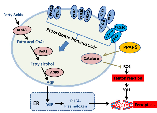
Peroxisomes are dynamic organelles involved in various cellular processes, including lipid metabolism, redox homeostasis, and intracellular metabolite transfer. Accumulating evidence suggests that peroxisomal homeostasis plays a crucial role in human health and disease, particularly in metabolic disorders and ferroptosis. The abundance and function of peroxisomes are regulated by a complex interplay between biogenesis and degradation pathways, involving peroxins, membrane proteins, and pexophagy. Peroxisome-dependent lipid metabolism, especially the synthesis of ether-linked phospholipids, has been implicated in modulating cellular susceptibility to ferroptosis, a newly discovered form of iron-dependent cell death. This review discusses the current understanding of peroxisome homeostasis, its roles in redox regulation and lipid metabolism, and its implications in human diseases. We also summarize the main mechanisms of ferroptosis and highlight recent discoveries on how peroxisome-dependent metabolism and signaling influence ferroptosis sensitivity. A better understanding of the interplay between peroxisomal homeostasis and ferroptosis may provide new insights into disease pathogenesis and reveal novel therapeutic strategies for peroxisome-related metabolic disorders and ferroptosis-associated diseases.

Keywords: Ferroptosis; Homeostasis; Lipid metabolism; Metabolic disorders; Peroxisome; Redox regulation; Therapeutic strategies.


Conflict of interest statement

The authors declare no competing interests.

Figures

Fig. 1
Fig. 1
Schematic overview of functional proteins in mammalian peroxisomes Peroxins can be categorized into three functional groups: peroxisomal membrane assembly, matrix protein import and receptor recycling, and peroxisomal division. Matrix protein import and receptor recycling: Proteins containing peroxisomal targeting signals (PTS1 or PTS2) are recognized by cytosolic receptors such as Pex5 or Pex7. Import into the matrix is facilitated by a membrane-docked complex such as Pex13/Pex14. The recycling of cargo-free PTS receptors relies on ubiquitination, which is mediated by an intricate interplay of Pex2, Pex10, and Pex12. Monoubiquitylated Pex5 is recruited to an exporting complex Pex1/Pex6, and then docked to membrane via Pex26. Pex5 is unfolded and extracted from the membrane by the ATPase activity of Pex1/Pex6. Membrane assembly: Cytosolic Pex19 recognizes proteins to be integrated into the peroxisomal membrane, and Pex3/Pex16 serve as membrane anchoring sites for Pex19. Peroxisomal division: Mammalian Pex11 (comprising α, β, and γ isoforms) serves as a membrane-remodeling PMP to assist in peroxisomal elongation and membrane curving prior to fission. The fission of peroxisomes relies on similar machinery to mitochondria, including Fis1, Mff, and GDAP1. Apart from peroxins, PMPs comprise multiple metabolite transporters, including ABCDs for fatty acid uptake, MCT1/2 for pyruvate or lactate transport, OCTN3, and other PMPs (such as PMP34) which are responsible for the transportation of cofactors such as CoA, NAD+, and FAD. Several lipid metabolism enzymes are also localized in the peroxisomal membrane, such as ASCL1/4, FAR1, and FALDH, coordinating lipid metabolism between peroxisomes and other organelles. The peroxisomal membrane also harbors MAVS for antiviral immunity and USP30 as an autophagy regulator. ABCD, ATP binding cassette subfamily D member; ACSL, acyl-CoA synthetase long chain family member; FALDH, fatty aldehyde dehydrogenase; FAR1, fatty acyl-CoA reductase 1; FIS1, fission mitochondrial 1; GDAP1, ganglioside-induced differentiation-associated protein 1; MAVS, mitochondrial antiviral signalling protein; MCT, monocarboxylate transporter; MFF, mitochondrial fission factor; OCTN3, organic cation/carnitine transporter 3; PEX, peroxin; PMP, peroxisomal membrane protein; SOD, superoxide dismutatse; USP30, ubiquitin-specific protease 30
Fig. 2
Fig. 2
Examples of pexophagy induction (1). Functional defects in the AAA exportomer complex (Pex1/Pex6/Pex26) cause the accumulation of Pex5 on the peroxisomal membrane, and excessive Pex5 monoubiquitination triggers pexophagy in an NBR1-dependent manner. (2). Oxidative stress induces Pex5 phosphorylation at the S141 residue by ATM kinase. Pex5 phosphorylation facilitates its monoubiquitination at the K209 residue by the E3 ligase activity of Pex2, Pex10 and Pex12, and the autophagic receptor p62 is responsible for targeting ubiquitinated peroxisomes to the autophagosome. (3). Under amino acid starvation, Pex2 causes the monoubiquitination of Pex5 and ABCD3, signaling the initiation of pexophagy in an NBR1-dependent manner. USP30 is the best-characterized deubiquitinase to antagonize Pex5 ubiquitination to maintain peroxisomal abundance upon amino acid deprivation. (4). Pex14 is implicated in starvation-induced pexophagy by directly interacting with LC3-II on the autophagosome membrane. PTS1-loaded Pex5 competitively binds to Pex14 under nutrient-rich conditions, while cargo-free Pex5 shows diminished affinity for Pex14 upon starvation, which favors Pex14 interaction with LC3-II for pexophagy. The interaction between TNKS1/2 and Pex14 also mediates pexophagy under starvation. ATG9A, Autophagy related 9 A; ATM, Ataxia-telangiectasia mutated; LC3, microtubule associated protein 1 light chain 3; NBR1, Neighbor of BRCA1 gene 1; P62, Nucleoporin 62; PST, peroxisomal targeting signals; TNKS1/2, Tankyrase 1/2; USP30, ubiquitin-specific protease 30
Fig. 3
Fig. 3
Mechanisms of positive and negative regulation in ferroptosis Iron homeostasis: Extracellular Fe3+ forms complex with TF and is imported through the TFRC complex into the cytosol. Fe3+ is reduced to Fe2+ which catalyzes the Fenton reaction to produce hydroxyl radicals (•OH) using ROS. Ferritin serves as an intracellular chaperone to sequester labile iron. Lipid metabolism: PUFAs are activated in the form of PUFA-CoA by ACSL4, which is further converted to PUFA-PL. PUFA-PL is incorporated into the phospholipid bilayer and can be oxidized by LOXs or hydroxyl radicals to produce lipid peroxides. GPX4-dependent reduction: GPX4 catalyzes the reduction of peroxidized lipids using GSH as the reducing agent. GSH biosynthesis: The system xc- antiporter mediates cystine import in exchange for glutamate. Cystine is converted to cysteine as a precursor for the biosynthesis of GSH, an antioxidant that counteracts lipid peroxides. FSP1-dependent axis: FSP1 catalyzes the conversion of CoQ10 to ubiquinol, which functions as a lipophilic antioxidant to neutralize lipid peroxidation. GCH1-dependent axis: GCH-1 synthesizes BH4 from BH2, and BH4 serves as a lipophilic antioxidant to suppress ferroptosis. ACSL4, acyl-CoA synthetase long-chain family member 4; BH2, dihydrobiopterin; BH4, tetrahydrobiopterin; CoQ10, coenzyme Q10; FSP1, ferroptosis suppression protein 1; GCH1, GTP cyclohydrolase 1; GPX4, glutathione peroxidase 4; LPCAT3, lysophosphatidylcholine acyltransferase 3; LOX, lipoxygenase; PUFA, polyunsaturated fatty acid; PUFA-PL, polyunsaturated phospholipid; ROS, reactive oxygen species; TF, transferrin; TFRC, transferrin receptor
Fig. 4
Fig. 4
An emerging role of peroxisome in ferroptosis regulation The biosynthesis of ether glycerolipids such as plasmalogens through peroxisome-dependent fatty acid metabolism and ER confers sensitivity towards ferroptosis. FAR1 is the rate-limiting enzyme that supplies fatty alcohol precursors for plasmalogen synthesis. Genetic ablation of FAR1 renders cells resistant to ferroptosis induction. Peroxins are peroxisome biogenesis factors required to maintain peroxisome abundance and the activity of plasmalogen biosynthesis. Depletion of Pex genes leads to peroxisome deficiency and ferroptosis resistance. Activation of the transcription factor PPARδ suppresses ferroptosis by upregulating catalase and neutralizing ROS. ACSL4, acyl-CoA synthetase long-chain family member 4; AGP, 1-O-alkyl-glycerol-3-phosphate; AGPS, alkylglycerone phosphate synthase; ER, endoplasmic reticulum; FAR1, fatty acyl-CoA reductase 1, PPARδ, peroxisome proliferator-activated receptor δ, PUFA, polyunsaturated fatty acid

References

    1. Farre JC et al. Peroxisome biogenesis, membrane contact sites, and quality control. EMBO Rep, 2019. 20(1). - PMC - PubMed
    1. Lodhi IJ, Semenkovich CF. Peroxisomes: a nexus for lipid metabolism and cellular signaling. Cell Metab. 2014;19(3):380–92. - DOI - PMC - PubMed
    1. Zhang J, et al. ATM functions at the peroxisome to induce pexophagy in response to ROS. Nat Cell Biol. 2015;17(10):1259–69. - DOI - PMC - PubMed
    1. Dixit E, et al. Peroxisomes are signaling platforms for antiviral innate immunity. Cell. 2010;141(4):668–81. - DOI - PMC - PubMed
    1. Knoblach B, et al. Peroxisomes exhibit compromised structure and matrix protein content in SARS-CoV-2-infected cells. Mol Biol Cell. 2021;32(14):1273–82. - DOI - PMC - PubMed

LinkOut - more resources