Comparative Study
. 2025 Jan;66(1):e13444.
doi: 10.1111/vru.13444. Epub 2024 Oct 4.

Ultrasonographic assessment of equine metacarpal cartilage thickness is more accurate than computed tomographic arthrography

Affiliations
Comparative Study

Ultrasonographic assessment of equine metacarpal cartilage thickness is more accurate than computed tomographic arthrography

Seamus Hoey et al. Vet Radiol Ultrasound. 2025 Jan.

Abstract

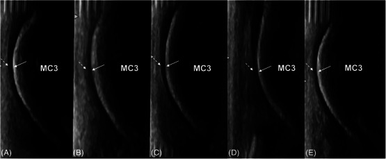
Articular cartilage can be directly imaged using ultrasonography. The fetlock is a common site of osteochondrosis, with the sagittal ridge of the third metacarpal bone most commonly affected. In osteochondrosis, cartilage thickening may be an initial finding. This postmortem study investigated the ability of ultrasonography to accurately measure the dorsodistal articular cartilage of the third metacarpal bone in young horses, compared to computed tomographic arthrography (CTA) and histological measurements. A total of 33 metacarpophalangeal joints from 18 horses between the ages of 12 days and 10 months old were imaged ultrasonographically and with CTA and sectioned and measured using histology. Imaging measurements were made by two observers. Despite overall weak agreement between ultrasonography and histology, the best agreement was at the distal aspect of the sagittal ridge of the third metacarpal bone. Interobserver agreement at this site was also moderate. CTA showed poor agreement overall with histology. Cartilage thickness decreased with age on ultrasonography, CTA, and histology. In conclusion, ultrasonography is a more accurate imaging modality than CTA in the assessment of cartilage in young horses.

Keywords: cartilage; computed tomography; equine; ultrasonography.


Conflict of interest statement

The authors declare that there is no conflict of interest.

Figures

FIGURE 1
FIGURE 1
Photograph of the dorsodistal aspect of the third metacarpal bone of a 3‐month‐old foal illustrating the sites where cartilage measurements were taken on ultrasonography, computed tomographic arthrography, and histology. The black squares identify the 10 site‐matched regions. These included proximal and distal sites in the medial condyle (1,2), the medial parasagittal groove (3,4), the sagittal ridge (5,6), the lateral parasagittal groove (7,8), and the lateral condyle (9,10).
FIGURE 2
FIGURE 2
Site‐matched ultrasonographic (A) and computed tomographic arthrography (CTA) (B) images of the sagittal ridge of the dorsodistal third left metacarpus with corresponding gross slab (C) and histological image (D) of a 1‐month‐old Thoroughbred colt. Magnified ultrasonographic (E) and CTA (F) images of the dorsodistal sagittal ridge. Dorsal is to the left and proximal to the top of the image. MC3: Third metacarpal bone. PrP: Proximal Phalanx. (A) Ultrasonographic image shows a homogeneous hypoechoic band (black arrow) superficial to a hyperechoic subchondral bone margin. (B) CTA image shows positive contrast filling of the metacarpophalangeal joint space (gray arrow), with defects in the contrast filling (dashed white arrow) corresponding with intra‐articular synovium. The articular cartilage appears as a homogeneously hypoattenuating band (black arrow) superficial to the subchondral bone (dashed black arrow). (C) Gross image. The articular cartilage (black arrow) is a white band (black arrow) overlying the subchondral bone of the epiphysis (dashed black arrow). (D) Histological image. Safranin O fast green stain of histological section. The pink stain represents the proteoglycan of hyaline articular cartilage (black arrow) and the blue stain represents the bone (dashed arrow). The bone is more compact under the articular surface, with more dispersed trabeculae deeper in the epiphysis. (E and F) The distal sites of measurement of the articular cartilage thickness are measured from the superficial margin to the articular cartilage‐subchondral bone interface (white arrows).
FIGURE 3
FIGURE 3
Illustration of the cartilage histological thickness measurement on a condyle at the distal third metacarpal bone from a 2‐month‐old foal. Dorsal is to the left and proximal to the top of the image. MC3: Third metacarpal bone. (A) Gross image of a longitudinal osteochondral slab cut from the condyle of the distal third metacarpal bone. Black rectangles reveal sites selected for cartilage thickness measurements. (B) Site‐matched histological section stained with Safranin O fast green. The pink stain reveals proteoglycan of cartilage, and the blue stain highlights bone. (C) Image J was employed, and three lines were made at three equidistant points (dashed lines) on digitalized images within a 1 mm Region of Interest at each site (black rectangles in B), and the mean of the three measurements was calculated for each site.
FIGURE 4
FIGURE 4
Ultrasonographic images of the dorsal aspect of the left third metacarpal bone of a 4‐month‐old Thoroughbred filly. Dorsal is to the left and proximal to the top of the image. MC3: Third metacarpal bone. Images show the subchondral bone margin (arrows) and superficial articular cartilage margin (dashed arrows) of the medial condyle (A), medial parasagittal groove (B), sagittal ridge (C), lateral parasagittal groove (D), and lateral condyle (E).
FIGURE 5
FIGURE 5
Computed tomographic arthrography images of the dorsal aspect of the left third metacarpal bone of a 4‐month‐old Thoroughbred filly. Dorsal is to the left and proximal to the top of the image. MC3: Third metacarpal bone. The synovial joint is filled with positive contrast material. Images show the subchondral bone margin (arrows) and superficial articular cartilage margin (dashed arrows) of the medial condyle (A), medial parasagittal groove (B), sagittal ridge (C), lateral parasagittal groove (D), and lateral condyle (E). There are gas bubbles (grey arrows) and dorsoproximal synovial reflections (white arrows) causing filling defects within the joint spaces. The peripheral synovial membrane and capsule are identified as a hypoattenuating margin (white dashed arrows).
FIGURE 6
FIGURE 6
Composite ultrasonographic (A) and corresponding computed tomographic arthrography (B), anatomical section (C), and histologic section (D) images of the sagittal ridge of the dorsodistal third metacarpus of a 2‐month‐old Thoroughbred filly with no history of lameness. Dorsal is to the left and proximal to the top of the image. MC3: Third metacarpal bone. There is good contrast filling of the metacarpophalangeal joint (black arrow). The articular cartilage (white arrows) is identified, with mild variable thickening of the distal articular cartilage on both ultrasonography and computed tomographic arthrography (dashed white arrows).
FIGURE 7
FIGURE 7
The effect of age on the histological cartilage thickness measurements of the distal aspect of the third metacarpal sagittal ridge. Age: age in weeks. srd: cartilage thickness at the distal site of the sagittal ridge measured in millimeters.
FIGURE 8
FIGURE 8
Ultrasonographic images of the dorsodistal aspect of the sagittal ridge of the right third metacarpal bone of a 12‐day‐old Thoroughbred colt (A) and the left third metacarpal bone of a 10‐month‐old Thoroughbred colt (B). Dorsal is to the left and proximal to the top of the image. MC3: Third metacarpal bone. PrP: Proximal phalanx. The articular cartilage thickness (arrows) is thicker in the 12‐day‐old foal at 1.8 mm compared to thinner cartilage thickness in the older 10‐month‐old horse at 0.9 mm.

Similar articles

References

    1. Merritt JS, Davies HMS, Burvill C, Pandy MG. Influence of muscle‐tendon wrapping on calculations of joint reaction forces in the equine distal forelimb. J Biomed Biotechnol. 2008;2008:1‐9. doi:10.1155/2008/165730 - DOI - PMC - PubMed
    1. Lecocq M, Girard CA, Fogarty U, Beauchamp G, Richard H, Laverty S. Cartilage matrix changes in the developing epiphysis: early events on the pathway to equine osteochondrosis? Equine Vet J. 2008;40(5):442‐454. doi:10.2746/042516408x297453 - DOI - PubMed
    1. Russell J, Matika O, Russell T, Reardon RJ. Heritability and prevalence of selected osteochondrosis lesions in yearling Thoroughbred horses. Equine Vet J. 2017;49(3):282‐287. doi:10.1111/evj.12613 - DOI - PMC - PubMed
    1. Ytrehus B, Carlson CS, Ekman S. Etiology and pathogenesis of osteochondrosis. Vet Pathol. 2007;44(4):429‐448. doi:10.1354/vp.44-4-429 - DOI - PubMed
    1. Olson EJ, Carlson CS. Bones, joints, tendons, and ligaments. In: Zachary JF, ed. Pathologic Basis of Veterinary Disease. 6th ed. Mosby; 2017;954‐1008. chap 16.

Publication types