Review
. 2024 Oct 5;81(1):415.
doi: 10.1007/s00018-024-05437-3.

Mechanisms for assembly of the nucleoplasmic reticulum

Affiliations
Review

Mechanisms for assembly of the nucleoplasmic reticulum

Michael McPhee et al. Cell Mol Life Sci. .

Abstract

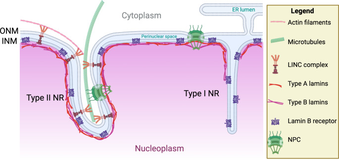
The nuclear envelope consists of an outer membrane connected to the endoplasmic reticulum, an inner membrane facing the nucleoplasm and a perinuclear space separating the two bilayers. The inner and outer nuclear membranes are physically connected at nuclear pore complexes that mediate selective communication and transfer of materials between the cytoplasm and nucleus. The spherical shape of the nuclear envelope is maintained by counterbalancing internal and external forces applied by cyto- and nucleo-skeletal networks, and the nuclear lamina and chromatin that underly the inner nuclear membrane. Despite its apparent rigidity, the nuclear envelope can invaginate to form an intranuclear membrane network termed the nucleoplasmic reticulum (NR) consisting of Type-I NR contiguous with the inner nuclear membrane and Type-II NR containing both the inner and outer nuclear membranes. The NR extends deep into the nuclear interior potentially facilitating communication and exchanges between the nuclear interior and the cytoplasm. This review details the evidence that NR intrusions that regulate cytoplasmic communication and genome maintenance are the result of a dynamic interplay between membrane biogenesis and remodelling, and physical forces exerted on the nuclear lamina derived from the cyto- and nucleo-skeletal networks.

Keywords: Calcium; DNA damage repair; Extracellular vesicles; Nuclear envelope; Nucleoplasmic reticulum; Phosphatidylcholine.


Conflict of interest statement

The authours have no financial or non-financial interests to disclose.

Figures

Fig. 1
Fig. 1
Type-I and Type-II nucleoplasmic reticulum. The Type-I NR are formed by invaginations of the INM at sites that are relatively lamina deficient, include the perinuclear space and are devoid of NPCs. The Type-II NR are invaginations of both the INM and ONM, have a cytoplasmic core as well as proteins associated with the nuclear envelope, such as lamins, LINC complexes and NPCs. Figure created using BioRender.com
Fig. 2
Fig. 2
CCT promotes membrane curvature and phosphatidylcholine synthesis to promote NR formation. (A) Phosphatidylcholine (PC) and phosphatidylethanolamine (PE), the predominant phospholipids of nuclear membranes, are synthesized by the CDP-choline and CDP-ethanolamine pathways, respectively. (B) The catalytic domain of CCTα (green) is activated by insertion of the Domain M amphipathic helix (blue) into the INM when enriched in anionic lipids and non-bilayer lipids or deficient in PC. CCTα translocation and activation is also accompanied by dephosphorylation of the C-terminus. (C) CCTα induces positive curvature in membrane tubules by insertion of Domain M helix (blue, shown in cross-section) into the lipid bilayer. (D) Cooperation between CCT insertion into the INM, the nuclear lamina and coupling of the ONM and INM via LINC complexes drives the extension of a Type-II NR tubule. Abbreviations: CK, choline kinase, CEPT1, choline/ethanolamine phosphotransferase 1; CHPT1, choline phosphotransferase 1; EK, ethanolamine kinase; ECT, phosphoethanolamine cytidylyltransferase; EPT1, ethanolamine phosphotransferase. Figure created using BioRender.com
Fig. 3
Fig. 3
Mechanisms for Type-II NR formation and function. (A) DNA damage signaling stimulates the formation of Type-II NR through the activation of DNA repair kinases ATM, ATR, and DNA dependent protein kinase, leading to ATAT1-KIF5B-LINC mediated extension of cytoplasmic microtubules into the nuclear envelope. (B) Extracellular vesicles (EVs) taken up by cells in late endosomes are escorted into Type-II NR by a Rab7, ORP3, and VAPA-dependent mechanism. Fusion of the EVs with late endosome membrane releases cargo in Type-II NR that are imported by NPC into nucleoplasm to affect transcription. (C) Calcium release through inositiol-3-phosphate receptors (IP3R) and ryanodine receptors (RyR) promotes calcium enrichment in the Type-II NR lumen. Calcium released into Type-II NR can diffuse through NPCs and, along with possible calcium release from Type-I NR, activate PKCγ and promote its translocation to the INM. Calcium uptake into the perinuclear space occurs by the channel proteins SERCA or ORA1-STIM1 and is regulated by the INM protein emerin. Figure created using BioRender.com
Fig. 4
Fig. 4
Type-I NR formation and nuclear lipid droplet biogenesis in hepatocytes. Promyelocytic leukemia protein (PML) isoform II localizes to the INM and excludes Lamin A and B, SUN1/2, and REEP3/4 to promote Type-I NR formation. In the case of SUN1/2, the absence of these proteins allows for uncoupling of the INM and ONM. ApoB-free ER luminal lipids droplets (eLDs) positive for apolipoprotein E (ApoE) and C3 (ApoC3) are precursors for very low-density lipoproteins (VLDL). ER stress causes these precursors to accumulate and migrate through the ER lumen into the perinuclear space and eventually into Type-I NR. There, PML II mediates the emergence of eLDs into the nucleoplasm to form a nuclear LDs, which retain PML structures to their surface and recruit lipid biosynthetic enzymes such as CCTα. Figure created using BioRender.com

Similar articles

Cited by

References

    1. Lin DH, Hoelz A (2019) The structure of the nuclear pore complex (an update). Annu Rev Biochem 88:725–783. 10.1146/annurev-biochem-062917-011901 - PMC - PubMed
    1. Murray-Nerger LA, Cristea IM (2021) Lamin post-translational modifications: emerging toggles of nuclear organization and function. Trends Biochem Sci 46:832–847. 10.1016/j.tibs.2021.05.007 - PMC - PubMed
    1. Starr DA, Fridolfsson HN (2010) Interactions between nuclei and the cytoskeleton are mediated by SUN-KASH nuclear-envelope bridges. Annu Rev Cell Dev Biol 26:421–444. 10.1146/annurev-cellbio-100109-104037 - PMC - PubMed
    1. Niethammer P (2021) Components and mechanisms of Nuclear Mechanotransduction. Annu Rev Cell Dev Biol 37:233–256. 10.1146/annurev-cellbio-120319-030049 - PMC - PubMed
    1. Bragulat-Teixidor H, Ishihara K, Szucs GM, Otsuka S (2024) The endoplasmic reticulum connects to the nucleus by constricted junctions that mature after mitosis. EMBO Rep 25:3137–3159. 10.1038/s44319-024-00175-w - PMC - PubMed

LinkOut - more resources