. 2024 Oct 5;81(1):414.
doi: 10.1007/s00018-024-05418-6.

Stabilization of RRBP1 mRNA via an m6A-dependent manner in prostate cancer constitutes a therapeutic vulnerability amenable to small-peptide inhibition of METTL3

Affiliations

Stabilization of RRBP1 mRNA via an m6A-dependent manner in prostate cancer constitutes a therapeutic vulnerability amenable to small-peptide inhibition of METTL3

Yuqing Feng et al. Cell Mol Life Sci. .

Abstract

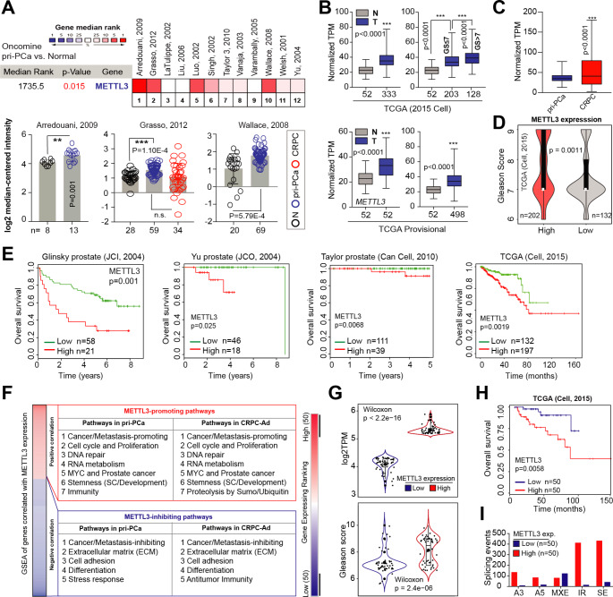
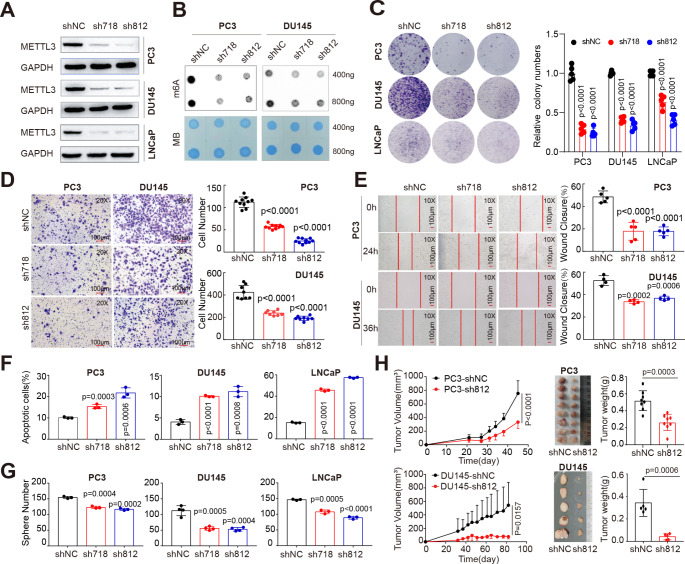
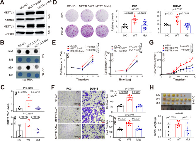
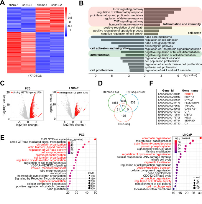
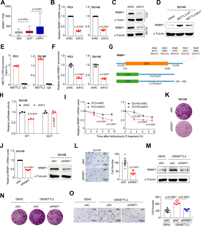
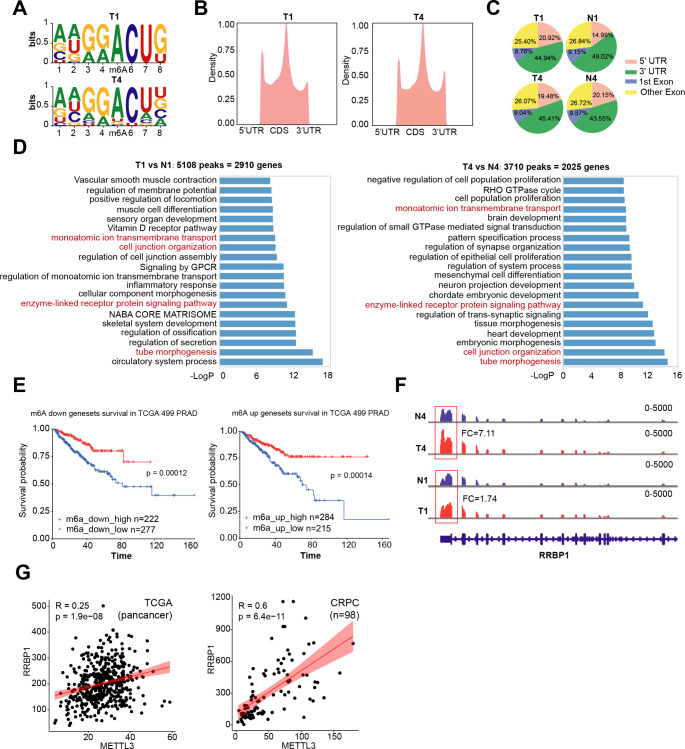
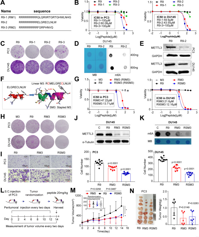
Mounting evidence has implicated the RNA m6A methylation catalyzed by METTL3 in a wide range of physiological and pathological processes, including tumorigenesis. The detailed m6A landscape and molecular mechanism of METTL3 in prostate cancer (PCa) remains ill-defined. We find that METTL3 is overexpressed in PCa and correlates with worse patient survival. Functional studies establish METTL3 as an oncoprotein dependent on its m6A enzymatic activity in both AR+ and AR- PCa cells. To dissect the regulatory network of m6A pathway in PCa, we map the m6A landscape in clinical tumor samples using m6A-seq and identify genome-wide METTL3-binding transcripts via RIP-seq. Mechanistically, we discover RRBP1 as a direct METTL3 target in which METTL3 stabilizes RRBP1 mRNA in an m6A-dependent manner. RRBP1 positively correlates with METTL3 expression in PCa cohorts and exerts an oncogenic role in aggressive PCa cells. Leveraging the 3D structural protein-protein interaction between METTL3 and METTL14, we successfully develop two potential METTL3 peptide inhibitors (RM3 and RSM3) that effectively suppress cancer cell proliferation in vitro and tumor growth in vivo. Collectively, our study reveals a novel METTL3/m6A/RRBP1 axis in enhancing aggressive traits of PCa, which can be therapeutically targeted by small-peptide METTL3 antagonists.

Keywords: METTL3; Peptide inhibitor; Prostate cancer; RRBP1; m6A.


Conflict of interest statement

We declare no competing interests.

Figures

Fig. 1
Fig. 1
Increased METTL3 expression in PCa correlates with multiple oncogenic pathways and worse patient survival. (A) Oncomine analysis showing increased METTL3 expression in PCa vs. normal or benign tissues. The bottom illustrates the details in three representative datasets. (B) Overexpression of METTL3 at mRNA level in both curated (2015 Cell) and noncurated pan-cancer PCa TCGA cohorts. (C) Comparison of METTL3 expression reveals an upregulation in CRPC vs. pri-PCa samples. D and E. High METTL3 mRNA levels correlate with increase GS (D) and worse patient overall survival (E) in indicated datasets. Survival p-value was determined using the Log-Rank test. F. GSEA of genes co-expressed with METTL3 in curated pri-PCa (TCGA) and CRPC (SU2C-PCF) cohorts. G-I. Fractionation of TCGA pri-PCa cohort (2015 Cell) into METTL3 high and low groups, and comparison of METTL3 expression (G, up), GS (G, bottom), patient’s survival outcome (H), and splicing landscape (I) showing METTL3 high group being more aggressive
Fig. 2
Fig. 2
Knock-down of METTL3 significantly inhibits PCa progression in vitro and in vivo.A. Western blot analysis showing METTL3 abundance in three PCa cell lines transduced with scrambled (shNC) or METTL3-specific hairpin shRNAs (sh718 and sh812) and probed with indicated antibodies. GAPDH served as a loading control. B. Dot blot assay showing the global m6A levels in indicated cells treated with different shRNAs. Methylene blue (MB) staining served as a loading control. C. Knocking down METTL3 inhibits clonal development in indicated PCa cells. Representative images and relative quantification are shown for each cell line. Experimental details are as follows: LNCaP (30 K/well for 14 days), DU145 (8 K/well for 9 days), PC3 (5 K/well for 9 days). D and E. Cell mobility evaluated by Trans-well (40 K PC3 per well for 24 h and 70 K DU145 per well for 30 h; D) and wound-healing (24 h for PC3 cells and 36 h for DU145 cells; E) assays showing that METTL3 knock-down inhibits cancer cell migration. F. Flow cytometry analysis showing an increased cellular apoptosis in indicated PCa cell lines upon METTL3 depletion. G. Knocking down METTL3 inhibits sphere formation in both AR + and AR- PCa cell lines. H. Depletion of METTL3 inhibits the growth of PC3 and DU145 xenograft tumors in vivo (n = 5 for each group). Shown are the tumor growth curves (left), endpoint tumor images (middle), and tumor weight (right) of indicated PCa models
Fig. 3
Fig. 3
METTL3 overexpression promotes PCa progression in a m6A-dependent way. (A) Western blot analysis showing overexpression of WT or catalytically dead (i.e., mutant) METTL3 in PCa cells. GAPDH served as a loading control. (B) Dot blot assay showing the global m6A levels in indicated cells treated with different overexpressing constructs. Methylene blue (MB) staining served as a loading control. (C) Relative m6A levels in indicated conditions detected via EpiQuik m6A quantification kit. D-F. Overexpression of WT, but not the mutant, METTL3 promotes CRPC clonal development (D), proliferation (E), and Trans-well migration (F) in both DU145 and PC3 cells in vitro. G and H. The m6A enzymatic activity is required for METTL3 in enhancing PCa development in vivo. Shown are the tumor growth curves (G), endpoint tumor images and tumor weight (H) of DU145 model. In D, 5 K PC3 and 8 K DU145 were initially seeded and visualized at day 10. In F, 40 K PC3 per well for 24 h and 70 K DU145 per well for 30 h were used
Fig. 4
Fig. 4
Identification of the genome-wide METTL3 targets in PCa cells. (A) Heatmap of differentially expressed genes (DEGs) identified in PC3 cells by RNA-seq (fold change (FC) ≥ 1.5 and FDR < 0.1). (B) GO analysis of DEGs as shown in A. (C) Volcano plot of METTL3-bound genes identified in AR + LNCaP and AR PC3 cells by RIP-seq (FC ≥ 2 and FDR < 0.1). (D) Overlap between DEGs and METTL3-bound genes in indicated contexts. (E) GO analysis of METTL3-bound genes showing that a significant proportion of enriched pathways are commonly enriched in both LNCaP and PC3 cells. (F) List of the 11 overlapped genes from D
Fig. 5
Fig. 5
Oncogenic RRBP1 is a direct and functional target of METTL3-mediated m6A signaling in promoting PCa aggressiveness. A. Overexpression of RRBP1 at mRNA level during PCa progression, as analyzed in TCGA (pri-PCa vs. N) and CRPC (vs. pri-PCa) cohorts. B. qPCR analysis showing a reduced RRBP1 expression in PC3 and DU145 cells upon METTL3 depletion. C and D. Western blot analysis of RRBP1 protein levels in indicated PCa cells with METTL3-KD (C) or METTL3 overexpression (D). Tubulin served as a loading control. E. RIP-qPCR analysis showing an enrichment of METTL3 binding at RRBP1 transcripts in PCa cells. F. m6A-qPCR analysis showing much reduced m6A modifications in RRBP1 transcripts in PCa cells upon METTL3-KD. G. Schematic of the potential m6A sites in the RRBP1 3’-UTR. Shown below is the experimental design of constructing luciferase reporters containing WT or mutant 3’-UTR sequences of RRBP1.H. Luciferase reporter assay using the WT or mutated 3’-UTR constructs in DU145 cells with or without METTL3-KD. The firefly luciferase activity was normalized to Renilla luciferase activity. I. mRNA stability assay showing the kinetics of RRBP1 expression in PCa cells with or without METTL3-KD after treatment with actinomycin D (10 µg/mL) for indicated time points. J. Efficient siRNA-mediated KD of RRBP1 both at the mRNA levels (left) and protein levels (right) in DU145 cells. Tubulin served as a loading control. K and L. Knocking down RRBP1 inhibits clonal development (8 K/well for 10 days, K) and migration (50 K/well for 30 h, L) in DU145 cells. M-O. RRBP1 is a key downstream effector of METTL3/m6A pathway in PCa. Reducing the RRBP1 protein into baseline level (M) significantly counteracts the pro-proliferative (N) and pro-migratory (O) effects of METTL3 overexpression in PCa cells. Experiments were performed in METTL3 overexpressing DU145 cells treated with or without siRNA targeting RRBP1. Empty vector-overexpressing cells (OE-NC) transfected with siNC was used as a baseline control
Fig. 6
Fig. 6
The clinical significance of the METTL3/RRBP1 axis in human PCa. A. Two paris of matched PCa and paracancer tissues were used for m6A-seq analysis. Shown are the concesus m6A RRACH motifs identified in two clinical prostate tumor samples. B and C. Statistics of m6A-seq peaks. Shown are the profiles of m6A peak density along mRNA transcript (B) and the genomic peak locations (C). D. GO analysis of genes bearing differential m6A peaks in tumor vs. nontumor tissues. Shown are the top 20 enriched pathways in two patient tumors, with a small proportion of GO pathways being commonly shared. E. Survival analysis of TCGA pan-cancer cohort based on genes that were co-upregulated or co-downregulated in both mRNA expression and m6A levels in tumor tissues. F. Distribution and differential enrichment of m6A peaks across RRBP1 transcripts in tumor (T) and normal (N) samples. G. Positive correlations between RRBP1 and METTL3 mRNA levels in TCGA pri-PCa (left) and CRPC (right) cohorts
Fig. 7
Fig. 7
Small peptides targeting METTL3-METTL14 interaction inhibites PCa in vitro and in vivo. (A) Sequence of peptides R9-1, R9-2, and R9-3. R9 denotes the cell penetrable arginine-9 motif. (B) CCK8 cell viability (3 days) assays in indicated cells treated with different peptides at escalating doses. Data represent mean ± SD from a representative experiment with four technical repeats and the experiment was replicated three times with similar results. (C) Colony formation assay in PC3 and DU145 cells treated with indicated peptides at IC50 dosages as revealed in B. 5 K PC3 and 8 K DU145 were initially seeded and visualized at day 10. D and E. Peptide R9-2 relative to control R9 treatment significantly reduces the global m6A levels (D) and METTL3 protein expression (E) in PC3 and DU145 cells. F. Schematic of devising a staple form of peptide R9-2. G. CCK8 cell viability (3 days) assays in indicated cells treated with different peptides at escalating doses. M3, linear form of the staple peptide. SM3, the stapled M3 peptide. H and I. PCa cells treated with RM3 and RSM3, compared to R9, peptides at IC50 markedly inhibits cell proliferation (5 K PC3 and 8 K DU145 for 8 days; H) and migration (40 K PC3 per well for 20 h and 70 K DU145 per well for 30 h; I) in vitro. J and K. Reduced METTL3 protein expression (J) and the global m6A levels (K) in DU145 cells treated with RM3 and RSM3, compared to R9 or vehicle, peptides at IC50. The relative global m6A status was detected via dot blot assay. L. Schematic of in vivo peptide treatment. M and N. Inhibitory effects of METTL3-targeting peptides on the growth of PC3 CRPC model in vivo. Shown are the tumor growth curve (M; insets present tumor randomizations), endpoint tumor image and tumor weight (N) of xenografts (n = 7 for each group)

Similar articles

Cited by

References

    1. Siegel RL, Miller KD, Wagle NS, Jemal A (2023) Cancer statistics, 2023. CA Cancer J Clin 73(1):17–48 - PubMed
    1. Wang H, Liu J, Zhu X, Yang B, He Z, Yao X (2023) AZGP1P2/UBA1/RBM15 Cascade mediates the fate determinations of prostate cancer stem cells and promotes therapeutic effect of Docetaxel in Castration-resistant prostate Cancer via TPM1 m6A modification. Res (Wash DC) 6:0252 - PMC - PubMed
    1. Shi H, Wei J, He C (2019) Where, when, and how: context-dependent functions of RNA methylation writers, readers, and Erasers. Mol Cell 74(4):640–650 - PMC - PubMed
    1. Han X, Wang M, Zhao YL, Yang Y, Yang YG (2020) RNA methylations in human cancers. Semin Cancer Biol 75:97–115. - PubMed
    1. Wang Z, Zhou J, Zhang H, Ge L, Li J, Wang H (2023) RNA m(6) a methylation in cancer. Mol Oncol 17(2):195–229 - PMC - PubMed

MeSH terms