. 2024 Oct 5;81(1):422.
doi: 10.1007/s00018-024-05452-4.

SENP1 mediates zinc-induced ZnT6 deSUMOylation at Lys-409 involved in the regulation of zinc metabolism in Golgi apparatus

Affiliations

SENP1 mediates zinc-induced ZnT6 deSUMOylation at Lys-409 involved in the regulation of zinc metabolism in Golgi apparatus

Chang-Chun Song et al. Cell Mol Life Sci. .

Abstract

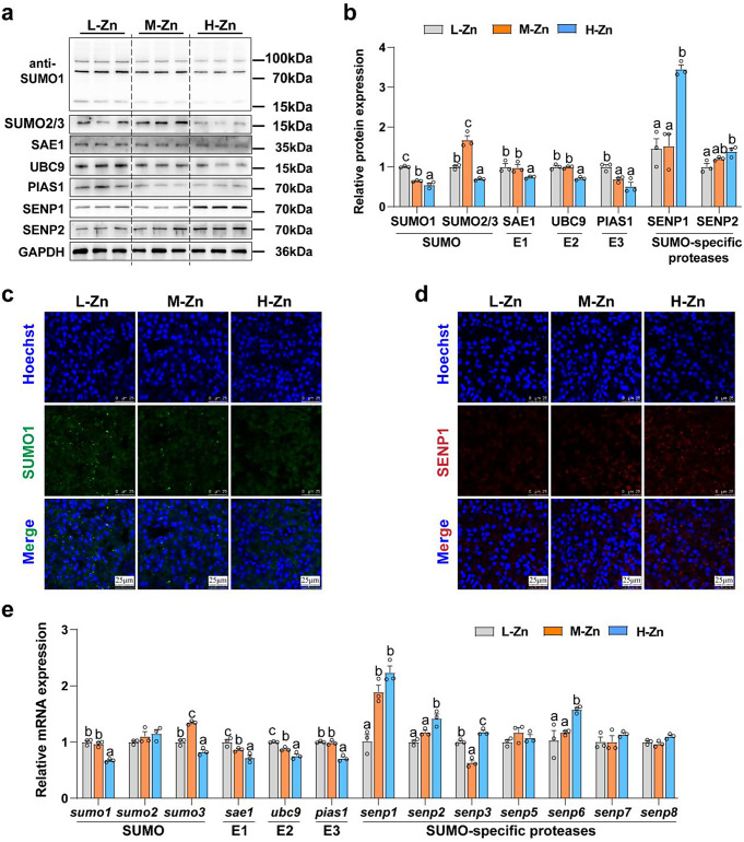
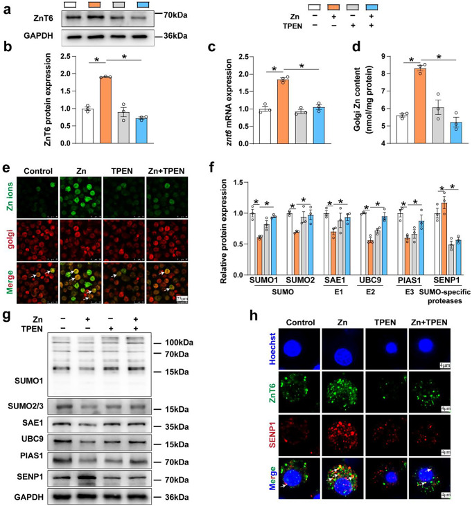
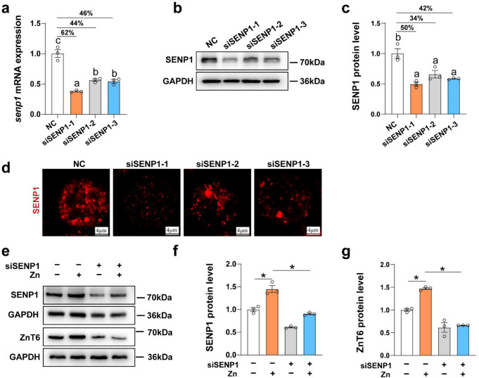
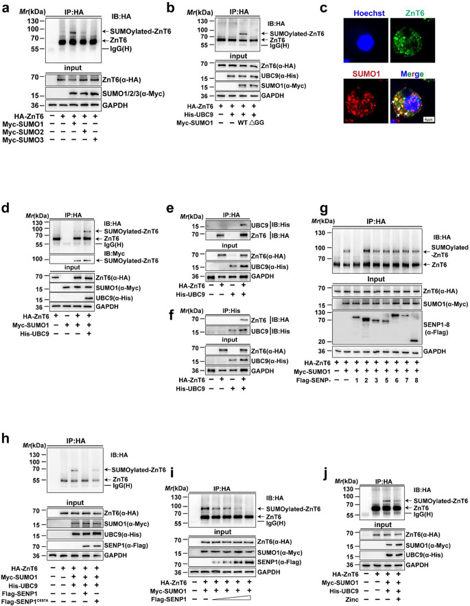
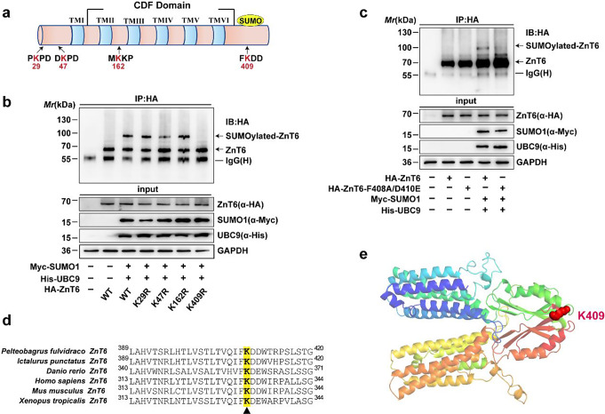
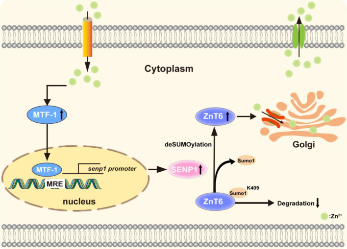
Zinc (Zn) transporters contribute to the maintenance of intracellular Zn homeostasis in vertebrate, whose activity and function are modulated by post-translational modification. However, the function of small ubiquitin-like modifier (SUMOylation) in Zn metabolism remains elusive. Here, compared with low Zn group, a high-Zn diet significantly increases hepatic Zn content and upregulates the expression of metal-response element-binding transcription factor-1 (MTF-1), Zn transporter 6 (ZnT6) and deSUMOylation enzymes (SENP1, SENP2, and SENP6), but inhibits the expression of SUMO proteins and the E1, E2, and E3 enzymes. Mechanistically, Zn triggers the activation of the MTF-1/SENP1 pathway, resulting in the reduction of ZnT6 SUMOylation at Lys 409 by small ubiquitin-like modifier 1 (SUMO1), and promoting the deSUMOylation process mediated by SENP1. SUMOylation modification of ZnT6 has no influence on its localization but reduces its protein stability. Importantly, deSUMOylation of ZnT6 is crucial for controlling Zn export from the cytosols into the Golgi apparatus. In conclusion, for the first time, we elucidate a novel mechanism by which SUMO1-catalyzed SUMOylation and SENP1-mediated deSUMOylation of ZnT6 orchestrate the regulation of Zn metabolism within the Golgi apparatus.

Keywords: MTF-1; SENP1; SUMOylation; Zinc; Zinc transporter; Zn homeostasis.


Conflict of interest statement

All the authors disclosed no conflict of interest.

Figures

Fig. 1
Fig. 1
Effects of dietary Zn levels on Zn metabolism in the liver of yellow catfish. (a), Zn content in the liver. (b), Zn content in the Golgi apparatus of the liver tissue. (c) and (d), mRNA levels of znt6 (c) and mtf-1 (d). (e-f) Western blot (e) and quantification (f) analysis of ZnT6, MTF-1 and n-MTF-1 (nucleus-MTF-1). (g), Immunofluorescent analysis of ZnT6. Scale bars, 75 μm. (h), Immunofluorescent analysis of MTF-1. Scale bars, 25 μm. Values represent the means ± SEM (n = 3 replicate tanks, and 6 fish were sampled from each tank). Significant variations at P < 0.05 are denoted by the Letter (a-c)
Fig. 2
Fig. 2
Effects of dietary Zn levels on SUMOylation progress in the liver of yellow catfish. (a and b), Western blot (a) and statistical analysis (b) of the protein involved in pro-SUMOylation (SUMO1, SUMO2/3, SAE1, UBC9 and PIAS1) and de-SUMOylation (SENP1 and SENP2). (c and d), Immunofluorescent analysis of SUMO1 (c) and SENP1 (d). Scale bars, 25 μm. (e), mRNA levels of genes involved in SUMOylation modification. Values represent the means ± SEM (n = 3 replicate tanks, and 6 fish were sampled from each tank). Significant variations at P < 0.05 are denoted by the letter (a-c)
Fig. 3
Fig. 3
TPEN attenuated Zn-induced increase in ZnT6 expression, decrease of SUMO level and the colocalization of ZnT6 and SENP1 in the hepatocytes of yellow catfish. (a and b), Western blot (a) and statistical analysis (b) of ZnT6. (c), mRNA level of znt6. (d), Zn content in Golgi apparatus. (e), Confocal microscopic image of Zn ions (green) co-localized with Golgi (red) by immunofluorescence staining. Scale bars, 25 μm. (f and g), Western blot and statistical analysis of the protein involved in pro-SUMOylation (SUMO1, SUMO2/3, SAE1, UBC9 and PIAS1) and de-SUMOylation (SENP1). (h), Confocal microscopy image of ZnT6 co-localized with SENP1 in yellow catfish hepatocytes detected by immunofluorescence staining using anti-ZnT6 (green) and anti- SENP1 antibodies (red). Scale bars, 4 μm. Values represent the means ± SEM (n = 3 independently biological experiments). The statistical significance (P value) was determined by Student’s t test. *P < 0.05
Fig. 4
Fig. 4
Zn induced the upregulation of SENP1 expression by promoting the DNA binding of MTF-1 to the senp1 promoter. (a-b), Western blot and statistical analysis of MTF-1 and n-MTF-1 (nucleus-MTF-1) in the hepatocytes of yellow catfish. (c-d), Immunofluorescent analysis of MTF-1 (d) and quantification of the co-localization between MTF-1 and nuclear (c). Scale bars, 10 μm. (e), MTF-1 binding sequence (MRE) located at − 589 bp to − 602 bp of senp1 promoter of yellow catfish. (f), Site mutagenesis of MTF-1 on the pGl3- senp1 -951/+99 vector. (g and h), EMSA analysis of predicted MTF-1 binding sequences (MRE) on the senp1 promoter. The 5′-biotin labeled double-stranded oligomers were incubated with nuclear protein in HEK-293T cell (g) or yellow catfish hepatocytes (h). Values represent the means ± SEM (n = 3 independently biological experiments). Asterisk (*) and hash sign (#) represent significant differences at P < 0.05 between the two groups, as determined using Student’s t-test
Fig. 5
Fig. 5
The SENP1 mediated Zn-induced increase of ZnT6 expression in the hepatocytes of yellow catfish. (a-c), qPCR (a), Western blot and statistical analysis (b and c) of the knockdown efficiency of siRNA targeting senp1 in hepatocytes. (d), Confocal microscopic image of the SENP1 protein detected by immunofluorescence staining. Scale bars, 4 μm. (e-g), Western blot and statistical analysis of SENP1 (e and f) and ZnT6 (e and g) protein expression. Values represent the means ± SEM (n = 3 independently biological experiments). The statistical significance (P value) was determined by Student’s t test. *P < 0.05
Fig. 6
Fig. 6
ZnT6 was mainly SUMOylated by SUMO1 and deSUMOylated by SENP1. (a), ZnT6 was mainly modified by SUMO1. HA-ZnT6 and Myc-SUMO1/2/3 were transfected into HEK-293T cells. The cell lysates were then IP with anti-HA antibody, followed by immunoblotting with the identical antibody. (b), Immunoprecipitation and immunoblot analysis of HEK-293T cells co-transfected with expressing vectors of HA-ZnT6, His-UBC9, Myc-SUMO1 and its indicated mutants. (c), Confocal microscopy image of ZnT6 co-localized with SUMO1 in yellow catfish hepatocytes detected by immunofluorescence staining using anti-ZnT6 (green) and anti-SUMO1 antibodies (red). Nuclear were stained blue. Scale bars, 4 μm. (d), UBC9 enhanced SUMOylation of ZnT6. HEK-293T cells were co-transfected with HA-ZnT6, Myc-SUMO1 and with or without His-UBC9, SUMOylated bands were then detected with anti-HA or anti-Myc antibody. (e and f), The interaction between ZnT6 and UBC9. The lysates of HEK-293T cells overexpression HA-ZnT6 and His-UBC9 were processed for IP with anti-HA (e) or anti-His (f) antibody, followed by immunoblot analysis with the reciprocal antibody. (g), SENP1, but not SENP2, 3, 5, 6, 7 and 8, deSUMOylated ZnT6. HA-ZnT6, Myc-SUMO1 and indicated isoforms of SENPs were transfected into HEK-293T cells, and then western blotting was performed to analyze the SUMOylation of ZnT6 with anti-HA antibody. (h), The deSUMOylated of ZnT6 is dependent on the catalytic activity of SENP1. HA-ZnT6, Myc-SUMO1 and His-UBC9 were transfected with wild-type SENP1 or the SENP1 C697A mutant into HEK-293T cells as indicated. Cell lysates were subjected to IP with anti-HA antibody, followed by immunoblot analysis using anti-HA antibody. (i), SENP1 down-regulated ZnT6 SUMOylation in a dose-dependent manner. HA-ZnT6 and Myc-SUMO1 were transfected into HEK-293T cells with varying amounts of Flag-SENP1, and then western blotting was performed to analyze the SUMOylation of ZnT6 using specified antibodies. (j), Zn incubation prevented ZnT6 from SUMOylation modification in HEK-293T cells. HA-ZnT6, Myc-SUMO1 and His-UBC9 were transfected into HEK-293T cells. After treatment with or without Zn (70 µM) for 24 h, the cell lysates were subjected to IP with anti-HA antibody, followed by immunoblotting with the same antibody to detect the SUMOylation of ZnT6 under Zn treatment. The band representing SUMOylation of ZnT6 was labeled with an arrow
Fig. 7
Fig. 7
Lysine 409 (K409) is the major SUMOylation site of ZnT6. (a), Prediction of four potential SUMOylation sites of ZnT6 in yellow catfish using SUMOplot software. (b), K409 was the major SUMOylation site in ZnT6. The lysates from HEK-293T cells overexpressing HA-ZnT6, HA-ZnT6 mutants (K29R/K47R/K162R/K409R), Myc-SUMO1 and His-UBC9 were IP with anti-HA antibody, and then immunoblot analysis was performed with the same antibody. (c), The mutant ZnT6-F408A/D410E further confirmed the SUMOylation of ZnT6. The lysates from HEK-293T cells overexpressing the indicated plasmids were IP with anti-HA antibody, and then immunoblot analysis was performed with the same antibody. (d), Sequences Alignment of ZnT6 from indicated species. The conserved lysine residue (K409) of SUMOylation modification is highlighted in yellow. (e), SUMOylation sites of yellow catfish ZnT6
Fig. 8
Fig. 8
SUMOylation of ZnT6 did not affect its localization, but reduced its protein stability, inhibited the increase of Zn content in Golgi apparatus induced by Zn. (a), Western blotting analysis was performed to determine the half-life of ZnT6 by using anti-HA antibody in HEK-293T cells overexpressing wild-type ZnT6 or the ZnT6-K409R with or without CHX incubation for different time. (b), Western blotting analysis was performed to determine the half-life of endogenous ZnT6 by using anti-ZnT6 antibody in yellow catfish hepatocytes treatment with SENP1 knockdown with or without CHX incubation for different time. (c and d), ZnT6 SUMOylation did not change its subcellular localization. (c) Subcellular localizations of nuclear (blue), ZnT6 (green) with Golgi (red) were determined by confocal microscopy. Scale bars, 4 μm. (d) HEK-293T cells overexpressing wild-type ZnT6 or the ZnT6-K409R mutant were immunostained with ZnT6 (green), Golgi (red) and Hoechst (blue). Scale bars, 20 μm. (e and f), ZnT6 SUMOylation inhibited Zn-induced increases of the Zn content in Golgi apparatus. (e) Subcellular localizations of nuclear (blue), Zn ions (green) and Golgi (red) were determined by confocal microscopy. Scale bars, 20 μm. (f) Zn content in Golgi apparatus. Values represent the means ± SEM (n = 3 independently biolological experiments). The statistical significance (P value) was determined by Student’s t test. *P < 0.05
Fig. 9
Fig. 9
A working model of MTF-1/SENP1 pathway-mediated ZnT6 deSUMOylation modification participating in the regulation of Zn metabolism in the Golgi apparatus

Similar articles

Cited by

References

    1. Wei CC, Luo Z, Hogstrand C, Xu YH, Wu LX, Chen GH et al (2018) Zinc reduces hepatic lipid deposition and activates lipophagy via Zn2+/MTF-1/PPARα and Ca2+/CaMKKβ/AMPK pathways. FASEB J 32:6666–6680 - PubMed
    1. Wu K, Luo Z, Hogstrand C, Chen GH, Wei CC, Li DD et al (2018) Zn stimulates the phospholipids biosynthesis via the pathways of oxidative and endoplasmic reticulum stress in the intestine of freshwater teleost yellow catfish. Environ Sci Technol 52(16):9206–9214 - PubMed
    1. Xu YC, Zheng H, Hogstrand C, Tan XY, Zhao T, Song YF et al (2023) Novel mechanism for zinc inducing hepatic lipolysis via the HDAC3-mediated deacetylation of β-catenin at lysine 311. J Nutr Biochem 121:109429 - PubMed
    1. Ibs KH, Rink L (2003) Zinc-altered immune function. J Nutr 133:1452S–6S - PubMed
    1. Foster M, Samman S (2010) Zinc and redox signaling: perturbations associated with cardiovascular disease and diabetes mellitus. Antioxid Redox Signal 13(10):1549–1573 - PubMed

MeSH terms

LinkOut - more resources