. 2024 Oct 5;81(1):421.
doi: 10.1007/s00018-024-05446-2.

DCAF13 promotes ovarian cancer progression by activating FRAS1-mediated FAK signaling pathway

Affiliations

DCAF13 promotes ovarian cancer progression by activating FRAS1-mediated FAK signaling pathway

Ze-Yi Tang et al. Cell Mol Life Sci. .

Abstract

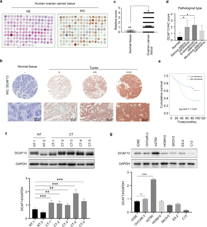
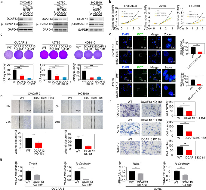
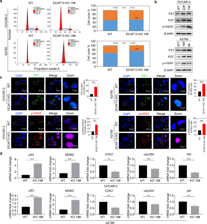
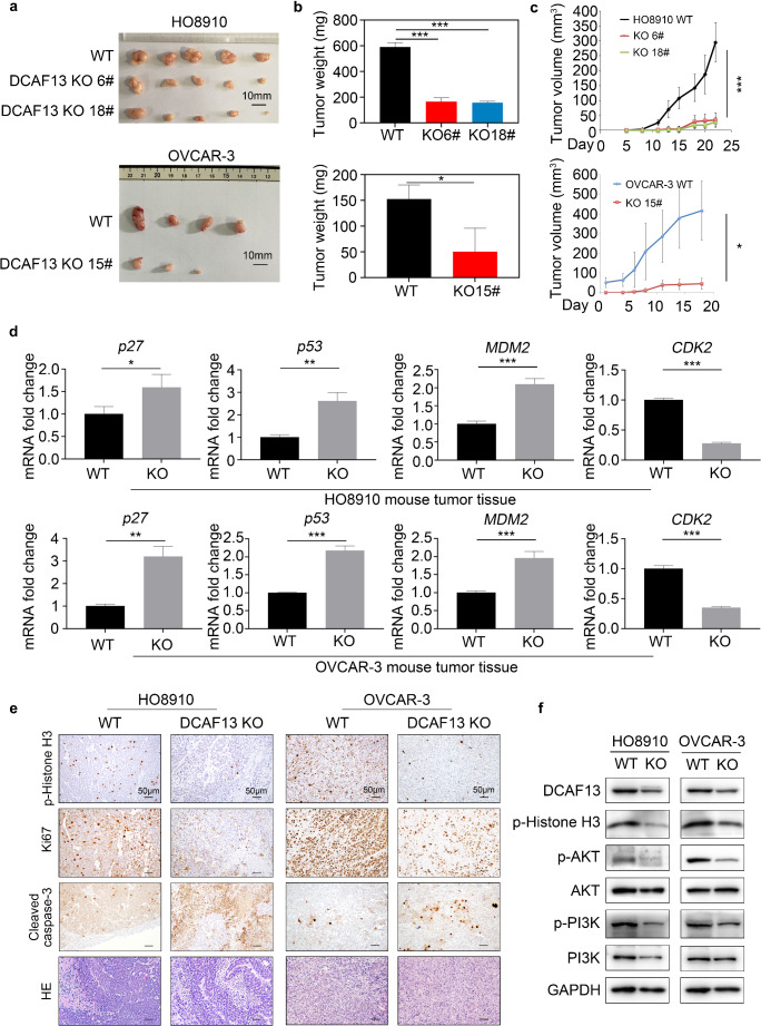
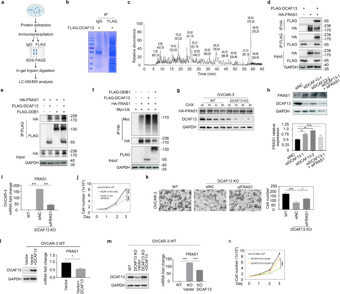
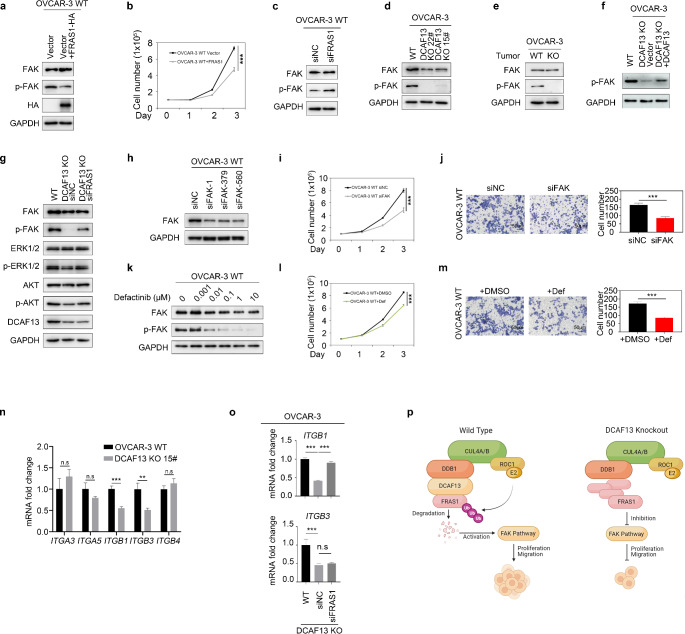
Cullin-RING ubiquitin ligase 4 (CRL4) is closely correlated with the incidence and progression of ovarian cancer. DDB1- and CUL4-associated factor 13 (DCAF13), a substrate-recognition protein in the CRL4 E3 ubiquitin ligase complex, is involved in the occurrence and development of ovarian cancer. However, its precise function and the underlying molecular mechanism in this disease remain unclear. In this study, we confirmed that DCAF13 is highly expressed in human ovarian cancer and its expression is negatively correlated with the overall survival rate of patients with ovarian cancer. We then used CRISPR/Cas9 to knockout DCAF13 and found that its deletion significantly inhibited the proliferation, colony formation, and migration of human ovarian cancer cells. In addition, DCAF13 deficiency inhibited tumor proliferation in nude mice. Mechanistically, CRL4-DCAF13 targeted Fraser extracellular matrix complex subunit 1 (FRAS1) for polyubiquitination and proteasomal degradation. FRAS1 influenced the proliferation and migration of ovarian cancer cell through induction of the focal adhesion kinase (FAK) signaling pathway. These findings collectively show that DCAF13 is an important oncogene that promotes tumorigenesis in ovarian cancer cells by mediating FRAS1/FAK signaling. Our findings provide a foundation for the development of targeted therapeutics for ovarian cancer.

Keywords: CRL4 E3 ubiquitin ligase; DCAF13; FAK; FRAS1; Ovarian cancer.


Conflict of interest statement

The authors declare no competing interests.

Figures

Fig. 1
Fig. 1
Expression pattern of DCAF13 in ovarian cancer tissues and ovarian cancer cells. a H&E staining and immunohistochemistry of DCAF13 protein expression in ovarian cancer tissue microarrays. b Immunohistochemistry for DCAF13 protein expression in normal ovarian tissue and ovarian cancer tissue in tissue microarray. The results were divided into negative (-), moderate (++) and strong positive (+++) staining pattern. c Statistical analysis of DCAF13 expression in normal ovarian tissue and ovarian cancer tissue of tissue microarray, *, P < 0.05. d Statistical analysis of DCAF13 expression in different pathological types of ovarian cancer tissue microarray, *, P < 0.05. e Kaplan-Meier analysis of cumulative survival rate of ovarian cancer tissue microarray, log-rank statistical test, P = 0.001. f Western blot (upper panel) and quantification (lower panel) of DCAF13 protein expression in patient-derived ovarian cancer tissues and paracancerous tissues (NT represents paracancerous tissue, n = 2; CT refers to cancerous tissue, n = 5). g Western blot (upper panel) and quantification (lower panel) of DCAF13 expression in ovarian cancer cells (OVCAR-3, A2780, HO8910, SKOV3, ES-2, and C13), and immortalized mouse ovarian surface epithelium (IOSE). GAPDH was used as the loading control
Fig. 2
Fig. 2
DCAF13 deletion inhibits ovarian cancer cell proliferation, clone formation, and metastatic ability. a Detection of DCAF13 knockout efficiency in OVCAR-3, A2780 and HO8910 by immunoblotting. Two independent clones (22# and 15# in OVCAR-3; 18# and 9# in A2780; 6# and 18# in HO8910) are shown. b Growth curve of DCAF13 knockout ovarian cancer cells in OVCAR-3, A2780 and HO8910. This experiment was replicated three times. The error bars represent SD. Two-way ANOVA test was applied. ***, P < 0.001. c Soft agar assay detection colony formation in DCAF13-deleted OVCAR-3, A2780 and HO8910 cells. Data are presented as mean ± SD. ***, P < 0.001. d Immunofluorescence analysis of Ki67 (green) levels in WT and DCAF13-deficient OVCAR-3 and A2780 cells. Nuclei are counterstained with DAPI (blue). Scale bar, 20 μm. The white box area is enlarged.e Wound-healing assay for the migration ability of WT and DCAF13-deleted cells. Data are presented as mean ± SD for n = 3 per cell line. Student’s t-test was applied. **, P < 0.01. Scale bar, 50 μm. f Representative images (left panel) and quantification (right panel) of Transwell assay results showed that DCAF13 deletion inhibits ovarian cancer cell migration. This experiment was replicated three times. The error bars represent SD. **, P < 0.01, two-way ANOVA test. Scale bar, 50 μm. g qRT-PCR detection of cell migration-related genes in WT and DCAF13-deleted cells in OVCAR-3 and A2780. The error bars represent SD. Student’s t-test was applied. **, P < 0.01; ***, P < 0.001
Fig. 3
Fig. 3
DCAF13 deletion affects the cell cycle and promotes cell senescence. a Flow cytometry was employed to analyze the changes in the cell cycle in DCAF13-deleted OVCAR-3 and A2780 cells. **, P < 0.01 (nonparametric test). b Western blot analysis of the expression of P21, P27 and p-H2AX in WT and DCAF13-deleted OVCAR-3 and A2780 cells. c Representative images (left panel) and quantification (right panel) of immunofluorescence analysis of P21 (green) and p-H2AX (red) levels in WT and DCAF13-deleted OVCAR-3 and A2780 cells. Cells were counterstained with DAPI (blue). Scale bar, 20 μm. The white box area is enlarged. **, P < 0.01; ***, P < 0.001 d qRT-PCR detection of cell cycle related genes in WT and DCAF13-deleted cells in OVCAR-3 and A2780. The error bars represent SD. Student’s t-test was applied. **, P < 0.01; ***, P < 0.001
Fig. 4
Fig. 4
DCAF13 deletion inhibits tumor growth in vivo. a Tumor size of HO8910 and OVCAR-3 wild-type and DCAF13 deletion ovarian cancer cells in nude mice. 5 × 106 cells were injected subcutaneously into flank of nude mice (n = 6). Some nude mice failed to form tumors. Scale bar, 10 mm. b Deletion of DCAF13 in HO8910 and OVCAR-3 cells inhibits tumor weight in vivo. *, P < 0.05; ***, P < 0.001, according to two-way ANOVA. c Tumor growth curve of nude mice. Tumor volume in each group was measured every 2–3 days. *, P < 0.05; ***, P < 0.001, according to two-way ANOVA. d qRT-PCR detection of cell cycle-related factors in tumors. The error bars represent SD. Student’s t-test was applied. *, P < 0.05; **, P < 0.01; ***, P < 0.001. e Histochemistry and immunohistochemical analysis of p-Histone H3, Ki67 and Cleaved Caspase-3 in wild-type and DCAF13 deletion xenografts. Scale bar, 50 μm. Objective magnification 20x. f Western blot analysis of DCAF13, p-Histone H3, p-AKT, AKT, p-PI3K and PI3K expression in WT and DCAF13-deleted HO8910 and OVCAR-3 mouse tumor tissue. GAPDH was used as control
Fig. 5
Fig. 5
FRAS1 is the key target of DCAF13 in ovarian cancer cells. a Schematic representation of immunoprecipitation combined with liquid chromatography-mass spectrometry. b Coomassie brilliant blue staining of proteins pulled with IgG antibody and FLAG antibody. FLAG-DCAF13 plasmid was overexpressed in this experiment. c The base peak of protein sample by mass spectrometry. d Co-immunoprecipitation results showing FRAS1 interacts with DCAF13. 293T cells transfected with plasmids encoding the indicated proteins were lysed and subjected to IP with anti-FLAG or anti-HA beads. Input lysates were immunoblotted with antibodies against HA and FLAG. e Co-immunoprecipitation results showing FRAS1 interacts with DDB1 and DCAF13. The overexpression of DDB1 enhances the interaction between FRAS1 and DCAF13. f Co-immunoprecipitation experiments showing overexpression of DCAF13 and DDB1 increase FRAS1 polyubiquitination. g The Cycloheximide (CHX)-chasing experiment revealed the FRAS1 stability. OVCAR-3 WT and DCAF13-deleted cells were transfected with HA-FRAS1 plasmid. 10 µM CHX was used to inhibit protein synthesis in OVCAR-3. At the specified time points after CHX treatment, the cells were lysed for immunoblotting. h Western blot shows the expression of FRAS1 in OVCAR-3 cells transfected with control siRNA (siNC), siDCAF13 or siFRAS1(upper panel) and relative quantitative analysis (lower panel). **, P < 0.01; ns, P > 0.05. i mRNA expression level of FRAS1 in WT or DCAF13 deficient OVCAR-3 cells transfected with control siRNA (siNC) or siFRAS1 ***, P < 0.001. j FRAS1 deletion rescues cell proliferation defects in DCAF13 deficient OVCAR-3 cells. ***, P < 0.001. k FRAS1 deletion rescues cell migration ability in OVCAR-3 DCAF13 KO cells. Representative images from the Transwell assay (left panel) and quantification analysis (right panel) were shown. *, P < 0.05, ***, P < 0.001. l Western blot results of DCAF13 protein expression (left panel) and qRT-PCR results of FRAS1 mRNA expression in wild-type OVCAR-3 transfected with DCAF13 plasmids. *, P < 0.05. m Western blot results of DCAF13 protein expression (left panel) and qRT-PCR results of FRAS1 mRNA expression in WT, DCAF13 KO and DCAF13 KO transfected with DCAF13 plasmids. ***, P < 0.001. n Overexpression of DCAF13 rescues the ability of cell proliferation. ***, P < 0.001
Fig. 6
Fig. 6
FRAS1 affects ovarian cancer cell proliferation and migration by mediating the FAK signaling pathway. a Western blot detection FAK, p-FAK, HA protein expression in cells. OVCAR-3 cells were transfected with HA-FRAS1 vector and or with control empty vector. GAPDH was used as control. b Cell proliferation assay of overexpressed HA-FRAS1 and control ovarian cancer cells. ***, P < 0.001. c Western blot results showing the FAK signaling pathway was activated after interfering with FRAS1. GAPDH was used as control. d Western blot analysis of FAK and p-FAK expression in WT and DCAF13-deleted OVCAR-3 cells. GAPDH was used as control. e Western blot analysis of FAK and p-FAK expression in WT and DCAF13-deleted OVCAR-3 mouse tumor tissue. f Western blot analysis of p-FAK expression in WT, DCAF13-deleted OVCAR-3 cells and DCAF13-deleted OVCAR-3 cell tansfected with DCAF13 plasmids. g Western blot analysis of p-FAK, p-ERK1/2 and p-AKT expression in WT, DCAF13-deleted OVCAR-3 cells, DCAF13 and FRAS1 double knockdown cells. h Western blot results for FAK siRNA interference efficiency. i FAK silencing inhibiting ovarian cancer cell proliferation. ***, P < 0.001. j FAK deletion inhibiting cell migration of ovarian cancer cells. Representative images from the Transwell assay (upper panel) and quantification analysis (lower panel) were shown. **, P < 0.01. k OVCAR-3 cells were exposed to increasing doses of the FAK inhibitor Defactinib (Def) for 24 h. Western blot analysis of FAK and p-FAK. l Ovarian cancer cell proliferation, after treatment with 1 µM Defactinib for 24 h. ***, P < 0.001. m Defactinib inhibiting cell migration of ovarian cancer cells. Representative images from the Transwell assay (upper panel) and quantification analysis (lower panel) were shown. **, P < 0.01. n Alterations in integrin expression in wild-type and DCAF13 deletion OVCAR-3 cells were analyzed by qRT-PCR. n.s, P > 0.05, ** P < 0.01, *** P < 0.001. o qRT-PCR detection the expression level of ITGB1 after interfering with FRAS1. *** P < 0.001. p A working model explaining how DCAF13 promotes ovarian cancer cell proliferation and migration. In wild-type ovarian cancer cells, DCAF13 ubiquitin degrades the FRAS1 substrate and activates the FAK signaling pathway, thus promoting the proliferation and migration of ovarian cancer cells. When DCAF13 is knocked out, FRAS1 accumulation leads to inhibition of the FAK signaling pathway and suppression of proliferation and migration of ovarian cancer cells

Similar articles

Cited by

References

    1. Sung H, Ferlay J, Siegel RL, Laversanne M, Soerjomataram I, Jemal A et al (2021) Global cancer statistics 2020: GLOBOCAN estimates of incidence and mortality worldwide for 36 cancers in 185 countries. CA Cancer J Clin 71(3):209–249. 10.3322/caac.21660 - PubMed
    1. Yang Y, Qi S, Shi C, Han X, Yu J, Zhang L et al (2020) Identification of metastasis and prognosis-associated genes for serous ovarian cancer. Biosci Rep 40(6):BSR20194324. 10.1042/BSR20194324 - PMC - PubMed
    1. Menon U, Gentry-Maharaj A, Burnell M, Singh N, Ryan A, Karpinskyj C et al (2021) Ovarian cancer population screening and mortality after long-term follow-up in the UK Collaborative Trial of Ovarian Cancer Screening (UKCTOCS): a randomised controlled trial. Lancet 397(10290):2182–2193. 10.1016/S0140-6736(21)00731-5 - PMC - PubMed
    1. Van Zyl B, Tang D, Bowden NA (2018) Biomarkers of platinum resistance in ovarian cancer: what can we use to improve treatment. Endocr Relat Cancer 25(5):R303–R318. 10.1530/ERC-17-0336 - PubMed
    1. Yang L, Xie HJ, Li YY, Wang X, Liu XX, Mai J (2022) Molecular mechanisms of platinum–based chemotherapy resistance in ovarian cancer. Oncol Rep 47(4):82–93. 10.3892/or.2022.8293 - PMC - PubMed

MeSH terms

LinkOut - more resources