. 2024 Oct 22;43(10):114819.
doi: 10.1016/j.celrep.2024.114819. Epub 2024 Oct 3.

PD-L1 restrains PD-1+Nrp1lo Treg cells to suppress inflammation-driven colorectal tumorigenesis

Affiliations

PD-L1 restrains PD-1+Nrp1lo Treg cells to suppress inflammation-driven colorectal tumorigenesis

Dakota B Poschel et al. Cell Rep. .

Abstract

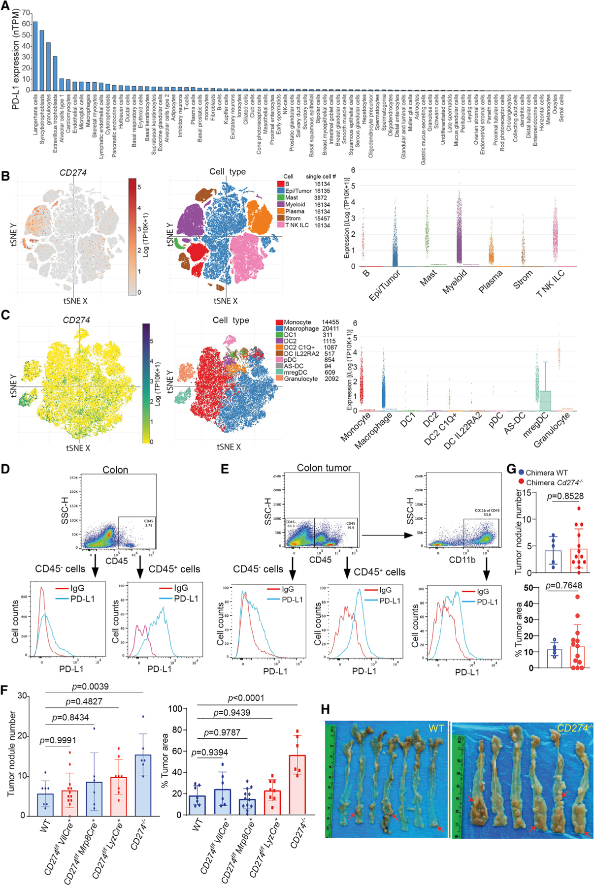
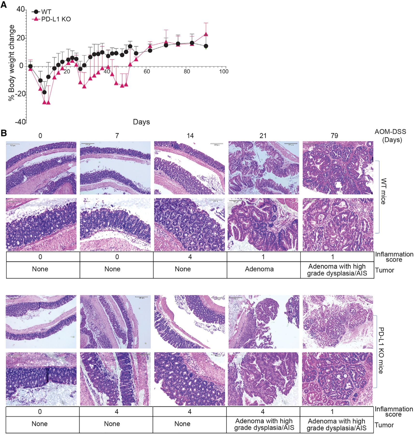
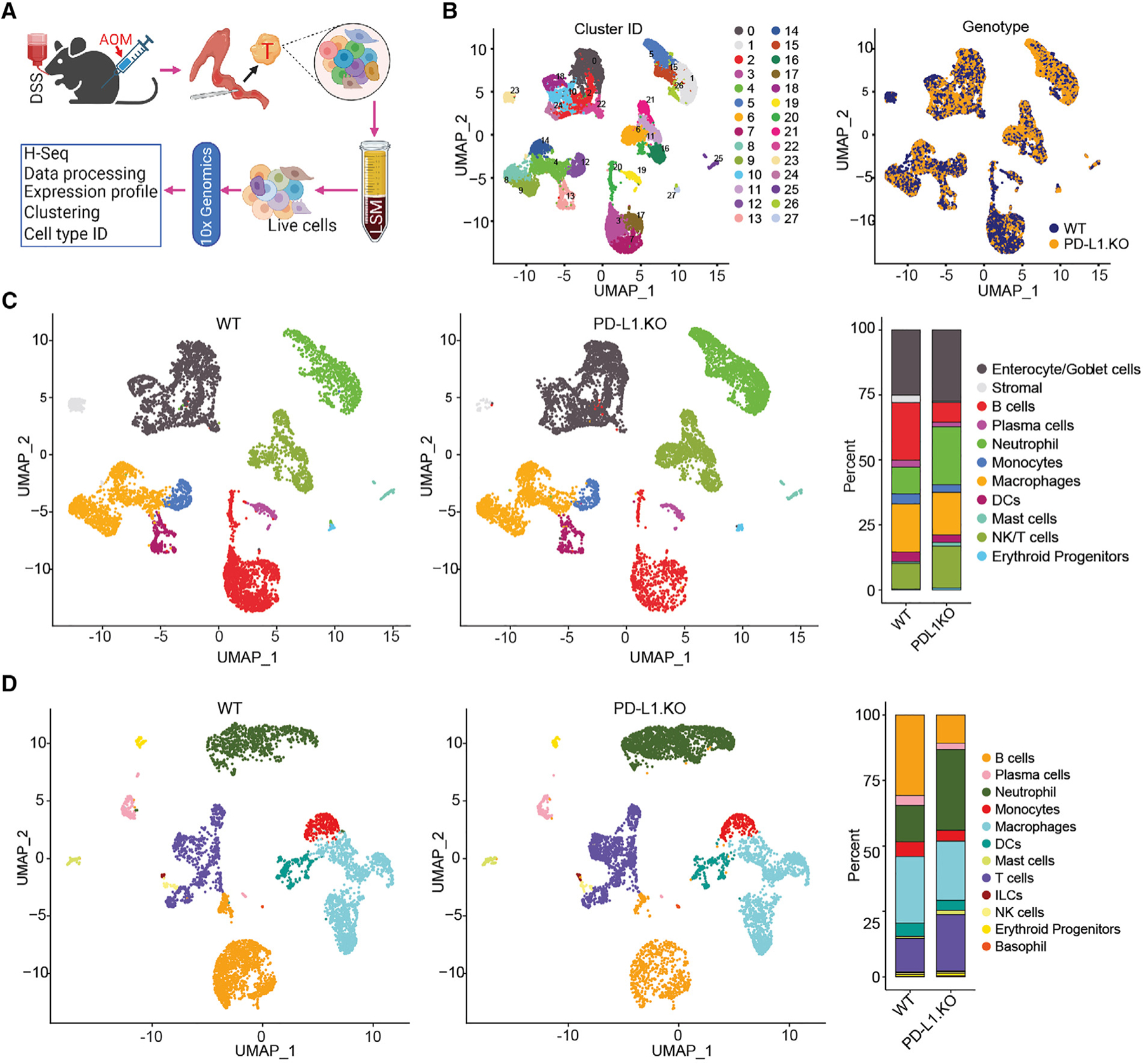
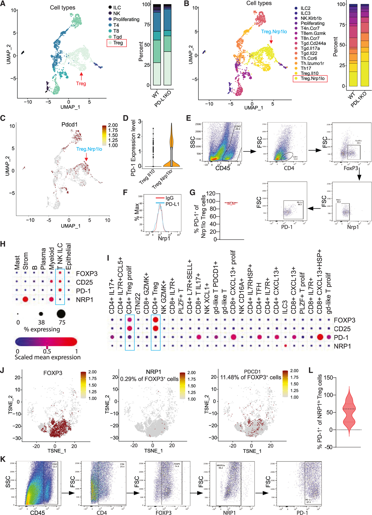
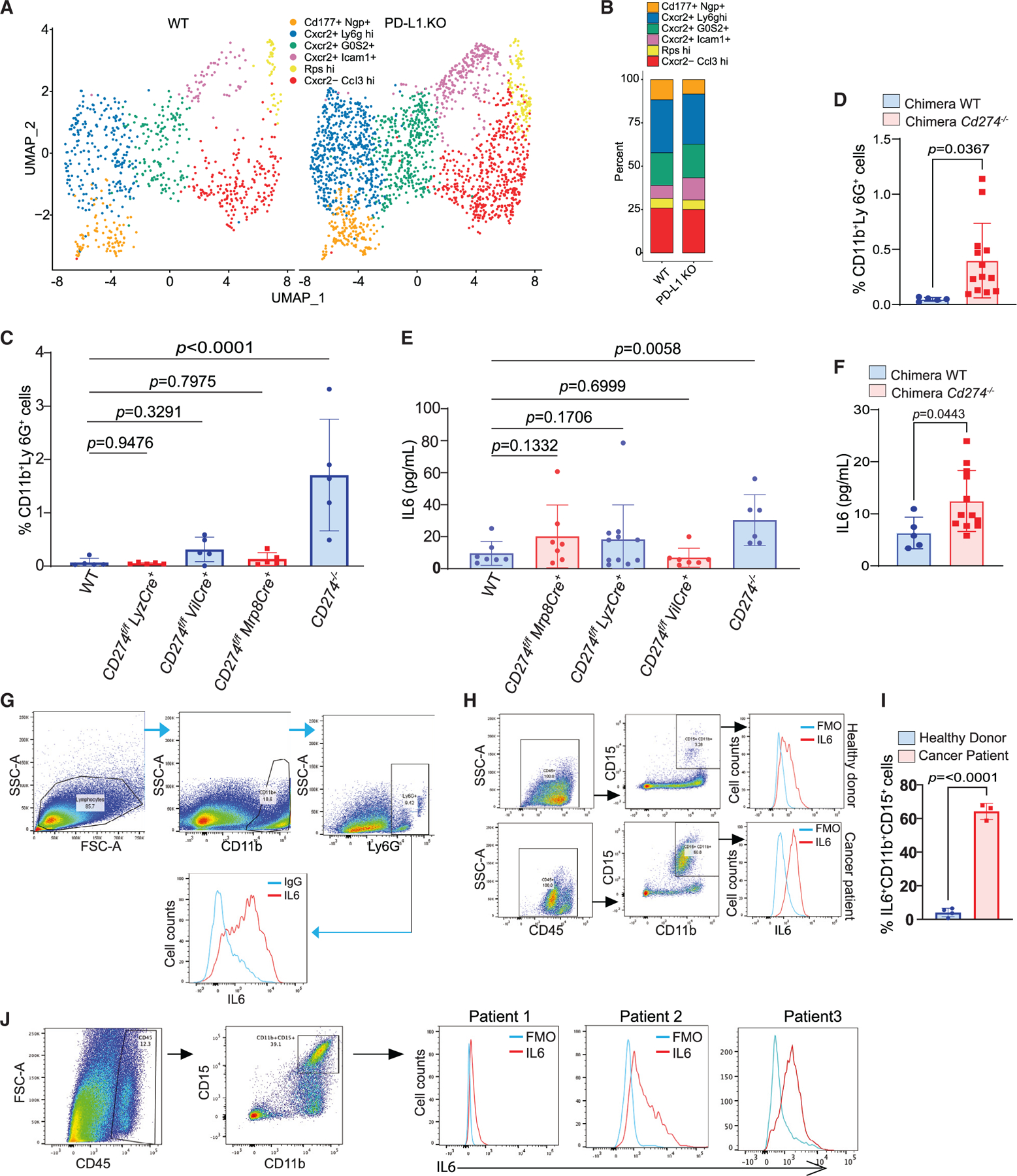
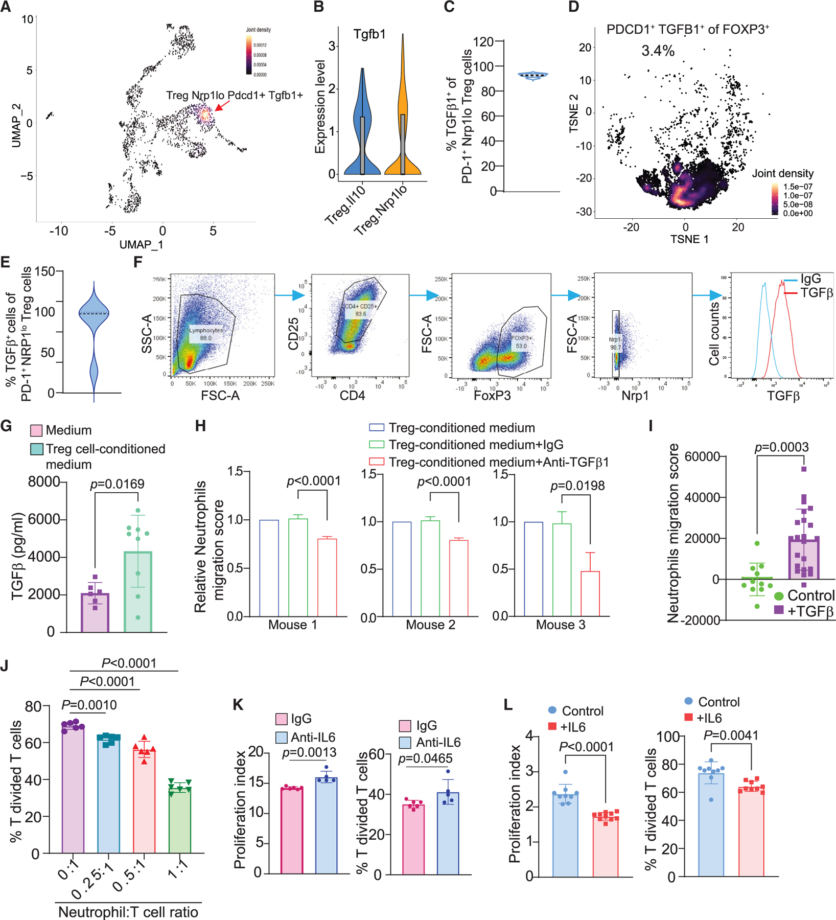
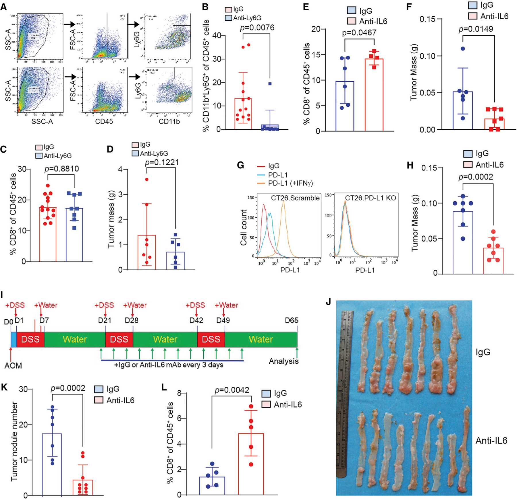
T cells function not only as an essential component of host cancer immunosurveillance but also as a regulator of colonic inflammation, a process that promotes colorectal cancer. Programmed death-ligand 1 (PD-L1) is a T cell-negative regulator, but its role in regulation of T cell functions in the context of colorectal cancer is unknown. We report that global deletion of Cd274 results in increased colonic inflammation, PD-1+ T cells, and inflammation-driven colorectal tumorigenesis in mice. Single-cell RNA sequencing (scRNA-seq) analysis revealed that PD-L1 suppresses subpopulations of programmed cell death protein 1 (PD-1)+Nrp1lo regulatory T (Treg) cells and interleukin (IL) 6+ neutrophils in colorectal tumor. Treg cells produce transforming growth factor (TGF) β to recruit IL6+ neutrophils. Neutrophils produce IL6 to inhibit activation of tumor-specific cytotoxic T lymphocytes (CTLs) and primary CTLs. Accordingly, IL6 blockade immunotherapy increases CTL activation and suppresses colon tumor growth in vivo. Our findings determine that PD-L1 restrains PD-1+Nrp1loTGFβ+ Treg cells to suppress IL6+ neutrophil tumor recruitment to sustain CTL activation to control inflammation-driven colorectal tumorigenesis.

Keywords: CP: Cancer; CP: Immunology.


Conflict of interest statement

Declaration of interests The authors declare no competing interests.

Figures

Figure 1.
Figure 1.. Loss of global PD-L1 promotes inflammation-driven colorectal tumorigenesis
(A) The human CD274 mRNA level in the indicated human cell types in the single-cell level. The CD274 transcript datasets were downloaded from the Human Protein Atlas and analyzed. (B and C) PD-L1 expression level in major cell types (B) and myeloid cell subpopulations (C) of human colon tumor tissues in the single-cell level. The human colon cancer patient scRNA-seq datasets (GEO: GSE178341) were analyzed for the indicated cell types. tSNE: t-distributed Stochastic Neighbor Embedding’ mregDC: mature dendritic cells enriched in immunoregulatory molecules (mregDCs). (D) PD-L1 expression in colon epithelial cells (CD45) and colon resident immune cells (CD45+) of tumor-free mice. Representative data of one of three mice. (E) PD-L1 expression in tumor cells (CD45), tumor-infiltrating total immune cells (CD45+), and myeloid cells (CD11b+) of colon tumor-bearing mice. Representative data of one of three tumor-bearing mice. (F) Colorectal tumor nodule number and tumor size in WT mice and mice with Cd274 deletion in the indicated cell types. Column, mean; bar, standard deviation (SD). p value was determined by Student’s t test. Each dot represents data from one mouse. (G) Colorectal tumor nodule number and tumor size in PD-L1 KO chimera mice and WT chimera control mice. Column, mean; bar, SD. p value was determined by Student’s t test. Each dot represents data from one mouse. (H) Colorectal tumorigenesis in WT mice and mice with Cd274 global deletion. The red arrows point to tumor nodules.
Figure 2.
Figure 2.. PD-L1 suppresses colonic inflammation during colon tumorigenesis
(A) Mouse body weight change kinetics after AOM-DSS treatment. (B) Colon tissues at the indicated time points were stained by H&E and analyzed for inflammation and tumor development. Bottom panels show magnified images (scale bar: 320 μM) of the top panels (scale bar: 130 μM) in both WT and PD-L1 KO panels. Inflammation scores are defined as grade 0, normal colonic mucosa; grade 1, loss of one-third of the crypts; grade 2, loss of two-thirds of the crypts; grade 3, lamina propria is covered with a single layer of epithelium, mild inflammatory cell infiltrate present; grade 4, erosions and marked inflammatory cell infiltration present. AIS, adenocarcinoma in situ.
Figure 3.
Figure 3.. Tumor cell and immune cell landscapes of WT and PD-L1 KO mouse colon tumor
(A) scRNA-seq experimental scheme. (B) UMAP plot of all cells isolated from colon tumor, colored by identified cell clusters (right). The cell cluster overlap of tumors by tumor-bearing mouse genotypes are shown at the right panel. (C) UMAP plot (left) and barplot (right) of identities of major cell subpopulations of total colon tumor cells as shown in (B). (D) UMAP plot (left) and barplot (right) of identities of the subpopulation of CD45+ cells in the colon tumor.
Figure 4.
Figure 4.. PD-L1 suppresses PD-1-expressing Nrp1lo Treg cell accumulation in colon tumor
(A) UMAP projection (left) and barplot (right) of T, NK, and ILC in colon tumor at day 106. Treg, T regulatory cells; Tgd, γδ T cells; T8, CD8+ T cells; T4, CD4+ T cells; proliferating, proliferating cells; NK, NK cells; and ILC, innate lymphoid cells. (B) UMAP projection (left) and barplot (right) of subpopulations of T, NK, and ILCs. (C) UMAP projection showing PD-1 expression level in the indicated cell subpopulations as shown in (B). (D) Expression of PD-1 in Treg subpopulations. (E–G) Flow cytometry analysis of AOM-DSS-induced mouse colon tumors. Shown are representative gating strategies of one of five mice (E), representative Nrp1 expression level in Treg cells of one of five mice (F), and quantification of PD-1+ cells in Nrp1lo Treg cells (G) (n = 5). (H and I) Human colon cancer patient scRNA-seq datasets (GEO: GSE178341) were analyzed for expression of the indicated genes in major cell types (H) and T cell subpopulations (I). The correlations are shown in dot plots. (J) TSNE projection showing NRP1 and PDCD1 expression in POXP3+ cells in human colon tumor as shown in (H) and (I). (K and L) Flow cytometry analysis of colon tumor tissues from human colon cancer patients. Shown are representative gating strategy of one of four mice (K) and quantification of PD-1+ cells in NRP1lo Treg cells (L) (n = 4).
Figure 5.
Figure 5.. Neutrophils express high levels of IL6 in mouse colorectal tumor
(A and B) UMAP projection (A) and barplot (B) of neutrophil subpopulations in colon tumor of WT and PD-L1 KO mice. (C) Quantification of neutrophils in spleens of WT and the indicated tissue-specific and global PD-L1 KO mice by flow cytometry. Column, mean; bar, SD. p value was determined by Student’s t test. Each dot represents data from one mouse. (D) Quantification of neutrophils in spleens of WT and PD-L1 KO chimera mice. Column, mean; bar, SD. p value was determined by Student’s t test. Each dot represents data from one mouse. (E) IL6 protein level in serum of WT and the indicated tissue-specific and global PD-L1 KO mice. Column, mean; bar, SD. p value was determined by Student’s t test. Each dot represents data from one mouse. (F) IL6 protein level in serum of WT and PD-L1 KO chimera mice. Column, mean; bar, SD. p value was determined by Student’s t test. Each dot represents data from one mouse. (G) IL6 protein level in neutrophils of the orthotopic CT26 colon tumor as analyzed by flow cytometry. Shown are representative data of one of five mice. (H) IL6 protein level in neutrophils in the peripheral blood of healthy donors (n = 3) and colorectal cancer patients (n = 3). Shown is a representative gating strategy of IL6 MFI of one donor of the three donors. (I) Quantification of neutrophil levels in the peripheral blood of healthy donors and colorectal cancer patients as shown in (H). Column, mean; bar, SD. p value was determined by Student’s t test. Each dot represents data from one donor. (J) IL6 protein level in human colon tumor. Shown are representative gating strategies (left) of one of three patients and IL6 MFI (right) in the tumor-infiltrating neutrophils.
Figure 6.
Figure 6.. Treg cells produce TGFb to recruit neutrophils that secret IL6 to inhibit T cell activation in mouse colorectal tumor
(A) Density plot of UMAP projection showing co-expression of Tgfb1 and PD-1 in Nrp1lo Treg cells in AOM-DSS-induced colon tumor. (B) Expression of Tgfb1 in Treg cell subpopulations. (C) Quantification of TGFβ1+ cells in PD-1+Nrp1lo Treg cells in AOM-DSS-induced mouse colon tumor as shown in Figure 4E. (D) Density plot of PD-1+TGFβ+ cells in FOXP3+ Treg cells in human colon tumor as shown in Figure 4J. (E) Quantification of TGFβ+ cells in PD-1+NRP1lo Treg cells in human colon tumor tissues (n = 4). The tumor tissues were analyzed by flow cytometry as shown in Figure 4K. (F) Naive T cells were isolated from mouse spleens and induced to differentiate into Treg cells in vitro and analyzed for TGFβ expression by flow cytometry. (G) Treg cells secrete TGFβ in vitro. Column, mean; bar, SD. p value was determined by Student’s t test. Each dot represents one replicate of measurement. (H) Neutrophils were isolated from three tumor-bearing mice and cultured in Transwells with in vitro differentiated Treg cells in the presence of TGFβ1 neutralization mAb. Neutrophil migration was quantified. Column, mean; bar, SD. p value was determined by Student’s t test. Shown are results from three individual mouse-derived neutrophils. (I) TGFβ protein induces migration of neutrophils in vitro. Column, mean; bar, SD. p value was determined by Student’s t test. Each dot represents one replicate of measurement. (J) Neutrophils inhibit T cell activation in vitro. Neutrophils and T cells were co-cultured at the indicated ratio in anti-CD3/CD28-coated plates for 3 days. Column, mean; bar, SD. p value was determined by Student’s t test. Each dot represents one replicate of measurement. (K and L) Neutrophils inhibit T cell activation through IL6. Neutrophils and T cells were co-cultured in a 1:1 ratio in the presence of immunoglobulin G (IgG) and IL6 neutralization mAb (K) and recombinant IL6 protein (L). Column, mean; bar, SD. p value was determined by Student’s t test. Each dot represents one replicate of measurement.
Figure 7.
Figure 7.. IL6 blockade immunotherapy activates T cells and suppresses colon tumor growth in vivo
(A–D) CT26 orthotopic tumor-bearing mice were treated with Ly6G neutralization mAb. Shown are flow cytometry analysis representative gating strategy of the tumor of one of 13 mice (A), quantification of tumor-infiltrating neutrophils (B), quantification of tumor-infiltrating CD8+ T cells (C), and tumor weight (D). Column, mean; bar, SD. p value was determined by Student’s t test. Each dot represents data from one mouse. (E and F) CT26 orthotopic tumor-bearing mice were treated with IL6 neutralization mAb. Shown are quantification of tumor-infiltrating CD8+ T cells by flow cytometry (E), and tumor weight (F). Column, mean; bar, SD. p value was determined by Student’s t test. Each dot represents data from one mouse. (G) Generation of PD-L1 KO colon tumor CT26 cells. (H) PD-L1 KO CT26 orthotopic tumor-bearing mice were treated with IL6 neutralization mAb and measured for tumor weight at the endpoint. Column, mean; bar, SD. p value was determined by Student’s t test. Each dot represents data from one mouse. (I–L) The AOM-DSS-induced colon tumor mice were treated with IL6 neutralization mAb. Shown are treatment scheme (I), tumor-bearing colons (J), colon tumor nodule number (K), and tumor-infiltrating CD8+ T cell level (L). Column, mean; bar, SD. p value was determined by Student’s t test. Each dot represents data from one mouse.

References

    1. Dong H, Strome SE, Salomao DR, Tamura H, Hirano F, Flies DB, Roche PC, Lu J, Zhu G, Tamada K, et al. (2002). Tumor-associated B7-H1 promotes T-cell apoptosis: a potential mechanism of immune evasion. Nat. Med 8, 793–800. 10.1038/nm730. - DOI - PubMed
    1. Christofides A, Katopodi XL, Cao C, Karagkouni D, Aliazis K, Yenyuwadee S, Aksoylar HI, Pal R, Mahmoud MAA, Strauss L, et al. (2023). SHP-2 and PD-1-SHP-2 signaling regulate myeloid cell differentiation and antitumor responses. Nat. Immunol 24, 55–68. 10.1038/s41590-022-01385-x. - DOI - PMC - PubMed
    1. Strauss L, Mahmoud MAA, Weaver JD, Tijaro-Ovalle NM, Christofides A, Wang Q, Pal R, Yuan M, Asara J, Patsoukis N, and Boussiotis VA (2020). Targeted deletion of PD-1 in myeloid cells induces anti-tumor immunity. Sci. Immunol 5, eaay1863. 10.1126/sciimmunol.aay1863. - DOI - PMC - PubMed
    1. Klement JD, Redd PS, Lu C, Merting AD, Poschel DB, Yang D, Savage NM, Zhou G, Munn DH, Fallon PG, and Liu K (2023). Tumor PD-L1 engages myeloid PD-1 to suppress type I interferon to impair cytotoxic T lymphocyte recruitment. Cancer Cell 41, 620–636.e9. 10.1016/j.ccell.2023.02.005. - DOI - PMC - PubMed
    1. Gordon SR, Maute RL, Dulken BW, Hutter G, George BM, McCracken MN, Gupta R, Tsai JM, Sinha R, Corey D, et al. (2017). PD-1 expression by tumour-associated macrophages inhibits phagocytosis and tumour immunity. Nature 545, 495–499. 10.1038/nature22396. - DOI - PMC - PubMed

Publication types

MeSH terms