

Effects of SZV-2649, a new multiple ion channel inhibitor mexiletine analogue
- PMID: 39369049
- PMCID: PMC11455950
- DOI: 10.1038/s41598-024-73576-5
Effects of SZV-2649, a new multiple ion channel inhibitor mexiletine analogue
Abstract
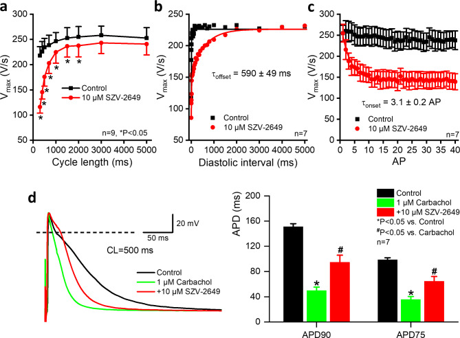
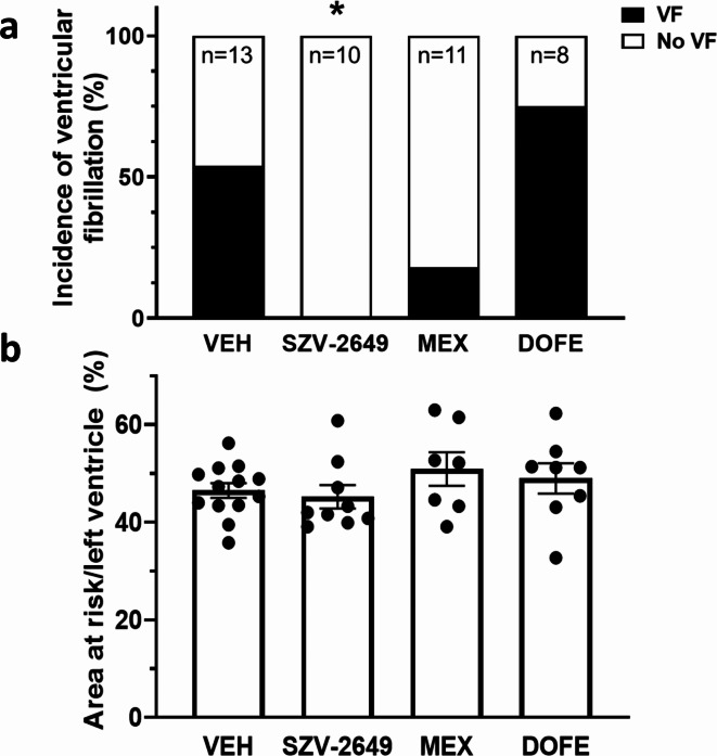
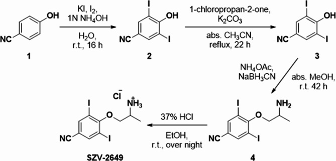
The antiarrhythmic and cardiac electrophysiological effects of SZV-2649 that contains a 2,6-diiodophenoxy moiety but lacks the benzofuran ring system present in amiodarone, were studied in mammalian cell line, rat and dog cardiac preparations. SZV-2649 exerted antiarrhythmic effects against coronary artery occlusion/reperfusion induced ventricular arrhythmias in rats and in acetylcholine- and burst stimulation induced atrial fibrillation in dogs. SZV-2649 inhibited hERG and GIRK currents in HEK cells (IC50: 342 and 529 nM, respectively). In canine ventricular myocytes, SZV-2649 (10 µM) decreased the densities of IKr, and Ito outward and INaL and ICaL inward currents. The compound (2.5-10 µM) elicited Class IB type Vmax reducing and Class III type action potential duration prolonging effects in dog right ventricular muscle preparations. In canine atrial muscle, SZV-2629 (2.5-10 µM) moderately prolonged action potential duration and this effect was greatly augmented in preparations pretreated with 1 µM carbachol. In conclusion, SZV-2649, has antiarrhythmic effects based on its multiple ion channel blocking properties. Since its chemical structure substantially differs from that of amiodarone, it is expected that SZV-2649 would exhibit fewer adverse effects than the currently used most effective multichannel inhibitor drug amiodarone and may be a promising molecule for further development.
© 2024. The Author(s).
Conflict of interest statement
The authors declare no competing interests.
Figures


References
-
- Goldenberg, I. et al. Prior history of atrial fibrillation and arrhythmic outcomes: data from the WEARIT-II prospective registry. J. Cardiovasc. Electrophysiol.2110.1111/jce.16215 (2024 Feb). Online ahead of print. - PubMed
MeSH terms
Substances
Grants and funding
- EFOP-3.6.2-16-2017-00006, the UNKP for young researchers, UNKP-23-5-SZTE-704/Ministry of Human Capacities Hungary
- EFOP-3.6.2-16-2017-00006, the UNKP for young researchers, UNKP-23-5-SZTE-704/Ministry of Human Capacities Hungary
- EFOP-3.6.2-16-2017-00006, the UNKP for young researchers, UNKP-23-5-SZTE-704/Ministry of Human Capacities Hungary
- EFOP-3.6.2-16-2017-00006, the UNKP for young researchers, UNKP-23-5-SZTE-704/Ministry of Human Capacities Hungary
- EFOP-3.6.2-16-2017-00006, the UNKP for young researchers, UNKP-23-5-SZTE-704/Ministry of Human Capacities Hungary
- EFOP-3.6.2-16-2017-00006, the UNKP for young researchers, UNKP-23-5-SZTE-704/Ministry of Human Capacities Hungary
- EFOP-3.6.2-16-2017-00006, the UNKP for young researchers, UNKP-23-5-SZTE-704/Ministry of Human Capacities Hungary
- EFOP-3.6.2-16-2017-00006, the UNKP for young researchers, UNKP-23-5-SZTE-704/Ministry of Human Capacities Hungary
- EFOP-3.6.2-16-2017-00006, the UNKP for young researchers, UNKP-23-5-SZTE-704/Ministry of Human Capacities Hungary
- KDP-2020/Ministry for Innovation and Technology, Cooperative Doctoral Programme
- RRF-2.3.1-21-2022-00001/Recovery and Resilience Facility (RRF)
- NKFIH K 135464, K 142738, K 147212 TKP2021-EGA-32, FK 138223, GINOP-2.3.2.-15-2016-00006, GINOP-2.3.2.-15-2016-00040/National Research Development and Innovation Office
- NKFIH K 135464, K 142738, K 147212 TKP2021-EGA-32, FK 138223, GINOP-2.3.2.-15-2016-00006, GINOP-2.3.2.-15-2016-00040/National Research Development and Innovation Office
- NKFIH K 135464, K 142738, K 147212 TKP2021-EGA-32, FK 138223, GINOP-2.3.2.-15-2016-00006, GINOP-2.3.2.-15-2016-00040/National Research Development and Innovation Office
- NKFIH K 135464, K 142738, K 147212 TKP2021-EGA-32, FK 138223, GINOP-2.3.2.-15-2016-00006, GINOP-2.3.2.-15-2016-00040/National Research Development and Innovation Office
- NKFIH K 135464, K 142738, K 147212 TKP2021-EGA-32, FK 138223, GINOP-2.3.2.-15-2016-00006, GINOP-2.3.2.-15-2016-00040/National Research Development and Innovation Office
- NKFIH K 135464, K 142738, K 147212 TKP2021-EGA-32, FK 138223, GINOP-2.3.2.-15-2016-00006, GINOP-2.3.2.-15-2016-00040/National Research Development and Innovation Office
- NKFIH K 135464, K 142738, K 147212 TKP2021-EGA-32, FK 138223, GINOP-2.3.2.-15-2016-00006, GINOP-2.3.2.-15-2016-00040/National Research Development and Innovation Office
- SZTE AOK-KKA 2021, SZGYA 2021, SZTE AOK-KKA 2022/The Albert Szent-Györgyi Medical School institutional grant
- SZTE AOK-KKA 2021, SZGYA 2021, SZTE AOK-KKA 2022/The Albert Szent-Györgyi Medical School institutional grant
- SZTE AOK-KKA 2021, SZGYA 2021, SZTE AOK-KKA 2022/The Albert Szent-Györgyi Medical School institutional grant
- HUN-REN TKI project/Hungarian Research Network
- HUN-REN TKI project/Hungarian Research Network
- HUN-REN TKI project/Hungarian Research Network
- bo_481_21/Hungarian Academy of Sciences, János Bolyai Research Scholarships
- RRF-2.3.1-21-2022-00003/National Heart Laboratory, Hungary
LinkOut - more resources
Full Text Sources
Miscellaneous
