. 2024 Oct 22;43(10):114809.
doi: 10.1016/j.celrep.2024.114809. Epub 2024 Oct 5.

The VLDLR entry receptor is required for the pathogenesis of multiple encephalitic alphaviruses

Affiliations

The VLDLR entry receptor is required for the pathogenesis of multiple encephalitic alphaviruses

Sathvik Palakurty et al. Cell Rep. .

Abstract

The very-low-density lipoprotein receptor (VLDLR) has been reported as an entry receptor for Semliki Forest (SFV) and Eastern equine encephalitis (EEEV) alphaviruses in cell cultures. However, the role of VLDLR in alphavirus pathogenesis and the extent to which other alphaviruses can engage VLDLR remains unclear. Here, using a surface protein-targeted CRISPR-Cas9 screen, we identify VLDLR as a receptor for Western equine encephalitis virus (WEEV) and demonstrate that it promotes the infection of multiple viruses in the WEE antigenic complex. In vivo studies show that the pathogenicity of WEEV, EEEV, and SFV, but not the distantly related Venezuelan equine encephalitis virus, is markedly diminished in VLDLR-deficient mice and that mice treated with a soluble VLDLR-Fc decoy molecule are protected against disease. Overall, these results expand our understanding of the role of VLDLR in alphavirus pathogenesis and provide a potential path for developing countermeasures against alphaviruses from different antigenic complexes.

Keywords: CP: Immunology; CP: Microbiology; Receptor; alphavirus; decoy; pathogenesis; therapy; tropism.


Conflict of interest statement

Declaration of interests M.S.D. is a consultant to or on the scientific advisory board for Inbios, IntegerBio, Akagera Medicines, Merck, GlaxoSmithKline, and Moderna. The Diamond laboratory has received unrelated funding support in sponsored research agreements from Emergent BioSolutions, Moderna, IntegerBio, and Vir Biotechnology. D.H.F. is a founder of Courier Therapeutics and has received unrelated funding support from Emergent BioSolutions and Mallinckrodt Pharmaceuticals.

Figures

Figure 1.
Figure 1.. A CRISPR-Cas9 screen identifies VLDLR as a factor that promotes WEEV infection
(A) Phylogenetic tree generated from structural protein sequences of indicated alphaviruses with members of the WEE complex highlighted in brown. (B) MAGeCK plot of enriched genes in surviving cells following two rounds of SINV-WEEV-GFP infection. (C) SINV-WEEV-GFP McMillan infection (MOI of 3, 8 h) in HAP-1 control, ΔVLDLR, and ΔVLDLR complemented with VLDLR-FLAG cells as assessed by GFP expression (pooled from 3 experiments performed in triplicate; all data points are shown). (D) SINV-WEEV-GFP McMillan infection in HAP-1 cells pre-treated with blocking anti-VLDLR antibody (1H5) for 1 h prior (pooled from 2–3 experiments performed in duplicate or triplicate; all data points are shown). (E–J) Infection of indicated viruses (MOI of 3, 24 h) as quantified by GFP expression (E, G, and H), WEEV-209 mAb (anti-WEEV E2) staining (F, H, and I), or DC2.112 (pan alphavirus anti-E1) staining (J) of K562 control, K562-VLDLR, K562-chicken MXRA8, or K562-mouse MXRA8 cells (pooled from 3–4 independent experiments performed in duplicate; all data points are shown). (K) Viral growth curves in the indicated cell lines after inoculation with SINV-WEEV McMillan, SINV-EEEV FL93, and RRV T48 (MOI of 0.1) as determined by focus-forming assay on Vero cells (pooled from 4 experiments; error bars indicate standard error of the mean). Dotted lines show the limit of detection (LOD), and column heights indicate mean values. Statistical analysis was performed on the mean values of the biological replicates: (C and E–H) one-way ANOVA with Tukey’s post-test and (D) Student’s t test (ns, not significant, **p < 0.01, ***p < 0.001, and ****p < 0.0001). See also Figures S1 and S2 and Tables S1 and S2.
Figure 2.
Figure 2.. VLDLR promotes attachment and internalization of WEEV via its ligand-binding domain
(A and B) Attachment of SINV-WEEV to the cell surface. Flow cytometry plots (A) and quantification (B) of anti-E2 (WEEV-209) staining of the surface of K562-, K562-chicken MXRA8-, and K562-VLDLR-expressing cells following incubation with SINV-WEEV at 4°C (pooled from 3 experiments performed in triplicate; all data points are shown). (C and D) Internalization of SINV-WEEV by cells. Flow cytometry plots (C) and quantification (D) of intracellular E2 (anti-WEEV-209) staining of K562-, K562- chicken MXRA8-, and K562-VLDLR-expressing cells following incubation with SINV-WEEV at 37°C (pooled from 3 experiments performed in triplicate). (E) Diagram of VLDLR and VLDLRΔLBD proteins (left); infection as assessed by GFP expression by flow cytometry (right) of K562 cells expressing VLDLR or VLDLRΔLBD after inoculation (MOI 3, 24 h) with SINV-WEEV-GFP McMillan (pooled from 3 experiments in duplicate). (F) Quantification of cell surface binding by indicated concentration of Fc-fusion proteins to Vero cells inoculated with indicated chimeric SINV (pooled from 2 experiments). (G) Binding of Fc-fusion proteins to immobilized VLPs. The indicated VLPs were captured on biosensors using mouse mAbs followed by incubation with 5 μg/mL of indicated Fc-fusion proteins (representative of two experiments). Column heights and bars indicate mean values. Statistical analysis was performed on the mean values of the biological replicates: (B and D) one-way ANOVA with Tukey’s post-test and (E) Student’s t test (***p < 0.001 and ****p < 0.0001). See also Figures S2 and S3.
Figure 3.
Figure 3.. VLDLR-LBD-Fc neutralizes WEEV McMillan infection and protects against lethal challenge
(A) SINV-WEEV-GFP McMillan and SINV-VEEV-GFP TrD infection as assessed by GFP expression following incubation with indicated soluble receptor decoy proteins prior to inoculation of HEK293T cells. Data are normalized to infection with no protein (pooled from 3 experiments). (B) SINV-WEEV-GFP McMillan infection following incubation with indicated decoy proteins (10 μg/mL, 3-fold dilutions) prior to inoculation of the indicated K562 cells (pooled from 3 experiments performed in duplicate; all data points are shown). (C and E) Survival (left) and weight loss (right) of 6-week-old C57BL6/J mice injected with VLDLR-LBD-Fc (n = 10, C and E) or LDLRAD3-LA1-Fc (n = 10, C and E) and 103 FFUs of WEEV McMillan (C) or 102 FFUs of VEEV ZPC738 (E). (D and F) Viral RNA levels in indicated tissues at 4 days post-infection (dpi) in mice inoculated with WEEV McMillan and co-administered (D: n = 10 for both groups) or administered separately as prophylaxis via an intraperitoneal injection (F: n = 8, VLDLR-LBD-Fc; n = 7; LDLRAD3-LA1-Fc) the indicated Fc-fusion protein (dotted lines show the limit of detection [LOD]; values at the LOD are plotted slightly below). Column heights indicate mean values, and bars indicate median values. Statistical analysis: (C and E) log-rank test and (D) Mann-Whitney test (*p < 0.05, **p < 0.01, and ****p < 0.0001). See also Figure S4.
Figure 4.
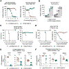
Figure 4.. VLDLR-deficient mice are protected from challenge with WEEV and other encephalitic alphaviruses that bind VLDLR
(A–D) Survival rates of indicated mice following subcutaneous inoculation with 103 FFUs of WEEV McMillan (A), intraperitoneal inoculation with 103 FFUs of SFV Kumba (B), subcutaneous inoculation with 2 × 10² FFUs of EEEV-MADV Argentina 1936 (C), or subcutaneous inoculation with 102 FFUs of VEEV ZPC738 (D). (E) Viral RNA levels as determined by RT-qPCR in the indicated perfused tissues at 5 dpi in mice inoculated subcutaneously with WEEV McMillan. (F) Viral burden as assessed by focus-forming assay in homogenates of the indicated brain regions at 36 h after intracranial infection of indicated mice with 102 FFUs of WEEV McMillan. Dotted lines show the LOD, and bars indicate median values. Data are pooled from two experiments for each panel as follows (A) n = 13–20 per group, (B) n = 6–7 per group, (C) n = 5–8 per group, (D) n = 5–7 per group, (E) n = 10 per group, and (F) n = 7–8 per group. Statistical analysis: (A–D) log-rank test and (E and F) Mann-Whitney test (ns, not significant, *p < 0.05, **p < 0.01, and ****p < 0.0001). See also Figures S4 and S5.

References

    1. Paessler S, Yun NE, Judy BM, Dziuba N, Zacks MA, Grund AH, Frolov I, Campbell GA, Weaver SC, and Estes DM (2007). Alpha-beta T cells provide protection against lethal encephalitis in the murine model of VEEV infection. Virology 367, 307–323. - PMC - PubMed
    1. Paessler S, Ni H, Petrakova O, Fayzulin RZ, Yun N, Anishchenko M, Weaver SC, and Frolov I (2006). Replication and clearance of Venezuelan equine encephalitis virus from the brains of animals vaccinated with chimeric SIN/VEE viruses. J. Virol 80, 2784–2796. - PMC - PubMed
    1. Reeves WC, Hutson GA, Bellamy RE, and Scrivani RP (1958). Chronic latent infections of birds with Western equine encephalomyelitis virus. Proc. Soc. Exp. Biol. Med 97, 733–736. - PubMed
    1. Deresiewicz RL, Thaler SJ, Hsu L, and Zamani AA (1997). Clinical and Neuroradiographic Manifestations of Eastern Equine Encephalitis. N. Engl. J. Med 336, 1867–1874. - PubMed
    1. Weaver SC, and Barrett ADT (2004). Transmission cycles, host range, evolution and emergence of arboviral disease. Nat. Rev. Microbiol 2, 789–801. - PMC - PubMed

Publication types

LinkOut - more resources