. 2024 Oct 6;7(1):1275.
doi: 10.1038/s42003-024-06985-x.

Cryo-EM analysis of Pseudomonas phage Pa193 structural components

Affiliations

Cryo-EM analysis of Pseudomonas phage Pa193 structural components

Stephano M Iglesias et al. Commun Biol. .

Abstract

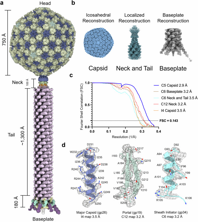
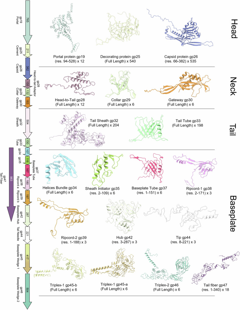
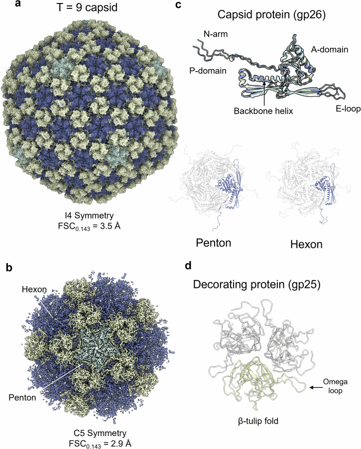
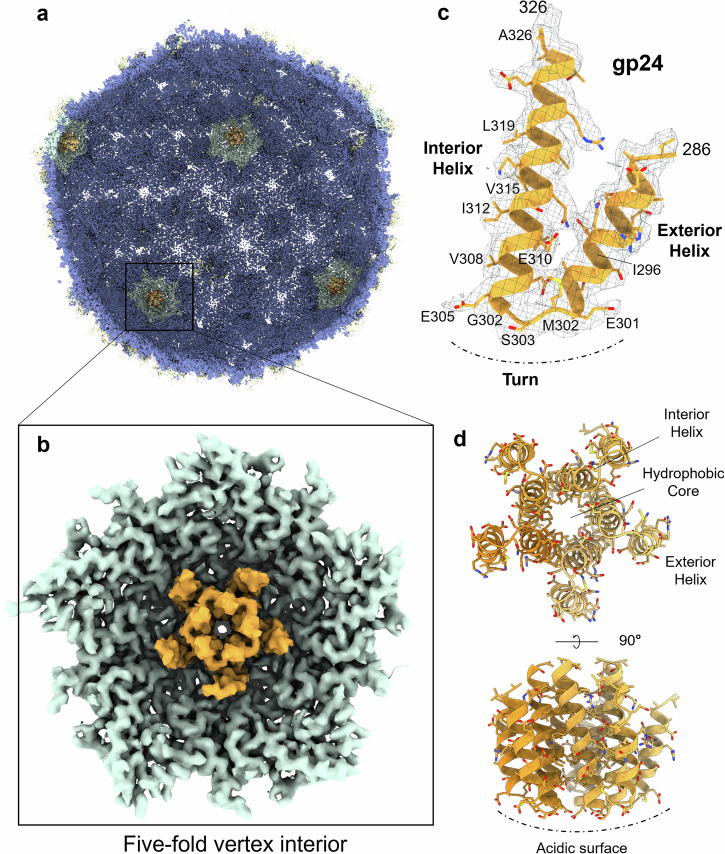
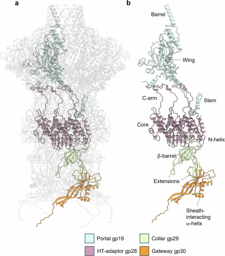
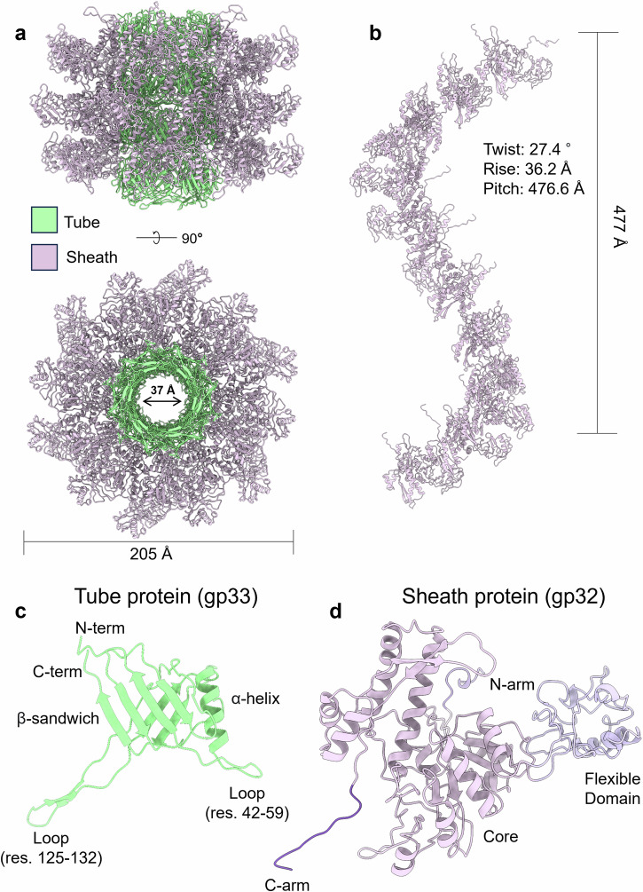
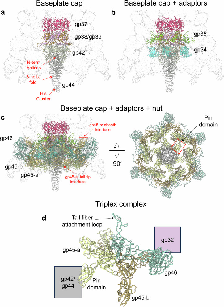
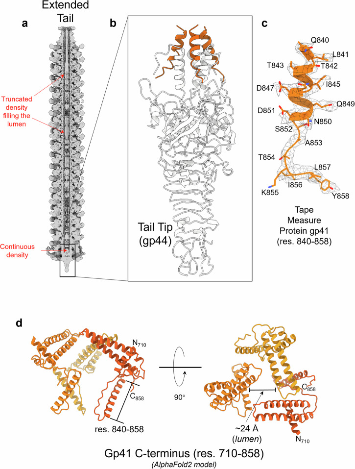
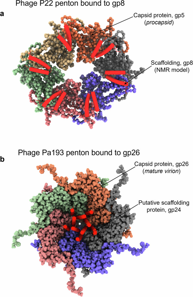
The World Health Organization has designated Pseudomonas aeruginosa as a critical pathogen for the development of new antimicrobials. Bacterial viruses, or bacteriophages, have been used in various clinical settings, commonly called phage therapy, to address this growing public health crisis. Here, we describe a high-resolution structural atlas of a therapeutic, contractile-tailed Pseudomonas phage, Pa193. We used bioinformatics, proteomics, and cryogenic electron microscopy single particle analysis to identify, annotate, and build atomic models for 21 distinct structural polypeptide chains forming the icosahedral capsid, neck, contractile tail, and baseplate. We identified a putative scaffolding protein stabilizing the interior of the capsid 5-fold vertex. We also visualized a large portion of Pa193 ~ 500 Å long tail fibers and resolved the interface between the baseplate and tail fibers. The work presented here provides a framework to support a better understanding of phages as biomedicines for phage therapy and inform engineering opportunities.


Conflict of interest statement

J.R., E.S., R.G., A.S., L.S., P.K., D.B., and S.L. are employees of Armata Pharmaceuticals Inc., a company involved in the development of bacteriophage therapies. LG and JW were contracted by Armata under a fee-for-service agreement. The other authors declare that the research was conducted in a way that is free of financial or commercial relationship that could be construed as conflict of interest.

Figures

Fig. 1
Fig. 1. Cryo-EM reconstruction of Pseudomonas phage Pa193.
a Composite map of the Pa193 virion with an extended tail. b 3D-reconstructions of phage Pa193 determined in this study: the capsid solved using icosahedral (I4 symmetry) reconstruction; the neck and tail determined using localized reconstruction; the baseplate determined picking the tail tip distal from the capsid with C6 symmetry imposed. c Fourier Shell Correlation (FSC) curves for all reconstructions determined in this study. All reconstructions were masked. d Representative densities corresponding to each cryo-EM SPA technique above. Major capsid protein residues 231–250 (purple) fit in the I4 icosahedral reconstruction; portal protein residues 183–188; 209–217 (green) fit into the C12 localized reconstruction; sheath initiator protein residues 92–106 (light blue) fit in the C6 baseplate reconstruction.
Fig. 2
Fig. 2. Structural atlas of Pseudomonas phage Pa193.
Ribbon diagrams of 19 of the 21 gene products identified and built de novo in this work: three in the capsid, four in the neck, three in the tail, and eleven in the baseplate. 19 ORFs were annotated and built as full-length and are shown in Fig. 2. The partial models for the putative scaffolding, gp24 (see Fig. 4), and tape measure protein, gp41 (see Fig. 9), are not displayed in this atlas.
Fig. 3
Fig. 3. Identification and annotation of Pa193 phage capsid and decorating proteins.
a Cryo-EM reconstruction of Pa193 capsid measured at 3.5 Å resolution and displayed at 3.5 σ. Major capsid protein assembles as pentons are colored (green) or hexons (blue), and decorating protein forms a trimer at every three-fold vertex (yellow). b Cryo-EM localized reconstruction of Pa193 capsid five-fold vertex at 2.9 Å resolution. The map is displayed at 4.5 σ and has the same protein color scheme as in panel (a). c Overlay of the Pa193 capsid protein gp26 conformers found in the penton (green), hexon (blue), and icosahedral 3-fold hexon (yellow) (penton:hexon, RMSD 5.5 Å; hexon:icosahedral 3-fold hexon, RMSD 0.001 Å). Ribbon diagrams of the capsid protein assembled in a penton or as part of a hexon are also shown. d Ribbon diagram of the decorating protein gp25 (residues 1–211) assembled as a trimer.
Fig. 4
Fig. 4. Identification of Pa193 putative scaffolding protein gp24.
a Cross-section view of Pa193 capsid map (I4 symmetry). b magnified view of a five-fold vertex visualized from the capsid interior, highlighting the helix-turn-helix density visible at the five-fold vertex (colored orange). c Magnified view of the putative scaffolding protein density calculated at 3.5 Å resolution and displayed at 1.5 σ, overlaid to the ModelAngelo model. d Cartoon representation of the gp24 pentamer.
Fig. 5
Fig. 5. Structure of Pa193 neck assembly.
a Overview of the Pa193 neck complex. Portal gp19 (green) binds to the HT-adapter gp28 (purple), which in turn binds to the collar gp29 (light green) and gateway gp30 (orange). b Ribbon diagram of the isolated neck protomer: gp19 (residues 94–528), gp28 (residues 1–155), gp29 (residues 1–132), and gp30 (residues 1–183).
Fig. 6
Fig. 6. Tertiary and quaternary structure of Pseudomonas phage Pa193 tail proteins.
a Side-view of a segment of Pa193 extended tail displayed as cartoon models. Four stacks of hexameric tube subunits (green) form the inner layer of the tail, while three layers of sheath proteins (magenta) assemble around the hexameric tube structure and comprise the outer layer of the tail. b Sheath protein (12 proteins in one complete turn) comprises a helical tail with a rise of 27.4 Å, a twist of 36.2°, and a pitch of 476.6 Å degrees. c Cartoon representation of the tail tube gp33 tertiary structure (light green) that comprises a β-sandwich (residues 9–16, 63–68, 97–114, 135–150), backbone α-helix (residues 72–85), and N-term (residues 1–8) extensions. d Cartoon representation of the sheath protein (gp32) tertiary structure including its flexible domain (residues 91–210), core (residues 23–90; 211–491), C-arm (residues 492–504), and N-arm (residues 1–22).
Fig. 7
Fig. 7. The composition and organization of the simple Pa193 baseplate.
a A semitransparent map of the Pa193 baseplate with overlayed ribbon diagrams of the baseplate cap comprising the baseplate tube gp37, ripcord-1 gp38, ripcord-2 gp39, tail hub gp42, and tail tip gp44. b The baseplate cap with the addition of two adapter proteins: the sheath initiator gp35 and helical bundle gp34. c The baseplate cap, with adapters and nut complex, comprises six copies of the triplex complexes (Baseplate Wedges gp45-a, gp45-b, and gp46). The left panel shows a bottom-up view of the Pa193 baseplate. The red square shows the position of one gp45-a Pin domain. d Ribbon diagram of Pa193 triplex complex formed by gp45-a, gp45-b and gp46.
Fig. 8
Fig. 8. Tail fiber structure and attachment to the baseplate.
a A cryo-micrograph of bacteriophage Pa193 reveals prominent and straight tail fibers. b Cartoon schematic of the baseplate assembly with the tail fibers (residues 1–340) attached to six vertices of the baseplate. c Cartoon representation of tail fiber gp47 AlphaFold2 trimeric assembly (residues 1–340) overlaid to the localized reconstruction (calculated at 3.3 Å resolution and displayed at 1.5 σ). d Zoom-in view of the interface between baseplate gp46 tail fiber attachment loop (green) and gp47 tail fiber loops (magenta). The red frame shows a zoom-in view of the hydrophobic core formed between gp46 (green) and gp47 (magenta).
Fig. 9
Fig. 9. Identification of the Pa193 tape measure protein C-termini.
a Cross-section of Pa193 tail calculated at 7.5 Å resolution and displayed at 3.0 σ. b Ribbon diagram of the tail tip gp44 (gray) bound to three TMP (gp41) C-termini (orange). c Zoomed-in view of the trimeric tape measure protein C-term (residues 840–858) overlaid to the C6 reconstruction (calculated at 3.3 Å resolution and displayed at 2.5 σ). d An AlphaFold2 model of Pa193 TMP C-terminal residues 710–858 predicted as a trimeric assembly. The position of residues 840–858 is shown.
Fig. 10
Fig. 10. Comparing P22 and Pa193 putative scaffolding proteins.
a A space-filling representation of phage P22 (PDB: 2XYY) (a) and Pa193 (b) pentons viewed from inside the procapsid and capsid, respectively. P22 scaffolding protein gp8 (PDB: 2GP8) and Pa193 putative scaffolding gp24 are shown as red helical hairpin.

Update of

References

    1. Qin, S. et al. Pseudomonas aeruginosa: pathogenesis, virulence factors, antibiotic resistance, interaction with host, technology advances and emerging therapeutics. Signal Transduct. Target Ther.7, 199 (2022). - PMC - PubMed
    1. Elborn, J. S. Cystic fibrosis. Lancet388, 2519–2531 (2016). - PubMed
    1. Morrison, C. B., Markovetz, M. R. & Ehre, C. Mucus, mucins, and cystic fibrosis. Pediatr. Pulmonol.54, S84–S96 (2019). - PMC - PubMed
    1. Law, S. M. & Gray, R. D. Neutrophil extracellular traps and the dysfunctional innate immune response of cystic fibrosis lung disease: a review. J. Inflamm. (Lond.)14, 29 (2017). - PMC - PubMed
    1. Breidenstein, E. B., de la Fuente-Nunez, C. & Hancock, R. E. Pseudomonas aeruginosa: all roads lead to resistance. Trends Microbiol19, 419–426 (2011). - PubMed

MeSH terms

Substances

LinkOut - more resources