

'Color Doppler stripes' make it difficult to diagnose the severity of valvular heart diseases: a report of two cases
- PMID: 39370511
- PMCID: PMC11457407
- DOI: 10.1186/s12947-024-00331-1
'Color Doppler stripes' make it difficult to diagnose the severity of valvular heart diseases: a report of two cases
Abstract
Background: Echocardiography remains the reference-standard imaging technique for assessing valvular heart disease (VHD), but artifacts like the 'color Doppler stripe' can complicate diagnosis. This artifact is not widely recognized and can mimic severe VHD, leading to potential misdiagnoses. We present two cases where color Doppler stripes mimicked severe VHD, highlighting the need for awareness and accurate interpretation in echocardiographic assessments.
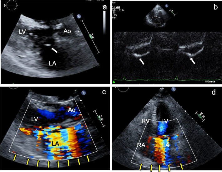
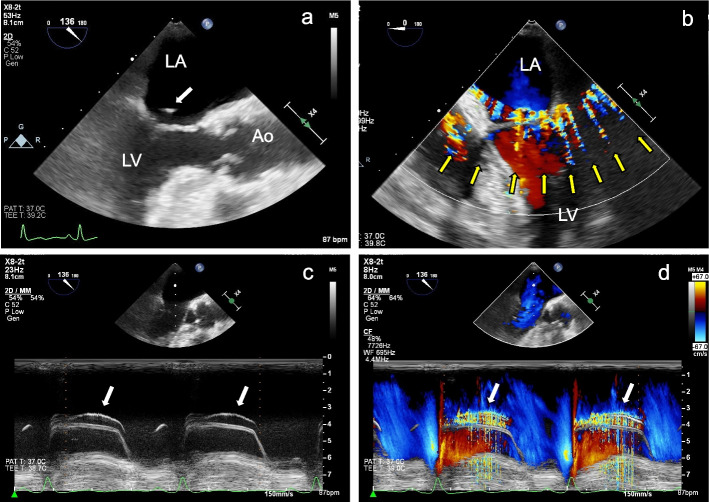
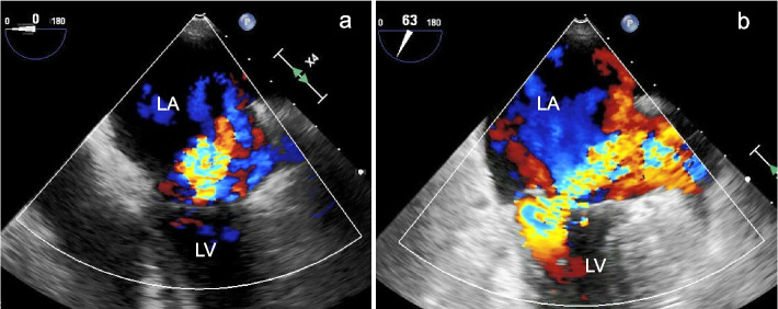
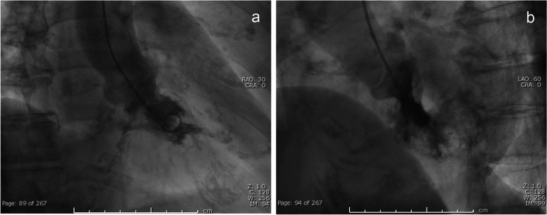
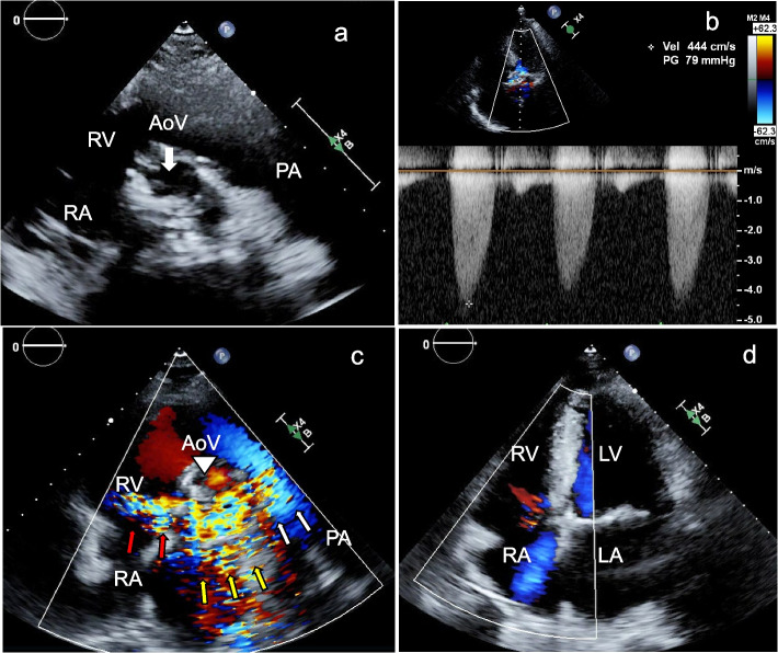
Case presentations: Case 1: An 85-year-old patient was referred for mitral valve surgery due to suspected severe mitral regurgitation (MR). Upon evaluation, transthoracic echocardiography (TTE) showed mitral valve prolapse (P3) and a high-echoic, vibrating structure attached to the mitral valve, indicative of chordal rupture. Color Doppler echocardiography revealed strong systolic signals in the left atrium, mimicking severe MR. Transesophageal echocardiography (TEE) also detected the vibrating structure and color Doppler stripes in the left atrium, left ventricle, and outside the cardiac chambers. The PISA method on TEE indicated moderate MR and left ventriculography showed Sellers grade II MR. The artifact was identified as color Doppler stripes caused by the vibrating high-echoic structure from the ruptured chorda. Case 2: A 64-year-old patient with severe aortic stenosis, end-stage kidney disease requiring hemodialysis, and a history of coronary bypass grafting presented for routine follow-up. B-mode echocardiography showed a severely calcified tricuspid aortic valve with a vibrating calcified nodule and restricted opening, corresponding to severe aortic stenosis. During systole, color Doppler signals were observed around the aortic, pulmonary, and tricuspid valves, mimicking significant pulmonary stenosis and tricuspid regurgitation. However, pulmonary stenosis was ruled out as the pulmonary valve opening was normal. Mild tricuspid regurgitation was confirmed in the apical view.
Conclusions: These cases highlight the diagnostic challenges posed by color Doppler stripes. Recognizing and understanding this artifact are crucial for the accurate diagnosis and management of VHD, ensuring appropriate treatment and patient outcomes.
Keywords: Artifact; Color Doppler stripe; Valvular heart disease.
© 2024. The Author(s).
Conflict of interest statement
The authors declare no competing interests.
Figures
References
-
- Nkomo VT, Gardin JM, Skelton TN, Gottdiener JS, Scott CG, Enriquez-Sarano M. Burden of valvular heart diseases: a population-based study. Lancet. 2006;368(9540):1005–11. - PubMed
-
- Coffey S, Roberts-Thomson R, Brown A, Carapetis J, Chen M, Enriquez-Sarano M, et al. Global epidemiology of valvular heart disease. Nat Rev Cardiol. 2021;18(12):853–64. - PubMed
-
- Vahanian A, Beyersdorf F, Praz F, Milojevic M, Baldus S, Bauersachs J, et al. 2021 ESC/EACTS Guidelines for the management of valvular heart disease. Eur Heart J. 2022;43(7):561–632. - PubMed
-
- Otto CM, Nishimura RA, Bonow RO, Carabello BA, Erwin JP 3rd, Gentile F, et al. 2020 ACC/AHA guideline for the management of patients with valvular heart disease: a report of the American College of Cardiology/American Heart Association Joint Committee on clinical practice guidelines. Circulation. 2021;143(5):e72–227. - PubMed
-
- Izumi C, Eishi K, Ashihara K, Arita T, Otsuji Y, Kunihara T, et al. JCS/JSCS/JATS/JSVS 2020 guidelines on the management of valvular heart disease. Circ J. 2020;84(11):2037–119. - PubMed
Publication types
MeSH terms
LinkOut - more resources
Full Text Sources
