

Mapping epidermal and dermal cellular senescence in human skin aging
- PMID: 39370688
- PMCID: PMC11709101
- DOI: 10.1111/acel.14358
Mapping epidermal and dermal cellular senescence in human skin aging
Abstract
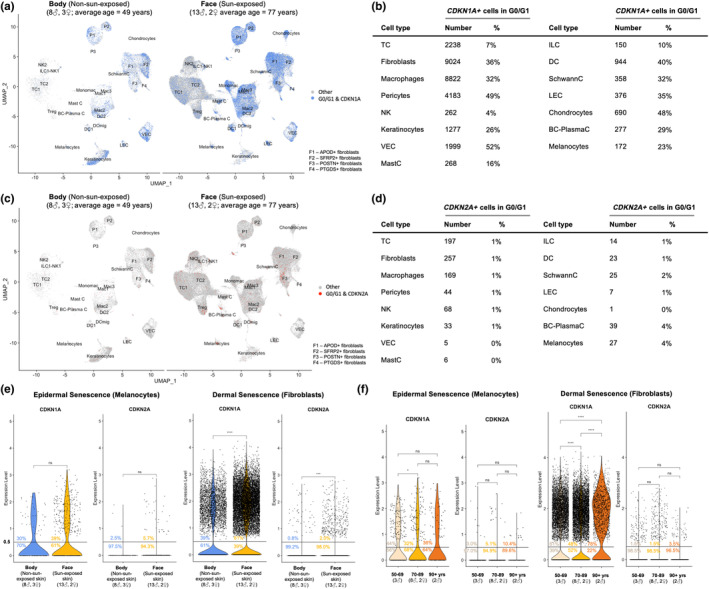
Single-cell RNA sequencing and spatial transcriptomics enable unprecedented insight into cellular and molecular pathways implicated in human skin aging and regeneration. Senescent cells are individual cells that are irreversibly cell cycle arrested and can accumulate across the human lifespan due to cell-intrinsic and -extrinsic stressors. With an atlas of single-cell RNA-sequencing and spatial transcriptomics, epidermal and dermal senescence and its effects were investigated, with a focus on melanocytes and fibroblasts. Photoaging due to ultraviolet light exposure was associated with higher burdens of senescent cells, a sign of biological aging, compared to chronological aging. A skin-specific cellular senescence gene set, termed SenSkin™, was curated and confirmed to be elevated in the context of photoaging, chronological aging, and non-replicating CDKN1A+ (p21) cells. In the epidermis, senescent melanocytes were associated with elevated melanin synthesis, suggesting haphazard pigmentation, while in the dermis, senescent reticular dermal fibroblasts were associated with decreased collagen and elastic fiber synthesis. Spatial analysis revealed the tendency for senescent cells to cluster, particularly in photoaged skin. This work proposes a strategy for characterizing age-related skin dysfunction through the lens of cellular senescence and suggests a role for senescent epidermal cells (i.e., melanocytes) and senescent dermal cells (i.e., reticular dermal fibroblasts) in age-related skin sequelae.
Keywords: cellular senescence; dermis; epidermis; single‐cell gene expression analysis; skin aging; skin pathology; spatial analysis.
© 2024 The Author(s). Aging Cell published by Anatomical Society and John Wiley & Sons Ltd.
Conflict of interest statement
J.L.K. and T.T. have financial interest related to this research, including patents, and pending patents covering senolytic drugs and their uses that are held by Mayo Clinic. This research has been reviewed by the Mayo Clinic Conflict of Interest Review Board and was conducted in compliance with Mayo Clinic and Cedars‐Sinai conflict of interest policies. M.D.L. is a co‐founder of Fibrodyne and has two patents related to skin fibroblasts. In the last twenty‐four months, D.B.A. has received personal payments or promises for same from: Novo Nordisk Foundation; and Zero Longevity Science (as stock options). D.B.A.'s institution, Indiana University, and the Indiana University Foundation have received funds or donations to support his research or educational activities from: Eli Lilly and Company; Pfizer, Inc.; and numerous other for‐profit and non‐profit organizations to support the work of the School of Public Health and the university more broadly.
Figures


References
-
- Ahlers, J. M. D. , Falckenhayn, C. , Holzscheck, N. , Solé‐Boldo, L. , Schütz, S. , Wenck, H. , Winnefeld, M. , Lyko, F. , Grönniger, E. , & Siracusa, A. (2021). Single‐cell rna profiling of human skin reveals age‐related loss of dermal sheath cells and their contribution to a juvenile phenotype. Frontiers in Genetics, 12, 797747. 10.3389/fgene.2021.797747 - DOI - PMC - PubMed
-
- Almet, A. A. , Yuan, H. , Annusver, K. , Ramos, R. , Liu, Y. , Wiedemann, J. , Sorkin, D. H. , Landén, N. X. , Sonkoly, E. , Haniffa, M. , Nie, Q. , Lichtenberger, B. M. , Luecken, M. D. , Andersen, B. , Tsoi, L. C. , Watt, F. M. , Gudjonsson, J. E. , Plikus, M. V. , & Kasper, M. (2023). A roadmap for a consensus human skin cell atlas and single‐cell data standardization. Journal of Investigative Dermatology, 143(9), 1667–1677. 10.1016/j.jid.2023.03.1679 - DOI - PMC - PubMed
-
- Blázquez‐Prieto, J. , Huidobro, C. , López‐Alonso, I. , Amado‐Rodriguez, L. , Martín‐Vicente, P. , López‐Martínez, C. , Crespo, I. , Pantoja, C. , Fernandez‐Marcos, P. J. , Serrano, M. , Sznajder, J. I. , & Albaiceta, G. M. (2021). Activation of p21 limits acute lung injury and induces early senescence after acid aspiration and mechanical ventilation. Translational Research, 233, 104–116. 10.1016/j.trsl.2021.01.008 - DOI - PMC - PubMed
-
- Chandra, A. , Lagnado, A. B. , Farr, J. N. , Doolittle, M. , Tchkonia, T. , Kirkland, J. L. , LeBrasseur, N. K. , Robbins, P. D. , Niedernhofer, L. J. , Ikeno, Y. , Passos, J. F. , Monroe, D. G. , Pignolo, R. J. , & Khosla, S. (2022). Targeted clearance of p21‐ but not p16‐positive senescent cells prevents radiation‐induced osteoporosis and increased marrow adiposity. Aging Cell, 21(5), e13602. 10.1111/acel.13602 - DOI - PMC - PubMed
-
- Chintala, S. , Li, W. , Lamoreux, M. L. , Ito, S. , Wakamatsu, K. , Sviderskaya, E. V. , Bennett, D. C. , Park, Y. M. , Gahl, W. A. , Huizing, M. , Spritz, R. A. , Ben, S. , Novak, E. K. , Tan, J. , & Swank, R. T. (2005). Slc7a11 gene controls production of pheomelanin pigment and proliferation of cultured cells. Proceedings of the National Academy of Sciences of the United States of America, 102(31), 10964–10969. 10.1073/pnas.0502856102 - DOI - PMC - PubMed
MeSH terms
Grants and funding
- 211276/E/18/Z/WT_/Wellcome Trust/United Kingdom
- National Cattlemen's Beef Association
- P30AG050886/NH/NIH HHS/United States
- P01AG062413/NH/NIH HHS/United States
- R01 AG076515/AG/NIA NIH HHS/United States
- Gordon and Betty Moore Foundation
- National Pork Board
- Pfizer, Inc
- P01 AG062413/AG/NIA NIH HHS/United States
- Novo Nordisk Fonden
- Noaber Foundation
- the Connor Fund
- 096540/Z/11/Z/WT_/Wellcome Trust/United Kingdom
- Weight Watchers
- R03AG082919-01/NH/NIH HHS/United States
- U54AG079754/NH/NIH HHS/United States
- U54 AG079754/AG/NIA NIH HHS/United States
- U24AG056053/NH/NIH HHS/United States
- WT_/Wellcome Trust/United Kingdom
- R01AG076515/NH/NIH HHS/United States
- Hevolution Foundation
- R37AG013825/NH/NIH HHS/United States
- R33AG061456/NH/NIH HHS/United States
- T32GM065841/NH/NIH HHS/United States
- R03 AG082919/AG/NIA NIH HHS/United States
LinkOut - more resources
Full Text Sources
Medical
