. 2024 Nov 1;32(6):793-800.
doi: 10.4062/biomolther.2024.033. Epub 2024 Oct 7.

Jolkinolide B Ameliorates Liver Inflammation and Lipogenesis by Regulating JAK/STAT3 Pathway

Affiliations

Jolkinolide B Ameliorates Liver Inflammation and Lipogenesis by Regulating JAK/STAT3 Pathway

Hye-Rin Noh et al. Biomol Ther (Seoul). .

Abstract

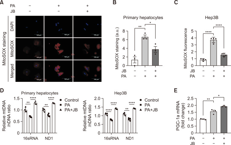
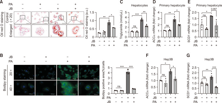
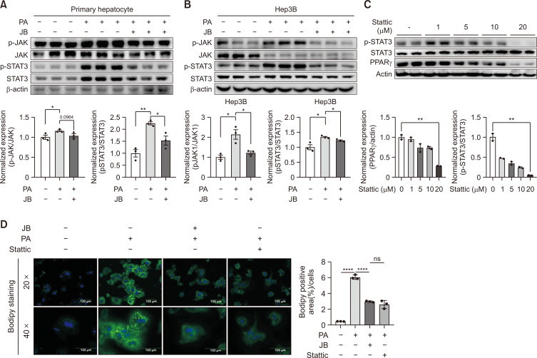
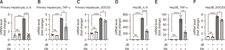
Hepatic dysregulation of lipid metabolism exacerbates inflammation and enhances the progression of metabolic dysfunction-associated steatotic liver disease (MASLD). STAT3 has been linked to lipid metabolism and inflammation. Jolkinolide B (JB), derived from Euphorbia fischeriana, is known for its pharmacological anti-inflammatory and anti-tumor properties. Therefore, this study investigated whether JB affects MASLD prevention by regulating STAT3 signaling. JB attenuated steatosis and inflammatory responses in palmitic acid (PA)-treated hepatocytes. Additionally, JB treatment reduced the mRNA expression of de-novo lipogenic genes, such as acetyl-CoA carboxylase and stearoyl-CoA desaturase 1. Interestingly, JB-mediated reduction in inflammation and lipogenesis was dependent on STAT3 signaling. JB consistently modulated mitochondrial dysfunction and the mRNA expression of inflammatory cytokines by inhibiting PA-induced JAK/STAT3 activation. This study suggests that JB is a potential therapeutic agent to prevent major stages of MASLD through inhibition of JAK/STAT3 signaling in hepatocytes.

Keywords: Inflammation; Jolkinolide B; Lipid accumulation; MASLD.


Conflict of interest statement

CONFLICT OF INTEREST

The authors have no conflicts of interest.

Figures

Fig. 1
Fig. 1
Jolkinolide B attenuates hepatocellular lipotoxicity. (A) JB structure. (B) Hepatocytes were treated with JB for each concentration (1, 5, and 10 μM), and cell viability was measured. (C) Cytotoxicity assay in hepatocytes were incubated with PA for 24 h with different concentrations (0, 1, 5, and 10 μM) of JB. Low control refers to cells under normal conditions, while the High control represents 100% cell death. (D) Whole cell lysates from primary hepatocytes and Hep3B cells were extracted and analyzed by immunoblotting of Bax/Bcl-2 after PA (200 μM) and JB (10 μM). The data are expressed as means ± sem. *p<0.05, **p<0.01, ***p<0.001, ****p<0.0001.
Fig. 2
Fig. 2
Jolkinolide B improves lipid-induced mitochondrial dysfunction. Hep3B and primary hepatocytes treat with PA (200 μM) and JB (10 μM). (A) mtROS was detected by Mitosox staining. The images were shown at 20× magnifications for each experimental condition (scale bar=100 μm). (B) Mitosox quantification was performed using the Image J software. (C) Expression of Mitosox red fluorescence for mtROS in Hep3B cells was measured with a microplate reader. (D) Relative mtDNA/nDNA ratio from primary hepatocytes and Hep3B cells under treated with PA (200 μM) and JB (10 μM) for 24 h. A comparison of ND1 and 16S rRNA DNA expression relative to HK DNA expression will give a measure of mtDNA copy number to nDNA copy number ratio. (E) The mRNA expression of PGC-1a in primary hepatocytes under PA (200 μM) and JB (10 μM) treatment for 24 h. Relative mRNA expression levels were normalized to GAPDH levels. The data are expressed as means ± sem. *p<0.05, **p<0.01, and ****p<0.0001.
Fig. 3
Fig. 3
Jolkinolide B inhibits PA-induced lipid accumulation and lipogenesis. (A) Primary hepatocyte lipid accumulation was presented by Oil Red O staining. Red-stained intracellular fat content was quantified with a fluorescence microscope. The images were shown at 20× magnifications for each experimental condition (scale bar=100 μm). (B) Primary hepatocyte lipid accumulation was presented by BODIPY staining. Lipid droplet-stained BODIPY was imaged with a fluorescence microscope. BODIPY expression was quantified using Image J. The images were shown at 20× and 40× magnifications for each experimental condition (scale bar=100 μm). (C) Cellular TG content measurement was performed in primary hepatocytes under PA and JB treatment. (D, E) Lipogenesis genes were measured using qRT-PCR in primary hepatocytes. (F, G) Expression of the lipogenic genes Acc and Scd1 determined by qRT-PCR in Hep3B cells. Relative mRNA expression levels were normalized to GAPDH levels. The data are expressed as means ± sem. *p<0.05, **p<0.01, and ***p<0.001.
Fig. 4
Fig. 4
JB alleviates hepatic steatosis by preventing JAK/STAT3 activity. JB alleviates hepatic steatosis by preventing JAK/STAT3 activity. (A) Relative protein expression levels of JAK and STAT3 and β-Actin were analyzed as immunoblot data concentrations. Values are presented as the means ± SEM and results are expressed by at least three independent experiments. (B) The protein expression levels of pSTAT3 and PPARgamma for each concentration of Stattic (0, 1, 5, 10, and 20 μM) were confirmed by immunoblot. (C) After 24 h of treatment with either Stattic or JB in the PA-treated group in primary hepatocytes. (D) lipid accumulation contents were compared through BODIPY staining. The images were shown at 20× and 40× magnifications for each experimental condition (scale bar=100 μm). Relative mRNA expression levels were normalized to mouse GAPDH levels. The data are expressed as means ± sem. *p<0.05, **p<0.01, and ****p<0.0001.
Fig. 5
Fig. 5
Jolkinolide B suppresses inflammatory responses. (A-F) Pro-inflammatory cytokines were measured using qRT-PCR. Expression of inflammatory cytokines IL-6, TNF-α, and SOCS3 determined by qRT-PCR shown as fold change compared with the control. Relative mRNA expression levels were normalized to mouse GAPDH levels. The data are expressed as means ± sem. *p<0.05, **p<0.01, ***p<0.001, and ****p<0.0001.

Similar articles

Cited by

References

    1. Abd El-Kader S. M., El-Den Ashmawy E. M. Non-alcoholic fatty liver disease: the diagnosis and management. World J. Hepatol. 2015;7:846–858. doi: 10.4254/wjh.v7.i6.846. - DOI - PMC - PubMed
    1. Chen X., Li L., Liu X., Luo R., Liao G., Li L., Liu J., Cheng J., Lu Y., Chen Y. Oleic acid protects saturated fatty acid mediated lipotoxicity in hepatocytes and rat of non-alcoholic steatohepatitis. Life Sci. 2018;203:291–304. doi: 10.1016/j.lfs.2018.04.022. - DOI - PubMed
    1. Flowers M. T., Ntambi J. M. Role of stearoyl-coenzyme A desaturase in regulating lipid metabolism. Curr. Opin .Lipidol. 2008;19:248–256. doi: 10.1097/MOL.0b013e3282f9b54d. - DOI - PMC - PubMed
    1. Geng Y., Faber K. N., de Meijer V. E., Blokzijl H., Moshage H. How does hepatic lipid accumulation lead to lipotoxicity in non-alcoholic fatty liver disease? Hepatol. Int. 2021;15:21–35. doi: 10.1007/s12072-020-10121-2. - DOI - PMC - PubMed
    1. Geng Z. F., Liu Z. L., Wang C. F., Liu Q. Z., Shen S. M., Liu Z. M., Du S. S., Deng Z. W. Feeding deterrents against two grain storage insects from Euphorbia fischeriana. Molecules. 2011;16:466–476. doi: 10.3390/molecules16010466. - DOI - PMC - PubMed

LinkOut - more resources