. 2024 Oct;96(10):e29946.
doi: 10.1002/jmv.29946.

A public, cross-reactive glycoprotein epitope confounds Ebola virus serology

Affiliations

A public, cross-reactive glycoprotein epitope confounds Ebola virus serology

Markus H Kainulainen et al. J Med Virol. 2024 Oct.

Abstract

Ebola disease (EBOD) in humans is a severe disease caused by at least four related viruses in the genus Orthoebolavirus, most often by the eponymous Ebola virus. Due to human-to-human transmission and incomplete success in treating cases despite promising therapeutic development, EBOD is a high priority in public health research. Yet despite almost 50 years since EBOD was first described, the sources of these viruses remain undefined and much remains to be understood about the disease epidemiology and virus emergence and spread. One important approach to improve our understanding is detection of antibodies that can reveal past human infections. However, serosurveys routinely describe seroprevalences that imply infection rates much higher than those clinically observed. Proposed hypotheses to explain this difference include existence of common but less pathogenic strains or relatives of these viruses, misidentification of EBOD as something else, and a higher proportion of subclinical infections than currently appreciated. The work presented here maps B-cell epitopes in the spike protein of Ebola virus and describes a single epitope that is cross-reactive with an antigen seemingly unrelated to orthoebolaviruses. Antibodies against this epitope appear to explain most of the unexpected reactivity towards the spike, arguing against common but unidentified infections in the population. Importantly, antibodies of cross-reactive donors from within and outside the known EBOD geographic range bound the same epitope. In light of this finding, it is plausible that epitope mapping enables broadly applicable specificity improvements in the field of serology.

Keywords: Ebola virus; epitope mapping; false positive reactions; glycoprotein; immunodominant site; serologic tests.


Conflict of interest statement

CONFLICT OF INTEREST STATEMENT

The authors declare no conflict of interest.

Figures

FIGURE 1
FIGURE 1
Frequent unspecific binding of IgG antibodies to adsorbed but not biotin-captured EBOV GP1,2 antigen. A trimeric EBOV GP1,2 antigen was either adsorbed to assay surface or captured via StrepTactin-biotin interaction. Left: serum samples from vaccinated individuals from Uganda (green; N = 11) were compared to serum samples from unvaccinated individuals from Uganda (gray; N = 54). Right: vaccinated individuals (teal; N = 22) and unvaccinated individuals (gray; N = 102) from the US. The numbers represent optical density values.
FIGURE 2
FIGURE 2
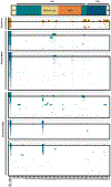
Identifying the epitope responsible for EBOV GP1,2 reactivity in unvaccinated, uninfected individuals. Sera from survivors, vaccinated individuals and unvaccinated individuals were tested against an array of 25-residue long, 15-residue overlapping peptides representing EBOV GP1,2 residues 1–655 out of 676. The schematic at the top shows the major features of the EBOV GP1,2 sequence. Individual donors are depicted in rows; antigen trimer and peptides, in columns. The heatmaps represent background-corrected absorbances between 0 and 4 units. Frequencies of positive signals are shown as columns above each panel with dashed line indicating 50% and solid line 100%. Ochre: samples collected in Liberia; teal: US; green: Uganda. SP, signal peptide; GP1 and GP2, glycoprotein subunit 1 and 2; MLD, mucin-like domain; TM, transmembrane domain. Donors from Uganda who were identified as unvaccinated are divided into two panels based on previously noted reactivity against biotin-captured antigen.
FIGURE 3
FIGURE 3
Verifying the role of P21 binding-antibodies in unspecific reactivity towards the EBOV GP1,2 trimer. Sera were depleted from antibodies binding peptide P21 (or a negative control peptide, P53) and tested for remaining reactivity against biotin-captured or adsorbed EBOV GP1,2 trimer. Six samples from vaccinees, out of which one bound P21, and eight from unvaccinated individuals, all of which bound P21, are depicted as groups of three bars each. Absorbances from a single dilution (1:200) represented with ranges of technical duplicates. OD, optical density.
FIGURE 4
FIGURE 4
Identifying residues critical for antibody binding within the P21 sequence. Top: P21 (first row) was shortened from both ends, and P21-reactive sera (and a negative control) were tested for loss of reactivity. Bottom: Each residue of the shortest reactive peptide identified above was changed to alanyl and sera were tested for loss of reactivity to identify residues critical for antibody binding. Each column represents an individual serum sample. The colors in the heatmaps represent background-corrected optical densities between 0 and 4 absorbance units. Green: serum samples from Uganda; teal: serum samples from US. v, sample from vaccinated individual that bound P21; b, blank; -, control sample that did not bind P21.
FIGURE 5
FIGURE 5
Generating a variant EBOV GP1,2 antigen to minimize unspecific binding. (A) Alignment of the identified epitope region in the glycoprotein sequence of EBOV (variant Kikwit; Uniprot P87666) and the other known viruses in the genus Orthoebolavirus: Bundibugyo virus (BDBV; Uniprot B8XCN0), Taï Forest virus (TAFV; Q66810), Bombali virus (BOMV; A0A4D5SG72), Reston virus (RESTV; Q66799), and Sudan virus (SUDV; Q7T9D9). Three residues critical for antibody binding are highlighted: two that could be changed to alanyls without impacting protein production in purple (Y214, T217), and one that could not in red (Y220). Graph created with BioRender.com. (B) Location of the epitope on the EBOV GP1,2 trimer. Image from the RSCB protein database (RCSB.org) of the crystal structure 6VKM (https://doi.org/10.2210/pdb6VKM/pdb) highlighting solved parts of GP1 in red, glycan cap within it in yellow, and the epitope in purple. Side and top views. (C) Biolayer interferometry graphs depicting binding of monoclonal antibodies to wild-type antigen (orange) or variant antigen (Y214A/T217A, purple).
FIGURE 6
FIGURE 6
Impact of Y214A/T217A changes on the performance of trimeric EBOV GP1,2 antigen. Serum samples identified as binders of the cross-reactive epitope, as non-binders of the cross-reactive epitope, or from vaccinated individuals were analyzed using the wild-type (WT) or the Y214A/T217A (Var) antigen. (A) Unvaccinated individuals and the absolute difference in background-corrected absorbances when comparing the WT and Var antigens in either biotin-captured or adsorption formats. (B) Heatmaps including a subset of samples from (A) illustrating the signal differences at various dilutions (background-corrected absorbances between 0 and 4). One panel enlarged. (C) Difference in WT and Var antigen-corrected absorbances in vaccinee samples. Differences were calculated for samples and dilutions for which both replicates of 2 repeated experiments fell within meaningful quantitation range of 0.35–3 absorbance units. (D) Heatmaps of samples from vaccinated individuals (those in C). In (A) and (C), difference to no-difference hypotheses were analyzed using one sample Wilcoxon signed-rank test. N.s., not significant; *, p < 0.05; **, p < 0.01; ***, p < 0.0001. Samples from US in teal, and from Uganda in green.

References

    1. Jacob ST, Crozier I, Fischer WA, et al. Ebola virus disease. Nat Rev Dis Primers. 2020;6(1):13. doi:10.1038/s41572-020-0147-3 - DOI - PMC - PubMed
    1. Judson SD, Munster VJ. The multiple origins of Ebola disease outbreaks. J Infect Dis. 2023;228(suppl 7):S465–S473. doi:10.1093/infdis/jiad352 - DOI - PMC - PubMed
    1. European Medicines Agency. EMA/CHMP/557387/2019 Summary of Opinion (Initial Authorisation) Ervebo Ebola Zaire Vaccine (rVSVΔG-ZEBOV-GP, live). 2019.
    1. U.S. Food & Drug Administration. BLA Approval BL 125690/0 (Ebola Zaire Vaccine, Live). 2019.
    1. European Medicines Agency. EMA/CHMP/138136/2020 Summary of Opinion (Initial Authorisation) Mvabea Ebola vaccine (MVA-BN-Filo [recombinant]). 2020.

MeSH terms