[Preprint]. 2024 Aug 21:2024.08.19.24312079.
doi: 10.1101/2024.08.19.24312079.

Biallelic variants in DAP3 result in reduced assembly of the mitoribosomal small subunit with altered intrinsic and extrinsic apoptosis and a Perrault syndrome-spectrum phenotype

Affiliations

Biallelic variants in DAP3 result in reduced assembly of the mitoribosomal small subunit with altered intrinsic and extrinsic apoptosis and a Perrault syndrome-spectrum phenotype

Thomas B Smith et al. medRxiv. .

Update in

Abstract

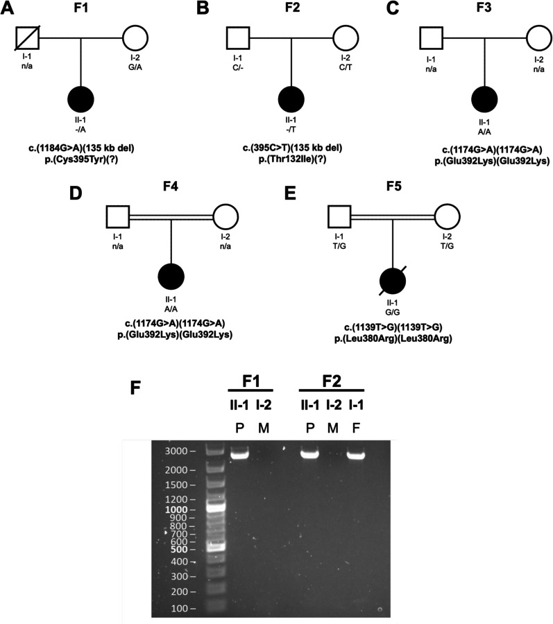
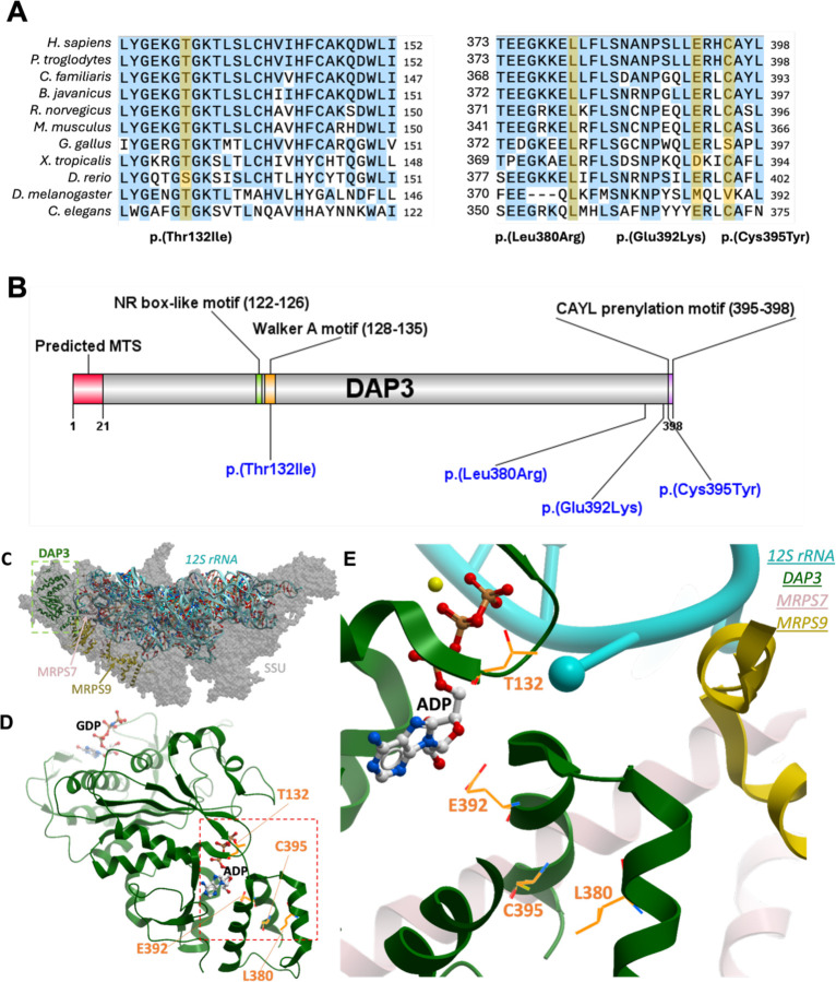
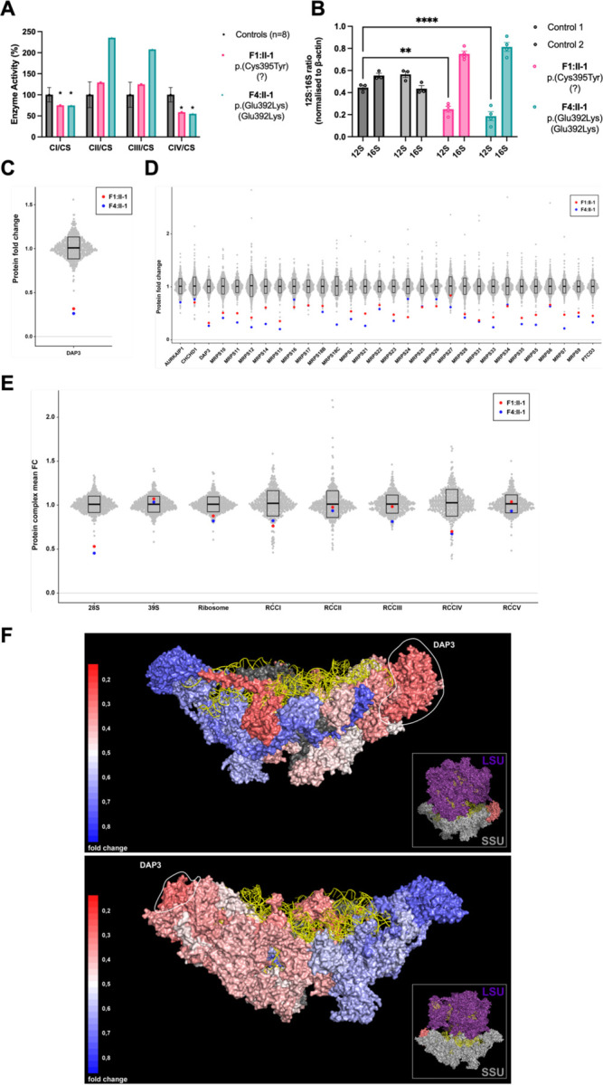
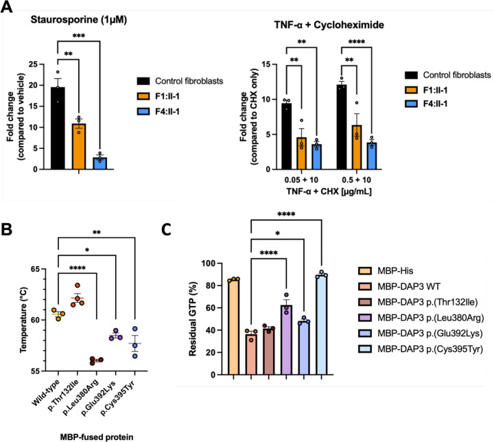
The mitoribosome synthesizes 13 protein subunits of the oxidative phosphorylation system encoded by the mitochondrial genome. The mitoribosome is composed of 12S rRNA, 16S rRNA and 82 mitoribosomal proteins encoded by nuclear genes. To date, variants in 12 genes encoding mitoribosomal proteins are associated with rare monogenic disorders, and frequently show combined oxidative phosphorylation deficiency. Here, we describe five unrelated individuals with biallelic variants in the DAP3 nuclear gene encoding mitoribosomal small subunit 29 (MRPS29), with variable clinical presentations ranging from Perrault syndrome (sensorineural hearing loss and ovarian insufficiency) to an early childhood neurometabolic phenotype. Assessment of respiratory chain function and proteomic profiling of fibroblasts from affected individuals demonstrated reduced MRPS29 protein levels, and consequently decreased levels of additional protein components of the mitoribosomal small subunit, associated with a combined complex I and IV deficiency. Lentiviral transduction of fibroblasts from affected individuals with wild-type DAP3 cDNA increased DAP3 mRNA expression, and partially rescued protein levels of MRPS7, MRPS9 and complex I and IV subunits, demonstrating the pathogenicity of the DAP3 variants. Protein modelling suggested that DAP3 disease-associated missense variants can impact ADP binding, and in vitro assays demonstrated DAP3 variants can consequently reduce both intrinsic and extrinsic apoptotic sensitivity, DAP3 thermal stability and DAP3 GTPase activity. Our study presents genetic and functional evidence that biallelic variants in DAP3 result in a multisystem disorder of combined oxidative phosphorylation deficiency with pleiotropic presentations, consistent with mitochondrial dysfunction.

Keywords: DAP3; MRPS29; Perrault syndrome; leukodystrophy; mitochondria; mitoribosomal small subunit; mitoribosome; ovarian insufficiency; rare disease; sensorineural hearing loss.


Conflict of interest statement

Declaration of interest The authors declare no competing interests.

Figures

Figure 1:
Figure 1:. Family pedigrees and characterization of the DAP3 deletion fusion product present in F1 and F2.
(A-E) Pedigrees for the five families, with known segregation and variant details listed. All variants are annotated against the DAP3 reference sequence NM_004632.4. (F) PCR analysis of F1 and F2 DNA using gel electrophoresis to detect a fusion product for the 135 kb deletion. P = proband, M = mother, F = father.
Figure 2:
Figure 2:. DAP3 variant residue conservation status, variant locations, and structural context.
(A) Evolutionary conservation of affected DAP3 residues, with a broad selection of species highlighted. Variant amino acids highlighted in black, with yellow signifying matching to the associated human residue. Sequences aligned using Jalview 2.11.2.7 . The DAP3 reference sequence used for these species are listed accordingly: H. sapiens: NP_001186778.1; P. troglodytes: XP_016802675.2; C. familiaris: XP_038527847.1; B. taurus: NP_001106765.1; R. norvegicus: NP_001011950.2; M. musculus: NP_001158005.1; G. gallus: XP_040546712.1; X. tropicalis: NP_001016002.1; D. rerio: NP_001092207.1; D. melanogaster: NP_523811.1; C. elegans: AAD20727.1. (B) Overview of DAP3 variant locations, with additional regions or domains of interest for additional context. MTS – mitochondrial targeting sequence, NR – nuclear receptor, CAYL – cysteine alanine tyrosine leucine (final 4 residues at the DAP3 C-terminus). (C) Cryo-EM structure of human mitochondrial ribosome small subunit at 2.40 Å resolution (PDB id: 7P2E), highlighting DAP3 (green), MRPS7 (rose) and MRPS9 (yellow) subunits. (D) Cartoon representation of DAP3 bound with GDP and ADP. (E) ADP binding site of DAP3 in proximity to the four sites of mutation (orange sticks).
Figure 3:
Figure 3:. Functional and proteomic analysis of F1 and F4 proband fibroblasts reveal DAP3 variants induce mitochondrial respiratory chain defects and decreased expression levels of small mitoribosomal subunit and OXPHOS components.
(A) Mitochondrial respiratory chain enzyme activities in control (black), F1:II-1 (pink) and F4:II-1 (blue) fibroblast samples. Mean enzyme activities in control fibroblasts (n = 8) are set at 100%. Error bars represent standard deviation between the controls. * indicates enzyme activity is beyond control standard deviation values. (B) MT-RNR1 (12S) and MT-RNR2 (16S) expression levels in fibroblast cDNA. Data expressed as a ratio using relative quantification (RQ) values. Error bars represent the SEM. N = 3–4, **p < 0.01, ****p < 0.0001, two-way ANOVA with Tukey’s multiple comparisons test, comparing 12S RQ value of controls to affected individuals. (C) DAP3 protein levels in affected individual fibroblasts expressed as protein fold change compared to the mean of 512 fibroblast samples. (D) Protein fold change of all components of the mitoribosomal SSU in affected individual fibroblasts, compared to 512 controls. (E) Grouped mean fold change of all proteins comprising mitoribosome subunits, whole mitoribosome, and OXPHOS components compared to the mean values of 512 controls. (F) Cryo-EM structure (PDB id: 6VLZ) of mitoribosomal SSU with individual subunits colored according to their mean fold change values (of individuals F1:II-1 and F4:II-1) compared to the mean of 512 controls. Colors ranging from weakly reduced (blue) to strongly reduced (red) and two subunits (MRPS18C and MRPS38) in dark grey as no mean fold change values could be calculated. 12S ribosomal RNA is colored in yellow, DAP3 is marked by a circle, and the small inset shows the relative position within the 55S ribosome.
Figure 4:
Figure 4:. Functional analysis of both affected individual fibroblasts and recombinant DAP3 protein establish DAP3 variants can diminish apoptotic sensitivity and destabilize DAP3 protein structure, impacting GTPase activity.
(A) Assessment of caspase-3/7 release following stimulation of intrinsic and extrinsic apoptotic pathways. Affected individual fibroblasts were challenged in duplicate with staurosporine for 4.5 hours, or TNF-α + cycloheximide (CHX) for 24 hours before addition of assay reagent. Data expressed as fold change in luminescence signal compared to DMSO-treated or CHX-treated fibroblasts, with error bars representing SEM. N = 3, **p < 0.01, ***p < 0.001, ****p < 0.0001, using one-way ANOVA with Dunnett’s multiple comparisons test (staurosporine) or two-way ANOVA with Dunnett’s multiple comparisons test (TNF-α), comparing affected individual fibroblasts to control. (B) Thermal stability of recombinant wild-type and variant MBP-DAP3 protein. Data points represent average Tm of triplicate reactions. Error bars represent SEM. N = 3–4, *p < 0.05, **p < 0.01, ****p < 0.0001, using one-way ANOVA with Dunnett’s multiple comparisons test, comparing wild-type to variants. (C) GTPase activity of recombinant wild-type and variant MBP-DAP3 protein. Data presented as mean luminescence produced by residual GTP, with error bars representing SEM. N = 3, *p < 0.05, ****p < 0.0001, one-way ANOVA with Dunnett’s multiple comparisons test, comparing wild-type protein activity to variants.
Figure 5:
Figure 5:. Lentiviral transduction of wild-type DAP3 increases protein levels of MRPS7, MRPS9 and OXPHOS components in F1:II-1 and F4:II-1 fibroblasts.
(A) Expression of DAP3 mRNA in control and fibroblasts from F1:II-1 after lentiviral transduction (LV) of DAP3 cDNA for 72 hours, or untransduced (UT). Each datapoint represents an averaged RQ value from triplicate reactions, using cDNA from independent transductions. Error bars represent SEM. N = 5, ****p < 0.0001, one-way ANOVA with Tukey’s multiple comparisons test. (B) Protein levels of MRPS7 and MRPS9 in control fibroblasts and fibroblasts from F1:II-1 and F4:II-1 after lentiviral transduction of DAP3 cDNA for 72 hours. Beta-actin was used as a loading control and for densitometric analysis. Blots are representative of results from 3 independent biological repeats. MRPS7 levels were unable to be quantified in fibroblasts from F4:II-1. (C) Protein levels of SDHB, COX II and NDUFB8 in control fibroblasts and fibroblasts from F1:II-1 and F4:II-1 after lentiviral transduction of DAP3 cDNA for 72 hours. Blots are representative of results from 3 independent biological repeats.

Similar articles

References

    1. Amunts A, Brown A, Toots J, Scheres SHW, Ramakrishnan V. The structure of the human mitochondrial ribosome. Science. 2015;348(6230):95–98. doi:10.1126/science.aaa1193 - DOI - PMC - PubMed
    1. Greber BJ, Bieri P, Leibundgut M, Leitner A, Aebersold R, Boehringer D, Ban N. Ribosome. The complete structure of the 55S mammalian mitochondrial ribosome. Science. 2015;348(6232):303–308. doi:10.1126/science.aaa3872 - DOI - PubMed
    1. Itoh Y, Andréll J, Choi A, Richter U, Maiti P, Best RB, Barrientos A, Battersby BJ, Amunts A. Mechanism of membrane-tethered mitochondrial protein synthesis. Science. 2021;371(6531):846–849. doi:10.1126/science.abe0763 - DOI - PMC - PubMed
    1. Kummer E, Leibundgut M, Rackham O, Lee RG, Boehringer D, Filipovska A, Ban N. Unique features of mammalian mitochondrial translation initiation revealed by cryo-EM. Nature. 2018;560(7717):263–267. doi:10.1038/s41586-018-0373-y - DOI - PubMed
    1. Bogenhagen DF, Ostermeyer-Fay AG, Haley JD, Garcia-Diaz M. Kinetics and Mechanism of Mammalian Mitochondrial Ribosome Assembly. Cell Rep. 2018;22(7):1935–1944. doi:10.1016/j.celrep.2018.01.066 - DOI - PMC - PubMed

Publication types

LinkOut - more resources