[Preprint]. 2024 Sep 22:2024.09.19.24314009.
doi: 10.1101/2024.09.19.24314009.

Cardiomyocyte-derived circulating extracellular vesicles allow a non-invasive liquid biopsy of myocardium in health and disease

Affiliations

Cardiomyocyte-derived circulating extracellular vesicles allow a non-invasive liquid biopsy of myocardium in health and disease

Michail Spanos et al. medRxiv. .

Abstract

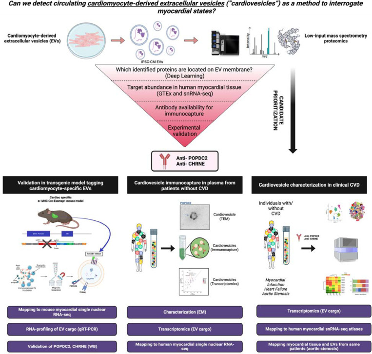
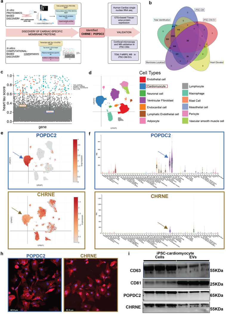
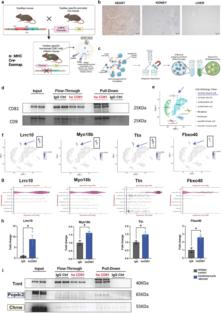
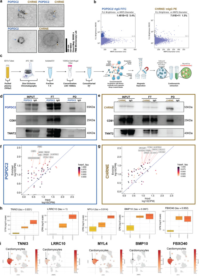
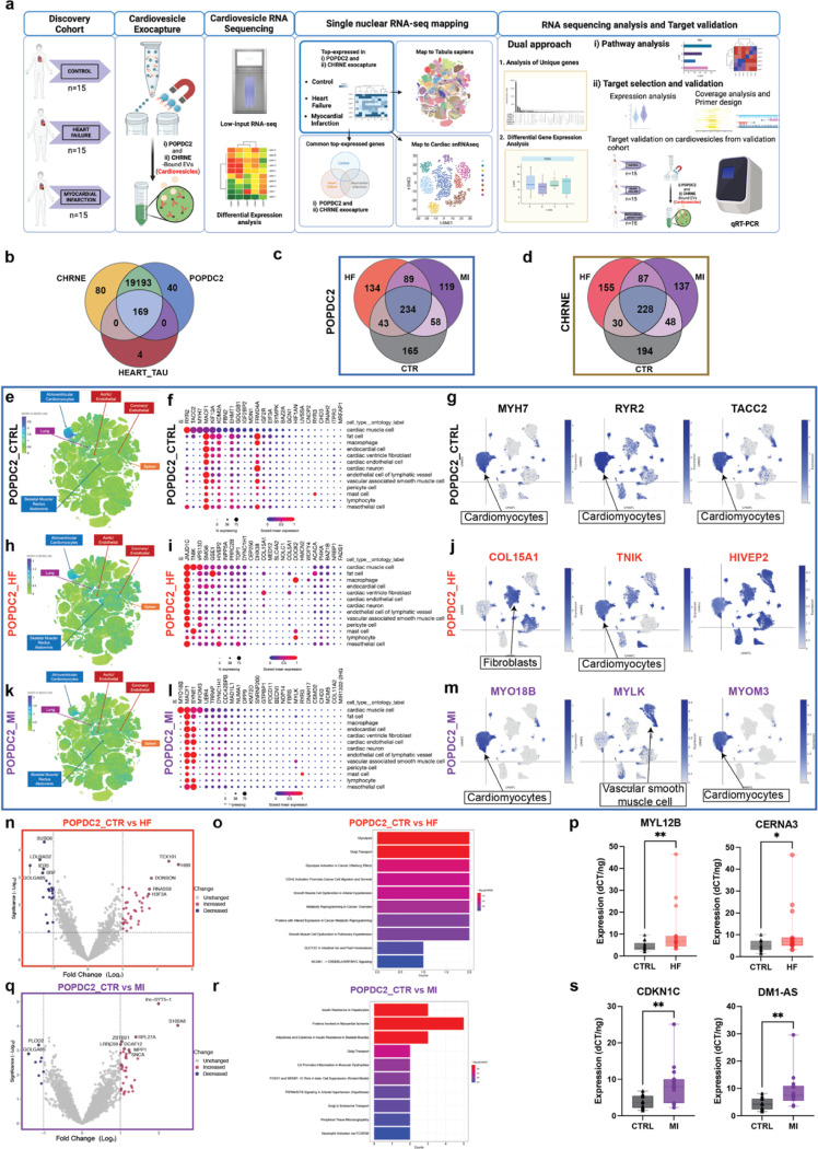
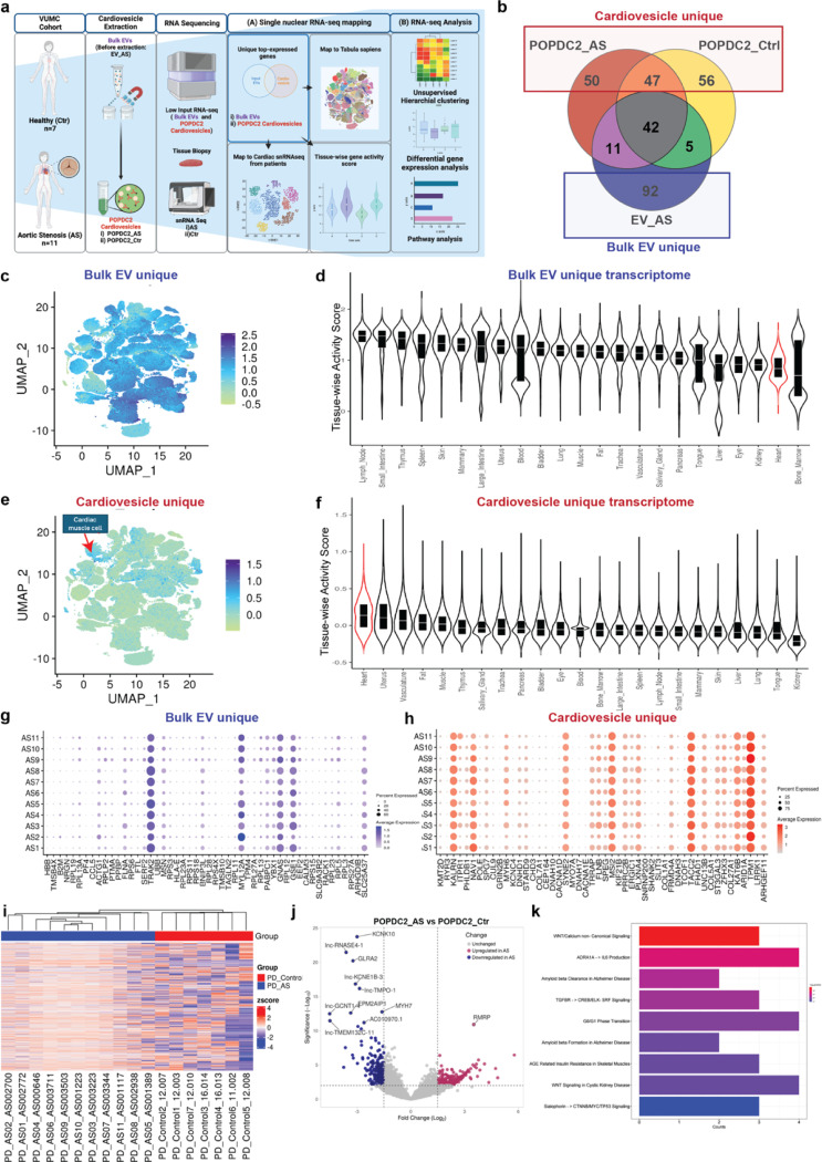
The ability to track disease without tissue biopsy in patients is a major goal in biology and medicine. Here, we identify and characterize cardiomyocyte-derived extracellular vesicles in circulation (EVs; "cardiovesicles") through comprehensive studies of induced pluripotent stem cell-derived cardiomyocytes, genetic mouse models, and state-of-the-art mass spectrometry and low-input transcriptomics. These studies identified two markers (POPDC2, CHRNE) enriched on cardiovesicles for biotinylated antibody-based immunocapture. Captured cardiovesicles were enriched in canonical cardiomyocyte transcripts/pathways with distinct profiles based on human disease type (heart failure, myocardial infarction). In paired myocardial tissue-plasma from patients, highly expressed genes in cardiovesicles were largely cardiac-enriched (vs. "bulk" EVs, which were more organ non-specific) with high expression in myocardial tissue by single nuclear RNA-seq, largely in cardiomyocytes. These results demonstrate the first "liquid" biopsy discovery platform to interrogate cardiomyocyte states noninvasively in model systems and in human disease, allowing non-invasive characterization of cardiomyocyte biology for discovery and therapeutic applications.


Figures

Figure 1.
Figure 1.. Study design to identify cardiovesicles for liquid biopsy in cardiovascular diseases.
Proteomics and bioinformatics-based approach to identify POPDC2 and CHRNE as cardiovesicular membrane proteins with validation in human RNA-seq datasets and in vitro experiments. Confirmation of cardiomyocyte EV-specific expression of POPDC2 and CHRNE proteins using the Exomap1 transgenic mouse model (humanized CD81 expression restricted to cardiomyocytes and its EVs). Characterization of the RNA cargo of cardiovesicles in healthy human plasma. Cardiovesicle RNA cargo characterization in cardiovascular disease cohorts with mapping to tissue single cell RNA sequencing data sets.
Figure 2.
Figure 2.. Discovery and experimental validation of POPDC2 and CHRNE as cardiomyocyte- EV (Extracellular Vesicle) membrane protein candidates.
a. Analytic scheme for identification and validation of cardiomyocyte EV membrane markers POPDC2 and CHRNE using step-wise proteomic-based and bioinformatic-based discovery followed by experimental validation in iPSC-CM cells and EVs. b. Venn diagram showing 35 targets that were identified using LC-MS/MS (Liquid Chromatography with tandem mass spectrometry) present in iPSC-CM (induced Pluripotent Stem Cell-derived Cardiomyocytes), iPSC-CM-EVs and prioritized by heart enrichment (from Human Protein Atlas data) and membrane-localization (Deep Transmembrane Helix Prediction). c. Scatter plot showing candidates from computational analysis obtained by mining GTEx for proteins with cardiac-specificity (tau) ≥ 0.6 (dots above the red dashed line) with red indicating protein-coding candidates present in the plasma membrane (predicted using Compartments). POPDC2 and CHRNE are indicated on the plot. d. UMAP (Uniform Manifold Approximation and Projection) legend of cardiac single nuclear RNA-seq dataset from Broad single cell Portal. e. POPDC2 and CHRNE UMAPs demonstrating their expression in cardiomyocytes. f. Tissue-wise transcriptomic expression of POPDC2 and CHRNE using GTEx showing elevated expression in heart tissue. h. Representative confocal images showing the expression of POPDC2 and CHRNE proteins in iPSC-CMs. i. POPDC2 and CHRNE protein expression shown in iPSC-CMs and iPSC-CM-EVs by western blotting (n=3 independent experiments).
Figure 3.
Figure 3.. Cardiomyocyte-EV Specific Expression of POPDC2 and CHRNE Proteins in Cardiac-Specific Cre-Driven EXOMAP1 Mouse Model.
a. Schematic detailing the generation of cardiac-specific EXOMAP1 transgenic mice. Exomap1 mice, which express HsCD81mNG (humanized CD81 fused with mNeonGreen) in a Cre recombinase-dependent manner, were crossed with alpha myosin heavy chain (αMHC)-Cre mice, ensuring HsCD81mNG expression on cardiomyocyte membranes and their secreted EVs. b. Cardiac-specific hsCD81 expression as observed in immunohistochemistry staining of heart (positive expression), and absent in kidney and liver. c. Overview of the ExoCapture-MSB method for EV immunocapture using biotin-streptavidin affinity and Streptavidin Magnetic Beads targeting cardiac-tissue specific EVs from αMHC-Cre Exomap1 mice. d. Confirmation of canonical EV markers CD81 and CD9 enrichment in cardiac-derived EVs via Western blot assay, with controls (PD: Pulled-Down, FT: Flow-Through). e. UMAP legend of cardiac single nuclear RNA-seq data from Tabula muris. f. and g. UMAPs and Violin plots demonstrating enrichment of cardiomyocyte-specific transcripts Lrrc10, Myo18b, Ttn, and Fbxo40 from Tabula muris. h. Bar plots showing significant differences in cardiomyocyte-specific transcripts between humanized CD81-positive EV pulldown and isotype control, with respective p-values using Mann Whitney t-test (Lrrc10, p = 0.0482; Myo18b, p=0.0105; Ttn p=0.0459; FBxo40- p=0.0103) n= 4 mice. i. Confirmation of POPDC2, CHRNE, and TNNI3 enrichment in cardiac-derived EVs via Western blot assay, validating the selective presence of these key cardiomyocyte markers, affirming their cardiac origin. N= 4 mice.
Figure 4.
Figure 4.. POPDC2+ and CHRNE+ Cardiovesicles from Human Plasma Demonstrate Cardiac-Specific Gene Expression Profiles.
a. Transmission Electron Microscopy images of POPDC2+ (blue arrows) and CHRNE+ (gold arrows) EVs in healthy human plasma, displaying both single positive (POPDC2+ CHRNE-; POPDC2- CHRNE+) and double positive (POPDC2+ CHRNE+) EVs, with gold particle sizing for POPDC2 (5 mm) and CHRNE (12 mm). b. Cardiovesicles exhibiting POPDC2+ and CHRNE+ signatures are visualized and quantified using Fluorescent-Microfluidic Resistive Pulse Sensing (F-MRPS) in plasma EVs (n=3 replicates). c. Detailed overview of the ExoCapture-MSB Method for immunocapture of cardiovesicles from 500 μL of Human Plasma, employing Pierce Streptavidin Magnetic Beads with biotin-streptavidin affinity. Western Blot analysis of POPDC2 (d) and CHRNE (e) immunocapture, highlighting enrichment for cardiomyocyte-specific protein Troponin. Transcriptomic enrichment of cardiac-specific transcripts, prioritized by tau score ≥ 0.9, in vesicles immunocaptured with Biotinylated POPDC2 (f) and CHRNE (g) antibody, respectively. with the expression of the bulk CD81 immunocaptured vesicles (Input) on the x-axis and the expression of the immunocaptured Cardiovesicles on the y-axis (Enriched). h. Box plots showing the expression of heart enriched genes (TNNI3, LRRC10, MYL4, BMP10, FBXO40) in vesicles immunocaptured with CD81, POPDC2, and CHRNE antibodies, confirming specific capture and analysis of Cardiovesicles. i. UMAP visualization from single nuclear RNA sequencing depicting plots (TNNI3, LRRC10, MYL4, BMP10, FBXO40) as highly cardiac-enriched transcripts within Cardiovesicles, affirming their cardiac origin. P<0.05 is utilized as the threshold of statistical significance. N= 3 replicates.
Figure 5.
Figure 5.. Cardiovesicle transcripts capture myocardial disease-state specific expression profiles in patients with myocardial infarction or heart failure with reduced ejection fraction.
a. Schematic detailing isolation and analysis of cardiovesicles from cardiovascular cohorts (90 samples), involving patients with heart failure (HF, 15), Type I myocardial infarction (MI, 15), and control (Ctrl, 15) using the ExoCapture-MSB method. Following isolation, RNA cargo from these vesicles were sequenced. Analysis included mapping of transcripts in cardiovesicles to single cell/nuclear atlases, and differential gene expression to identify targets significantly dysregulated in HF and MI compared to Control. b. Venn diagram illustrating a 99% overlap among all POPDC2+ and CHRNE+ cardiovesicle transcripts across the three groups (Ctrl, HF, and MI), including 169 heart-enriched transcripts with tau score >0.6 from GTEx. Venn diagrams of the top 500 enriched transcripts (by the highest mean expression for each group) for c. POPDC2+ and d. CHRNE+ cardiovesicles in HF and MI relative to control demonstrating shared as well as unique transcripts between the cohorts. Top 25 enriched transcripts (mean expression threefold greater than standard deviation) from POPDC2+ Ctrl, HF, and MI patients were mapped onto the multiorgan single-cell transcriptomic atlas dataset (Tabula Sapiens) and a single-nuclear dataset of human control (no cardiovascular disease), dilated cardiomyopathy. Cardiovesicle transcripts from control patients mapped to the multi-organ atlas is shown as a target UMAP in e, with summary dot-plots and individual target UMAPs from the heart single nuclear dataset shown in f and g respectively. The same representations for the cardiovesicles from HF are shown in h, i, and j, while those from MI are depicted in k, l and m. Volcano plots displaying differentially expressed transcripts (with log FC + 1 and p < 0.01) in POPDC2+ cardiovesicles from patients with HF compared to control patients (n) with pathway enrichment analysis (o). Validation in the MGH validation cohort (n=30) for selected transcripts differentially expressed in HF-POPDC2+ EVs (MYL12B, p=0.0091, CERNA3, p=0.0178) using qRT-PCR normalized to internal and spike-in controls. Similar analysis including volcano plot for differentially expressed transcripts in POPDC2+ EVs in patients with MI compared to controls (q), pathway enrichment analysis (r), and experimentally validation of select transcripts in the MGH validation cohort (CDKN1A, p=0.0019, DM1-AS, p=0.0025, n= 30. Significance levels are marked as * p < 0.01, ** p < 0.001, *** p < 0.0001, calculated using the Kruskal-Wallis test.
Figure 6.
Figure 6.. Molecular profiling of cardiovesicles from patients with aortic stenosis reveals enrichment of transcripts expressed in cardiomyocytes and relevant pathways in myocardial remodeling
a. Schematic overview detailing the isolation and transcriptomic analysis of cardiovesicles from the AS cardiovascular cohort (718 samples), including Bulk EVs (unprocessed EVs, no cardiovesicle extraction) from AS patients (EV_AS, n=11) and POPDC2+ cardiovesicles from AS patients (POPDC2_AS, n=11) and controls (POPDC2_Ctr, n=7). Analysis of the top abundant transcripts, prioritized by mean expression, of EV_AS, POPDC2_AS, and POPDC2_Ctr, was conducted to identify genes unique to EV_AS (labeled as ‘Bulk EV_unique’) and those unique to POPDC2+ cardiovesicles (POPDC2_AS and POPDC2_Ctr; labeled as Cardiovesicle_unique). These were mapped onto single nuclear RNA-seq datasets and differential expression analysis was performed to identify and validate targets significantly dysregulated in AS compared to Control. b. Venn diagram analysis of the top 150 abundant transcripts (prioritized by mean expression) of EV_AS, POPDC2_AS, and POPDC2_Ctr showing Bulk EV_unique and Cardiovesicle_unique transcripts. Distribution of Bulk EV_Unique and (c) and cardiovesicle_unique transcripts (e) represented as UMAPs of the multiorgan single cell transcriptomic atlas dataset (Tabula Sapiens), with their respective tissue-wise transcriptional activity score showing other tissues in Bulk EV_unique (d) and heart in Cardiovesicle_unique gene sets (f). Dot plots g. and h. show the expression counts of Bulk EV_unique and Cardiovesicle_unique transcripts in cardiomyocytes of each individual AS patient, respectively. i. Unsupervised hierarchical clustering illustrates the transcriptional differences between AS (POPDC2_AS) and Ctrl (POPDC2_Ctr) POPDC2+ cardiovesicles. j. Volcano plots represent differentially expressed genes between POPDC2_AS and POPDC2_Ctr k. Pathway enrichment analysis reveals the differentially expressed genes between AS and Ctr POPDC2+ cardiovesicles. log FC cutoff +1, and FDR <1% have been used as statistical significance cut-offs for the differential gene expression analysis

References

    1. Wells Q. S. et al. Accelerating Biomarker Discovery Through Electronic Health Records, Automated Biobanking, and Proteomics. J. Am. Coll. Cardiol. 73, 2195–2205 (2019). - PMC - PubMed
    1. Helgason H. et al. Evaluation of Large-Scale Proteomics for Prediction of Cardiovascular Events. JAMA 330, 725–735 (2023). - PMC - PubMed
    1. Rasooly D. et al. Genome-wide association analysis and Mendelian randomization proteomics identify drug targets for heart failure. Nat. Commun. 14, 3826 (2023). - PMC - PubMed
    1. Royer P. et al. Plasma proteomics for prediction of subclinical coronary artery calcifications in primary prevention. Am. Heart J. 271, 55–67 (2024). - PubMed
    1. Bom M. J. et al. Predictive value of targeted proteomics for coronary plaque morphology in patients with suspected coronary artery disease. EBioMedicine 39, 109–117 (2019). - PMC - PubMed

Publication types

LinkOut - more resources