[Preprint]. 2024 Sep 16:2024.09.16.24313748.
doi: 10.1101/2024.09.16.24313748.

Allelic effects on KLHL17 expression likely mediated by JunB/D underlie a PDAC GWAS signal at chr1p36.33

Affiliations

Allelic effects on KLHL17 expression likely mediated by JunB/D underlie a PDAC GWAS signal at chr1p36.33

Katelyn E Connelly et al. medRxiv. .

Update in

Abstract

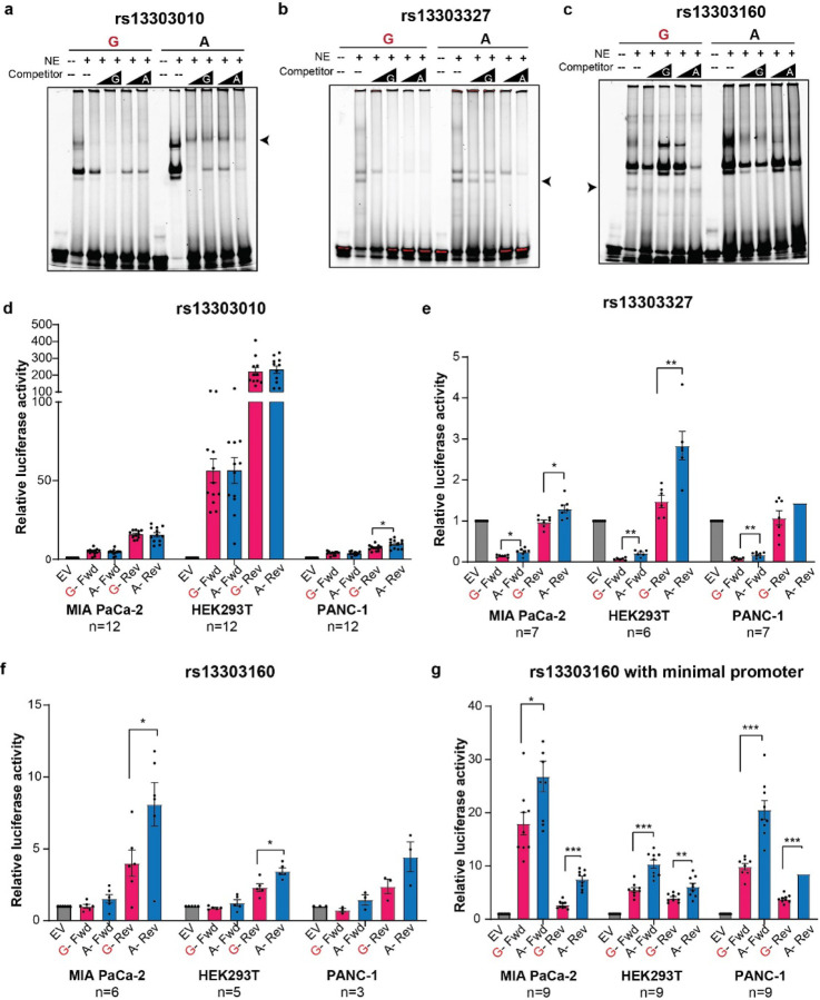
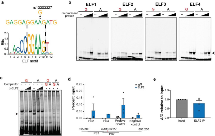
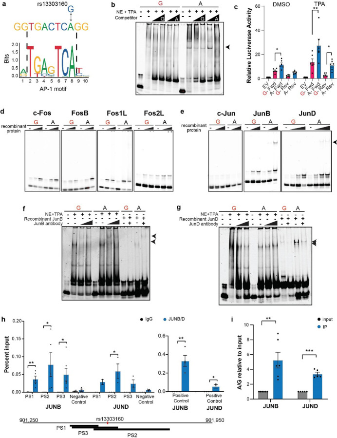
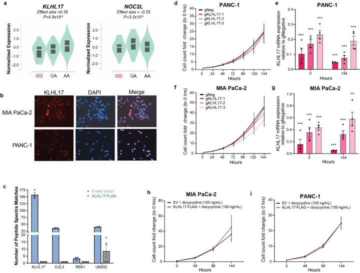
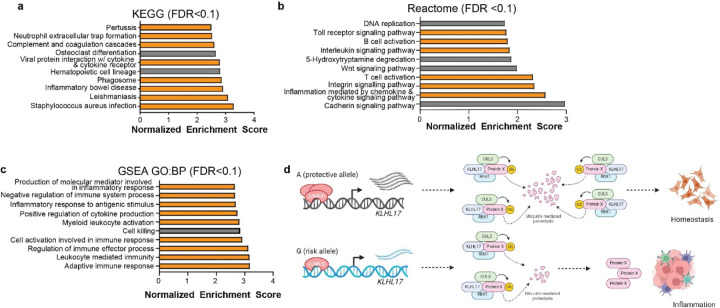
Pancreatic Ductal Adenocarcinoma (PDAC) is the third leading cause of cancer-related deaths in the U.S. Both rare and common germline variants contribute to PDAC risk. Here, we fine-map and functionally characterize a common PDAC risk signal at 1p36.33 (tagged by rs13303010) identified through a genome wide association study (GWAS). One of the fine-mapped SNPs, rs13303160 (r2=0.93 in 1000G EUR samples, OR=1.23, P value=2.74x10⁻⁹) demonstrated allele-preferential gene regulatory activity in vitro and allele-preferential binding of JunB and JunD in vitro and in vivo. Expression Quantitative Trait Locus (eQTL) analysis identified KLHL17 as a likely target gene underlying the signal. Proteomic analysis identified KLHL17 as a member of the Cullin-E3 ubiquitin ligase complex in PDAC-derived cells. In silico differential gene expression analysis of the GTExv8 pancreas data suggested an association between lower KLHL17 (risk associated) and pro-inflammatory pathways. We hypothesize that KLHL17 may mitigate inflammation by recruiting pro-inflammatory proteins for ubiquitination and degradation thereby influencing PDAC risk.


Conflict of interest statement

Competing Interests statement The authors declare no competing interests.

Figures

Figure 1:
Figure 1:. Overview of chr1p36.33 PDAC risk locus.
a) Locus Zoom plot of the variants identified in the meta-analysis, colors are indicative of the LD r2 in reference to the lead SNP b) UCSC genome browser view of 1p36.33 with ChromHMM and ATAC-seq annotations in PDAC and normal-derived duct epithelial cell lines c) Zoomed in UCSC browser showing the candidate functional variants and nearby genes
Figure 2:
Figure 2:. Identification of allele-preferential binding and activity using EMSA and luciferase reporter assays
a-c) Representative EMSA with PANC-1 nuclear extract and fluorescently labelled 31bp oligonucleotides with the variant, rs13303010, rs13303327, rs13303160, respectively centered in the middle. Competitor is the same sequence with no fluorescent label in excess (50, 100X); d-f) Luciferase reporter assay using rs13303010, rs13303327, and rs13330160, respectively and the surrounding sequence as a promoter to the luciferase gene in three cell lines, number of biological replicates are indicated below the cell line name; g) Luciferase assay for rs13303160 and surrounding sequence as an enhancer upstream of a minimal promoter and luciferase gene in three cell lines. The number of replicates is indicated below the cell line name. The risk allele is colored in red. For luciferase, the forward and reverse orientation of the sequence was used. Error bars represent the standard error of the mean (SEM) and significance was determined by an unpaired, two-tailed t-test; * P <0.05; ** P < 0.01, *** P < 0.001.
Figure 3:
Figure 3:. Allele-preferential binding of ELF to rs13303327 in vitro and in vivo
a) in silico TF binding motif prediction for rs13303327; b) EMSA with increasing amounts of recombinant ELF proteins and fluorescently labeled oligonucleotide; c) EMSA supershift with an antibody against ELF2 using PANC-1 nuclear lysate; d) ChIP-qPCR for ELF2 in Hs766T PDAC cell line with primers near or encompassing the SNP. The positive control is a documented region from an ELF2 ChIP-seq in K562 and negative control is from a quiescent region of 1p36.33. Percent input enrichment was quantitatively determined using a standard curve derived from input DNA as described in the ActiveMotif protocol); e) TaqMan genotyping of enriched ChIP-qPCR DNA; A to G ratio was calculated in relative to the input ratio. For ChIP-qPCR, all IPs were performed in quadruplicate, but IPs that did not enrich are not shown. Error bars represent SEM. Unpaired, two-tailed t-tests were performed; nothing was deemed significant (P <0.05)
Figure 4:
Figure 4:. Allele-preferential binding of AP1 proteins to rs13303160 in vitro and in vivo.
a) in silico TF binding predictions; b) EMSA using TPA-stimulated nuclear HeLa extract and fluorescently labeled oligonucleotide. Arrow indicates the allele-preferential binding; c) Luciferase reporter assay using DMSO and TPA stimulation and the rs13303160 sequence as an enhancer in the PANC-1 cell line; luciferase activity reported relative to the Empty Vector (EV). Unpaired t-tests were performed on the relative luciferase activity of the A/G ratio compared to A/A; d) Representative EMSAs with increasing amounts of recombinant Fos proteins (from left to right: c-Fos; FosB; Fos1L; Fos2L); e) Representative EMSAs with increasing amounts of recombinant Jun proteins (from left to right: c-Jun, JunB, JunD). Arrow indicates the allele-specific binding; f, g) Supershift EMSA with antibodies against JunB and JunD respectively using both TPA-stimulated nuclear lysate and recombinant protein; Arrows denote the shift in the bands; h) ChIP-qPCR in SW1990 PDAC cells for JunB and JunD using 3 primer sets (PS) surrounding the SNP. Positive controls are from a JunB ChIP-seq performed in the CFPAC1 PDAC cell line. Negative control is from a quiescent region on 1p36.33; i) TaqMan genotyping assay for rs13303160 using immunoprecipitated DNA from the ChIP. The ratio of A to G was determined relative to the quantity of A and G alleles in the input DNA. For all graphs, error bars represent the SEM. Unpaired two-tailed t-tests were performed; * P <0.05; ** P <0.01; *** P <0.001.
Figure 5:
Figure 5:. Analysis of the effects of altered KLHL17, a Cullin3-E3 complex member, expression on cellular growth of PDAC cells in vitro.
a) Pancreas GTEx v8 eQTLs for rs13303010; b) Immunofluorescence for KLHL17 in the MIA PaCa-2 and PANC-1 KLHL17 overexpressing cell line; c) Peptide Spectra Matches for Cullin3-E3 members identified by KLHL17-FLAG immunoprecipitation and mass spectrometry analysis; d,f) Cell counts normalized to 0 Hours for CRISPRi-mediated knockdown of KLHL17 in PANC-1 and MIA PaCa-2 cells, respectively (left panel). sgNeg is the negative control targeting a sequence within the same topologically associated domain (TAD). e,g) qPCR analysis of CRISPRi gRNA efficiency; expression is relative to the sgNeg control and internal HPRT control (right panel) and measured at the start and end of the growth assay; h, i) Cell counts normalized to 0 hours for doxycycline-inducible KLHL17-FLAG overexpressing and empty vector (EV) control MIA PaCa-2 and PANC-1 cell lines, respectively. For panels c-g, error bars indicate the SEM; For panels d-g, unpaired, two-tailed t-tests were performed, ** P <0.01, *** P <0.001
Figure 6:
Figure 6:. Gene Set Enrichment Analysis (GSEA) of differentially expressed genes from an in silico KLHL17 knockdown
a) GSEA using the KEGG functional database and the significantly (FDR < 0.05) differentially expressed genes when KLHL17 expression is lower; b) GSEA using the Reactome dataset; c) GSEA using Biological Processes Gene Ontology dataset. For all three analyses only gene sets with an FDR < 0.1 are shown. Bars in orange indicate gene sets associated with inflammation or inflammation-related diseases. No gene sets were negatively enriched at this FDR threshold; d) Working hypothesis for the function of rs13303160 and KLHL17 in PDAC risk

References

    1. Rahib L. et al. Projecting Cancer Incidence and Deaths to 2030: The Unexpected Burden of Thyroid, Liver, and Pancreas Cancers in the United States. Cancer Research 74, 2913, doi:10.1158/0008-5472.CAN-14-0155 (2014). - DOI - PubMed
    1. Cronin K. A. et al. Annual report to the nation on the status of cancer, part 1: National cancer statistics. Cancer 128, 4251–4284, doi:10.1002/cncr.34479 (2022). - DOI - PMC - PubMed
    1. Capasso M. et al. Epidemiology and risk factors of pancreatic cancer. Acta Biomed 89, 141–146, doi:10.23750/abm.v89i9-S.7923 (2018). - DOI - PMC - PubMed
    1. Klein A. P. et al. Genome-wide meta-analysis identifies five new susceptibility loci for pancreatic cancer. Nature communications 9, 556–556, doi:10.1038/s41467-018-02942-5 (2018). - DOI - PMC - PubMed
    1. Amundadottir L. et al. Genome-wide association study identifies variants in the ABO locus associated with susceptibility to pancreatic cancer. Nat Genet 41, 986–990, doi:10.1038/ng.429 (2009). - DOI - PMC - PubMed

Publication types