Review
. 2024 Aug 20;4(9):639-656.
doi: 10.1016/j.jacasi.2024.07.003. eCollection 2024 Sep.

Widespread Use of Imaging-Guided PCI in Asia: Time for Extended Application

Affiliations
Review

Widespread Use of Imaging-Guided PCI in Asia: Time for Extended Application

Yongcheol Kim et al. JACC Asia. .

Abstract

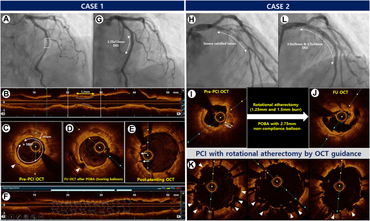
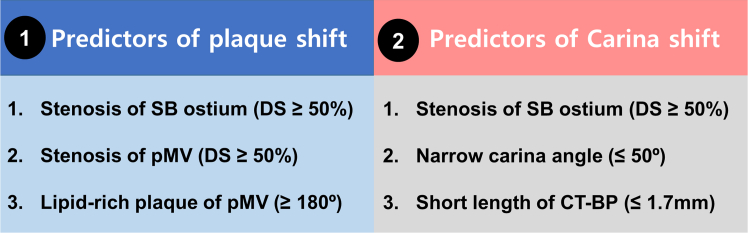
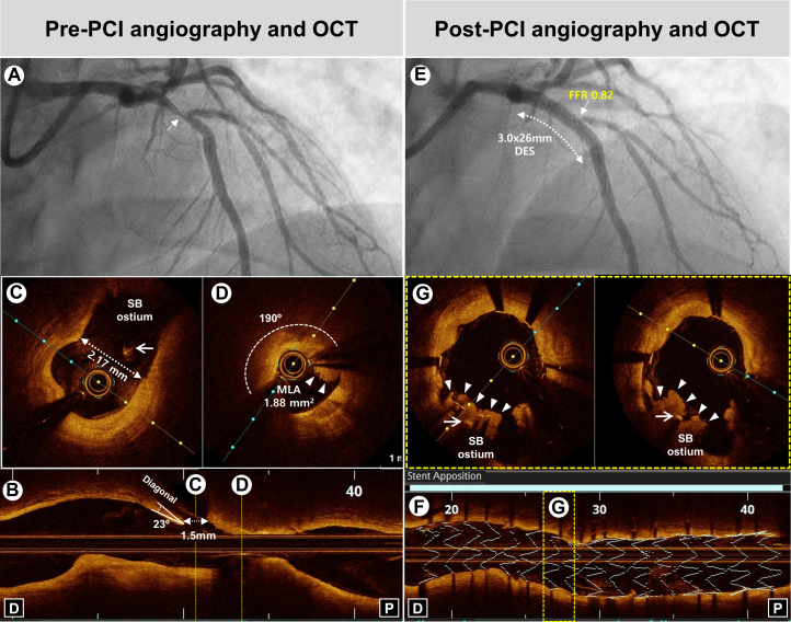
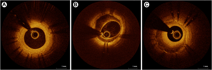
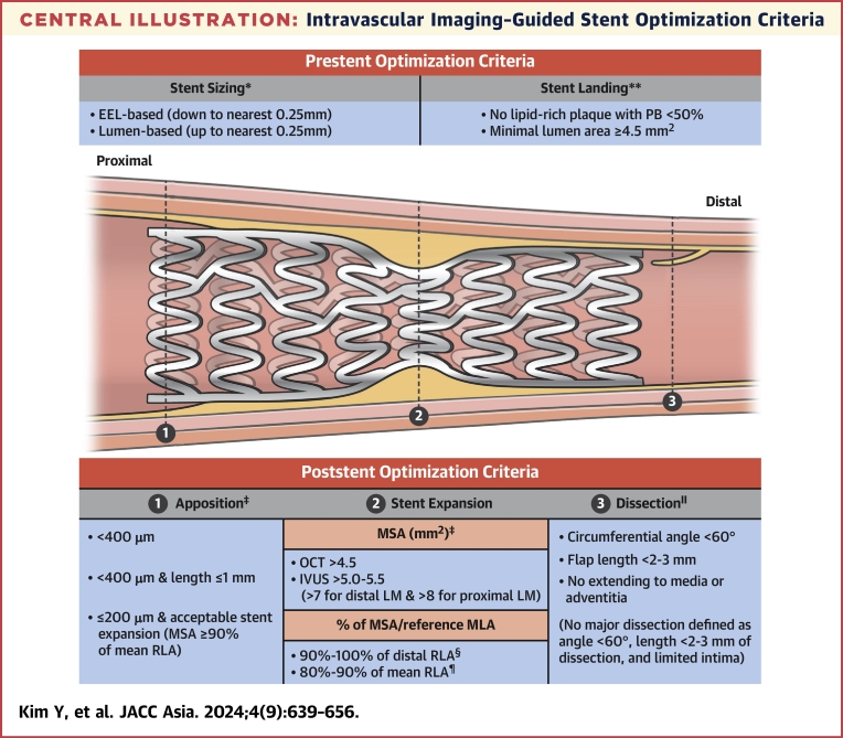
In recent years, a wealth of clinical data has emerged regarding intravascular imaging involving either intravascular ultrasound or optical coherence tomography. This surge in data has propelled the adoption of intravascular imaging-guided percutaneous coronary intervention (PCI) in daily clinical practice. The findings of current randomized clinical trials regarding imaging guidance have lent strong support to the benefits of intravascular imaging-guided PCI. This holds especially true for the diagnosis and treatment of complex lesions, such as left main disease, diffuse long lesions, chronic total occlusion, severely calcified lesions, bifurcations, and in-stent restenosis, as well as in high-risk patients such as those with acute myocardial infarction or chronic kidney disease. During intravascular imaging-guided PCI, operators attempt to achieve stent optimization for maximized benefits of imaging guidance. This paper provides a comprehensive review on the updated clinical data of intravascular imaging-guided PCI and intravascular ultrasound/optical coherence tomography-derived stent optimization criteria.

Keywords: interventional; optical coherence; percutaneous coronary intervention; tomography; ultrasound.


Conflict of interest statement

This work was supported by the Imaging and Physiology with Cardiovascular Disease (IPOP) and the Korean Society of Interventional Cardiology (KSIC). The authors have reported that they have no relationships relevant to the contents of this paper to disclose.

Figures

None
Graphical abstract
Figure 1
Figure 1
OCT-Guided PCI With DES Implantation for the Severe Calcified Lesions Case 1. (A) Angiogram showing severe stenosis in the mid-portion of the LAD in a 67-year-old woman presenting with non–ST-segment elevation myocardial infarction. (B and C) Pre-PCI OCT assessment of a longitudinal circumferential calcified plaque with OCT-based calcium score of 3 points (maximal calcium angle >180° [2 points], calcium length >5.0 mm [1 point], and maximal calcium thickness [0 point]). (D) Post-ballooning OCT showing a calcium fracture after balloon angioplasty using a scoring balloon (arrowhead). (E) Post-stent cross-sectional OCT showing the presence of cracks (arrows). (F) 3-dimensional stent image and apposition index demonstrating a well-expanded stent without malapposition. (G) Final angiography showing good distal flow without residual stenosis after stent implantation, Case 2. (H) Angiogram showing diffuse severe stenosis with heavy calcification in the proximal to mid-portion of the LAD in a 54-year-old man presenting with stable angina. (I) Pre-PCI OCT assessment of diffuse circumferential plaques with thick calcifications leading to OCT-based calcium score of 4 points (maximal calcium angle >180° [2 points], calcium length >5.0 mm [1 point], and maximal calcium thickness [1 point]). (J) Post-rota and ballooning OCT showing good modification of the calcified plaque by rotational atherectomy and noncompliant balloon inflation. (K) Post-stent OCT showing multiple calcium fractures (arrowheads). (L) The final angiogram showing good distal flow without residual stenosis. DES = drug-eluting stent(s); LAD = left anterior descending artery; OCT = optical coherence tomography; PCI = percutaneous coronary intervention; POBA = plain old balloon angioplasty.
Figure 2
Figure 2
Risk Factors of SB Compromise The predictors of plaque shift and carina shift are summarized at the boxes (① and ②). CT-BP = carina tip and branching point; DS = diameter stenosis; MV = main vessel; pMV = proximal main vessel; SB = side branch.
Figure 3
Figure 3
Representative Case of Bifurcation PCI by OCT Guidance (A) Baseline angiography showing severe stenosis (arrow) at the bifurcation lesion of the proximal LAD. (B) Pre-PCI OCT showing narrow carina angle (23°) and short carina tip to branching point (1.5 mm). (C and D) Cross-sectional OCT images demonstrating MLA of 1.88 mm2 with a large lipid-rich plaque (190°) and intraluminal thrombus (arrowheads) in proximal MV without the stenosis of SB ostium (diameter of SB ostium: 2.17 mm) (arrow: guidewire of SB). (E) Post-PCI angiography showing successful PCI using SB wire jailing technique in the proximal LAD with severe stenosis and TIMI 3 flow in the SB ostium, demonstrating functional nonsignificance (FFR: 0.82). (F) 3-dimensional stent image and apposition index demonstrating stent optimization. (G) Cross-sectional OCT images demonstrating plaque shift (arrowheads) to SB ostium (arrow: guidewire of SB). FFR = fractional flow reserve; MLA = minimal lumen area; SB = side branch; other abbreviations as in Figures 1 and 2.
Figure 4
Figure 4
Representative Optical Coherence Tomography Images of Neointimal Tissue Homogeneous (uniform optical properties without focal variation in the backscattering patterns) (A), heterogeneous (focally changing optical properties and various backscattering patterns) (B), and layered (concentric layers with different optical properties) (C) types.
Central Illustration
Intravascular Imaging–Guided Stent Optimization Criteria Summary of pre- and post-stenting optimization criteria derived from various randomized trials regarding intravascular imaging–guided percutaneous coronary intervention. EEL = external elastic lamina; IVUS = intravascular ultrasound; LM = left main; MSA = minimal stent area; OCT = optical coherence tomography; PB = plaque burden; RLA = reference lumen area. ∗Data from RENOVATE-COMPLEX PCI (Randomized Controlled Trial of Intravascular Imaging Guidance versus Angiography-Guidance on Clinical Outcomes after Complex Percutaneous Coronary Intervention), OCTIVUS (Optical Coherence Tomography versus Intravascular Ultrasound-Guided Percutaneous Coronary Intervention), ILUMIEN IV (Optimal Coherence Tomography Guided Coronary Stent Implantation Compared to Angiography: a Multicenter Randomized Trial in PCI), and OCCUPI (Optical Coherence Tomography-Guided Coronary Intervention in Patients With Complex Lesions: a Randomized Controlled Trial). ∗∗Data from OPINION (Optical Frequency Domain Imaging vs. Intravascular Ultrasound in Percutaneous Coronary Intervention), ULTIMATE (Intravascular Ultrasound Guided Drug Eluting Stents Implantation in “All-Comers” Coronary Lesions), MISTIC-1 (The Multimodality Imaging Study in Cardiology Cohort 1), RENOVATE-COMPLEX-PCI, OCTIVUS, ILUMIEN IV, and OCCUPI. †Data from RENOVATE-COMPLEX-PCI, ILUMIEN IV, and OCCUPI. ‡Data from CTO-IVUS (Chronic Total Occlusion Intervention With Drug-eluting Stents), ULTIMATE, RENOVATE-COMPLEX-PCI, ILUMIEN IV, and OCCUPI. §Data from CTO-IVUS, IVUS-XPL (Impact of Intravascular Ultrasound Guidance on Outcomes of Xience Prime Stents in Long Lesions), ULTIMATE, and OCCUPI. ¶Data from OPINION, MISTIC-1, RENOVATE-COMPLEX-PCI, OCTIVUS, ILUMIEN IV, and OCCUPI. ‖Data from ULTIMATE, RENOVATE-COMPLEX-PCI, OCTIVUS, ILUMIEN IV, and OCCUPI.

References

    1. Mintz G.S., Popma J.J., Pichard A.D., et al. Limitations of angiography in the assessment of plaque distribution in coronary artery disease: a systematic study of target lesion eccentricity in 1446 lesions. Circulation. 1996;93:924–931. - PubMed
    1. Hong S.J., Kim B.K., Shin D.H., et al. Effect of intravascular ultrasound-guided vs angiography-guided everolimus-eluting stent implantation: the IVUS-XPL randomized clinical trial. JAMA. 2015;314:2155–2163. - PubMed
    1. Holm N.R., Andreasen L.N., Neghabat O., et al. OCT or angiography guidance for PCI in complex bifurcation lesions. N Engl J Med. 2023;389:1477–1487. - PubMed
    1. Neumann F.J., Sousa-Uva M., Ahlsson A., et al. 2018 ESC/EACTS guidelines on myocardial revascularization. Eur Heart J. 2019;40:87–165. - PubMed
    1. Writing Committee Members. Lawton J.S., Tamis-Holland J.E., et al. 2021 ACC/AHA/SCAI guideline for coronary artery revascularization: a report of the American College of Cardiology/American Heart Association Joint Committee on Clinical Practice Guidelines. J Am Coll Cardiol. 2022;79:e21–e129. - PubMed

LinkOut - more resources