Reappraisal of the Variations in the Origin of the Obturator Artery With Clinical and Developmental Perspectives: A Cadaveric Analysis
- PMID: 39371812
- PMCID: PMC11454843
- DOI: 10.7759/cureus.68728
Reappraisal of the Variations in the Origin of the Obturator Artery With Clinical and Developmental Perspectives: A Cadaveric Analysis
Abstract
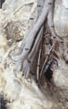
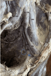
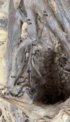
Background The obturator artery (OA), typically originating from the anterior division of the internal iliac artery (ADIIA), shows significant variability in its origin. Such variations can present clinical challenges during pelvic surgeries, potentially causing unnoticed bleeding and complicating effective treatment. This study aims to thoroughly document the diverse anatomical variations of the OA and explore their implications for surgical practice. Materials and methods Forty-eight hemipelvis specimens from adult human cadavers were dissected. The origin of each OA was meticulously documented, photographed, and analyzed descriptively. Results In 38 specimens (79.2%), the OA originated from the IIA. It branched off at various levels from either the ADIIA or the posterior division of the IIA (PDIIA), either individually or in combination with other named branches. In nine cases (18.8%), the OA originated directly from the external iliac artery (EIA), either as a distinct branch or alongside the inferior epigastric artery (IEA). Additionally, one specimen (2%) exhibited a dual origin involving both the ADIIA and the IEA. Conclusion These findings highlight the frequent anatomical variations in the origin and pathway of the OA. Understanding these variations is crucial for accurately assessing pelvic anatomical relationships, which is essential for effective surgical planning and ensuring procedural safety. This knowledge is particularly important during vascular and surgical procedures, as it can impact the risk of bleeding and the effectiveness of treatment strategies.
Keywords: anatomical variations; obturator artery; pelvic cavity; pelvic vasculature; surgical procedures.
Copyright © 2024, Patra et al.
Conflict of interest statement
Human subjects: Consent was obtained or waived by all participants in this study. Animal subjects: All authors have confirmed that this study did not involve animal subjects or tissue. Conflicts of interest: In compliance with the ICMJE uniform disclosure form, all authors declare the following: Payment/services info: All authors have declared that no financial support was received from any organization for the submitted work. Financial relationships: All authors have declared that they have no financial relationships at present or within the previous three years with any organizations that might have an interest in the submitted work. Other relationships: All authors have declared that there are no other relationships or activities that could appear to have influenced the submitted work.
Figures


References
LinkOut - more resources
Full Text Sources
