Review
. 2024 Sep 20:18:1448791.
doi: 10.3389/fnhum.2024.1448791. eCollection 2024.

Entorhinal cortex-hippocampal circuit connectivity in health and disease

Affiliations
Review

Entorhinal cortex-hippocampal circuit connectivity in health and disease

Melissa Hernández-Frausto et al. Front Hum Neurosci. .

Abstract

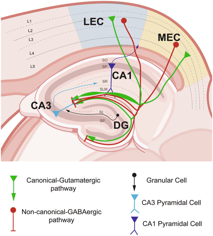
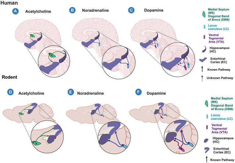
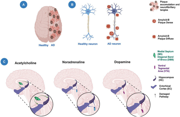
The entorhinal cortex (EC) and hippocampal (HC) connectivity is the main source of episodic memory formation and consolidation. The entorhinal-hippocampal (EC-HC) connection is classified as canonically glutamatergic and, more recently, has been characterized as a non-canonical GABAergic connection. Recent evidence shows that both EC and HC receive inputs from dopaminergic, cholinergic, and noradrenergic projections that modulate the mnemonic processes linked to the encoding and consolidation of memories. In the present review, we address the latest findings on the EC-HC connectivity and the role of neuromodulations during the mnemonic mechanisms of encoding and consolidation of memories and highlight the value of the cross-species approach to unravel the underlying cellular mechanisms known. Furthermore, we discuss how EC-HC connectivity early neurodegeneration may contribute to the dysfunction of episodic memories observed in aging and Alzheimer's disease (AD). Finally, we described how exercise may be a fundamental tool to prevent or decrease neurodegeneration.

Keywords: Alzheimer’s disease; aging; entorhinal cortex; episodic memory; exercise; hippocampus; neuromodulation.


Conflict of interest statement

The authors declare that the research was conducted in the absence of any commercial or financial relationships that could be construed as a potential conflict of interest. The author(s) declared that they were an editorial board member of Frontiers, at the time of submission. This had no impact on the peer review process and the final decision.

Figures

Figure 1
Figure 1
Entorhinal cortex–hippocampal circuit through the canonical-glutamatergic and non-canonical GABaergic pathways in non-pathological conditions. Schematic representation of the entorhinal cortex inputs from lateral- (LEC) and medial entorhinal cortex (MEC) into the hippocampal area. In green, the canonical–glutamatergic pathway, showing the variability of the density of projections in the stratum lacunosum moleculare (SLM) layer of the hippocampus (HC). In red, the non-canonical–GABAergic pathway originated from LEC and MEC running into the HC area, please note the differential distribution among the SLM layer. Figure partially created with BioRender and based on Hernández-Frausto et al., 2023.
Figure 2
Figure 2
Acetylcholine, Noradrenaline and Dopamine inputs to the entorhinal cortex-hippocampal circuit in humans and rodents. Schematic representation of known (solid lines) and unknown (dashed lines) modulatory inputs to the entorhinal cortex (EC) and hippocampus (HC) in humans (A–C) and rodents (C–F). (A–C) in humans, acetylcholine released from the medial septum (MS) and diagonal band of Broca (DBB) (A) and noradrenaline released from the Locus Ceruleus (LC) (B) modulate both the EC and HC areas. (C) Dopamine released from the Ventral Tegmental Area (VTA) modulates hippocampal processing, however, it is unknown whether it does so in the EC. In humans, it is unknown whether EC and HC receive dopaminergic modulation from the LC as is observed in rodents. (D–F) In rodents, the entorhinal cortex-hippocampal circuit is modulated by the cholinergic input from MS and DBB (D), noradrenergic input from LC (E), and dopaminergic inputs from the VTA and LC (F). Created with BioRender.
Figure 3
Figure 3
Neuronal degeneration and neuromodulation alterations in Alzheimer’s disease. (A) Schematic representation of a human brain showing a healthy hemisphere (left) and a hemisphere in Alzheimer’s disease (AD) pathology (right). The right hemisphere shows the presence of plaque accumulation and neurofibrillary tangles. (B) Schematic representation of a healthy neuron (left) and a neuron with neurodegeneration (right) showing the presence of amyloid plaques and axonal damage. (C) The modulatory cholinergic and noradrenergic inputs from the Medial Septum/Diagonal Band of Broca (MS/DBB) and Locus Ceruleus (LC), respectively, to the entorhinal cortex-hippocampal circuit are damaged (faded lines) during the temporal course of AD. The modulatory dopaminergic input from the Ventral Tegmental Area (VTA) to the hippocampus is also altered (faded lines). Created with BioRender.

References

    1. Ahmad S. O., Park J. H., Stenho-Bittel L., Lau Y. S. (2009). Effects of endurance exercise on ventral tegmental area neurons in the chronic 1-methyl-4-phenyl-1,2,3,6-tetrahydropyridine and probenecid-treated mice. Neurosci. Lett. 450, 102–105. doi: 10.1016/j.neulet.2008.11.065, PMID: - DOI - PubMed
    1. Ahnaou A., Moechars D., Raeymaekers L., Biermans R., Manyakov N. V., Bottelbergs A., et al. . (2017). Emergence of early alterations in network oscillations and functional connectivity in a tau seeding mouse model of Alzheimer’s disease pathology. Sci. Rep. 7, 14189–14114. doi: 10.1038/s41598-017-13839-6, PMID: - DOI - PMC - PubMed
    1. Aizawa K., Ageyama N., Yokoyama C., Hisatsune T. (2009). Age-dependent alteration in hippocampal neurogenesis correlates with learning performance of macaque monkeys. Exp. Anim. 58, 403–407. doi: 10.1538/expanim.58.403, PMID: - DOI - PubMed
    1. Amani M., Lauterborn J. C., Le A. A., Cox B. M., Wang W., Quintanilla J., et al. . (2021). Rapid aging in the perforant path projections to the rodent dentate gyrus. J. Neurosci. 41, 2301–2312. doi: 10.1523/JNEUROSCI.2376-20.2021, PMID: - DOI - PMC - PubMed
    1. Amaral D. G., Scharfman H. E., Lavenex P. (2007). The dentate gyrus: fundamental neuroanatomical organization (dentate gyrus for dummies). Prog. Brain Res. 163, 3–22. doi: 10.1016/S0079-6123(07)63001-5, PMID: - DOI - PMC - PubMed

LinkOut - more resources