. 2024 Sep 20:15:1448717.
doi: 10.3389/fimmu.2024.1448717. eCollection 2024.

Epigenetic regulation in epithelial cells and innate lymphocyte responses to S. Typhi infection: insights into IFN-γ production and intestinal immunity

Affiliations

Epigenetic regulation in epithelial cells and innate lymphocyte responses to S. Typhi infection: insights into IFN-γ production and intestinal immunity

Rosângela Salerno-Goncalves et al. Front Immunol. .

Abstract

Infection by Salmonella enterica serovar Typhi (S. Typhi), the cause of enteric fevers, is low in high-income countries but persistent in low- and middle-income countries, resulting in 65,400-187,700 deaths yearly. Drug resistance, including in the United States, exacerbates this issue. Evidence indicates that innate lymphocytes (INLs), such as natural killer (NK) cells, and unconventional T lymphocytes (e.g., Mucosal-associated invariant T (MAIT) cells and T-cell receptor gamma delta (TCR-γδ) cells) can impact the intestinal epithelial barrier, the primary site of exposure to S. Typhi. Moreover, INL production of IFN-γ is central in controlling S. Typhi infection. However, the impact of epithelial cells (EC) on the secretion of IFN-γ by INLs and the relationship between these events and epigenetic changes remains unknown. Epigenetic modifications in host cells are fundamental for their differentiation and function, including IFN-γ production. Herein, using a human organoid-derived polarized intestinal epithelial cell monolayer, we investigated the role of H3K4me3 and H3K27me3 epigenetic marks in intestinal immunity, focusing on the function of EC, NK, MAIT, and TCR-γδ cells in response to S. Typhi. This study builds on our previous findings that MAIT subsets exhibiting specific IFN-γ pattern signatures were associated with protection against typhoid fever and that S. Typhi infection regulates changes in chromatin marks that depend on individual cell subsets. Here, we show that cultures exposed to S. Typhi without EC exhibit a significant increase in NK and MAIT cells, and, to a lesser extent, TCR-γδ cells, expressing IFN-γ and H3K4me3 but not H3K27me3 marks, contrasting with cultures where EC is present. The influence of EC on INL H3K4me3 marks might be indirectly mediated through the modulation of IL-18 secretion via the Histone Deacetylase 6 gene during S. Typhi infection.

Keywords: bacteria; epigenetic; gut; human; salmonella.


Conflict of interest statement

The authors declare that the research was conducted in the absence of any commercial or financial relationships that could be construed as a potential conflict of interest. The author(s) declared that they were an editorial board member of Frontiers, at the time of submission. This had no impact on the peer review process and the final decision

Figures
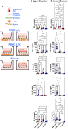
Figure 1
Figure 1
Crosstalk between migrating immune cells and intestinal epithelial cells and the consequences for host responses to S. Typhi. (A) Cartoon of the experimental design. (A-C) Cultures were left untreated (⬤, media only) or exposed to S. Typhi strain Ty2 (⬤) in the presence (PBMC only, or PBMC & EC [epithelial cells]) or absence of PBMC (EC only). After 16 hours, supernatants were harvested and used to determine cytokine/chemokine secretion in the upper (B) and lower compartment (C). Mixed-effects models were used to compare groups. Data are representative of 7 independent experiments. P values < 0.05 were considered statistically significant. The levels of significance are: *, 0.01 to 0.05; **, 0.001 to 0.01; ***, 0.0001 to 0.001; ****<0.0001.
Figure 2
Figure 2
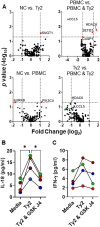
PBMC effects on epithelial cell responses to S. Typhi. HODIM were left untreated (NC) or exposed to S. Typhi strain Ty2 (Ty2) in the presence or absence of PBMC. After 16 hours, epithelial cells and supernatants were harvested and used to determine (A) anti-bacterial and epigenome profile by qRT-PCR. In some experiments, EC & PBMC co-cultures exposed to S. Typhi were treated with H3K27 demethylase inhibitor GSK-J4 (10μM) and the supernatants used to measure (B) IL-18 (upper chamber) and (C) IFN-γ (lower chamber) secretion. Dots are color coded per experiment. Data are representative of 4 independent experiments. *, P values < 0.05 were considered statistically significant.
Figure 3
Figure 3
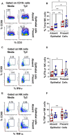
Epithelial cell effects on NK cell responses to S. Typhi. HODIM were left untreated (⬤, media only) or exposed to S. Typhi strain Ty2 (⬤) in the presence or absence of epithelial cells. After 16 hours, PBMC from the lower chamber were harvested to perform EpiTOF analyses (A-F). Levels of total NK cells (A, B), or their subsets: IFN-γ+ (C, D) and TNF-α+ NK cells (E, F). Mixed-effects models were used to compare groups. Data are representative of 6 independent experiments. P values < 0.05 were considered statistically significant. P values < 0.05 were considered statistically significant. The levels of significance are: *, 0.01 to 0.05; **, 0.001 to 0.01; ***, 0.0001 to 0.001; ****<0.0001.
Figure 4
Figure 4
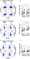
Epithelial cell effects on TCR-γδ cell responses to S. Typhi. HODIM were left untreated (⬤, media only) or exposed to S. Typhi strain Ty2 (⬤) in the presence or absence of epithelial cells. After 16 hours, PBMC from the lower chamber were harvested to perform EpiTOF analyses (A-F). Levels of total NK cells (A, B), or their subsets: IFN-γ+ (C, D) and TNF-α+ NK cells (E, F). Mixed-effects models were used to compare groups. Data are representative of 6 independent experiments. P values < 0.05 were considered statistically significant. P values < 0.05 were considered statistically significant. The levels of significance are: *, 0.01 to 0.05; **, 0.001 to 0.01; ***, 0.0001 to 0.001; ****<0.0001.
Figure 5
Figure 5
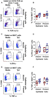
Epithelial cell effects on MAIT cell responses to S. Typhi. HODIM were left untreated (⬤, media only) or exposed to S. Typhi strain Ty2 (⬤) in the presence or absence of epithelial cells. After 16 hours, PBMC from the lower chamber were harvested to perform EpiTOF analyses (A-F). Levels of total NK cells (A, B), or their subsets: IFN-γ+ (C, D) and TNF-α+ NK cells (E, F). Mixed-effects models were used to compare groups. Data are representative of 6 independent experiments. P values < 0.05 were considered statistically significant. P values < 0.05 were considered statistically significant. The levels of significance are: *, 0.01 to 0.05; **, 0.001 to 0.01; ***, 0.0001 to 0.001; ****<0.0001.
Figure 6
Figure 6
Epithelial cell effects on H3K4me3 and H3K27me3 marks in INLs exposed to S. Typhi. HODIM were left untreated (⬤, media only) or exposed to S. Typhi strain Ty2 (⬤) in the presence or absence of EC. After 16 hours, PBMC from the lower chamber were harvested to perform EpiTOF analyses (A-F). Levels of H3K4me3 and H3K27me3 marks in IFN-γ+ INLs (NK cells (A), TCR-γδ cells (C), and MAIT cells (E). Data are representative of 6 independent experiments. In some experiments, EC-PBMC co-cultures exposed to S. Typhi were treated with H3K27 demethylase inhibitor GSK-J4 (10μM) (⬤) or not (0) and concomitant changes in H3K4me3 and H3K27me3 marks in IFN-γ+ INLs (NK cells (B), TCR-γδ cells (D), and MAIT cells (F) were analyzed. Mixed-effects models were used to compare groups. Data are representative of 4 independent experiments. P values < 0.05 were considered statistically significant. P values < 0.05 were considered statistically significant. The levels of significance are: *, 0.01 to 0.05; **, 0.001 to 0.01; ***, 0.0001 to 0.001; ****<0.0001.

References

    1. Typhoid fever and paratyphoid fever, centers for disease control and prevention. (2024). Available at: https://wwwnc.cdc.gov/travel/yellowbook/2024/infections-diseases/typhoid....
    1. G.T.A.P. Collaborators . The global burden of typhoid and paratyphoid fevers: a systematic analysis for the Global Burden of Disease Study 2017. Lancet Infect Dis. (2019) 19:369–81. doi: 10.1016/S1473-3099(18)30685-6 - DOI - PMC - PubMed
    1. Antillon M, Warren JL, Crawford FW, Weinberger DM, Kurum E, Pak GD, et al. . The burden of typhoid fever in low- and middle-income countries: A meta-regression approach. PloS Negl Trop Dis. (2017) 11:e0005376. doi: 10.1371/journal.pntd.0005376 - DOI - PMC - PubMed
    1. Crump JA. Updating and refining estimates of typhoid fever burden for public health action. Lancet Global Health. (2014) 2:e551–3. doi: 10.1016/S2214-109X(14)70306-7 - DOI - PMC - PubMed
    1. Mogasale V, Maskery B, Ochiai RL, Lee JS, Mogasale VV, Ramani E, et al. . Burden of typhoid fever in low-income and middle-income countries: a systematic, literature-based update with risk-factor adjustment. Lancet Global Health. (2014) 2:e570–80. doi: 10.1016/S2214-109X(14)70301-8 - DOI - PubMed

MeSH terms

LinkOut - more resources