. 2024 Sep 20:15:1286942.
doi: 10.3389/fimmu.2024.1286942. eCollection 2024.

Cytosolic nucleic acid sensors and interferon beta-1 activation drive radiation-induced anti-tumour immune effects in human pancreatic cancer cells

Affiliations

Cytosolic nucleic acid sensors and interferon beta-1 activation drive radiation-induced anti-tumour immune effects in human pancreatic cancer cells

Sylvia Kerschbaum-Gruber et al. Front Immunol. .

Abstract

Introduction: Pancreatic ductal adenocarcinoma (PDAC) remains a leading cause of cancer-related deaths worldwide with limited treatment options due to extensive radiation and chemotherapy resistance. Monotherapy with immune checkpoint blockade showed no survival benefit. A combination of immunomodulation and radiotherapy may offer new treatment strategies, as demonstrated for non-small cell lung cancer. Radiation-induced anti-tumour immunity is mediated through cytosolic nucleic acid sensing pathways that drive the expression of interferon beta-1 (IFNB1) and proinflammatory cytokines.

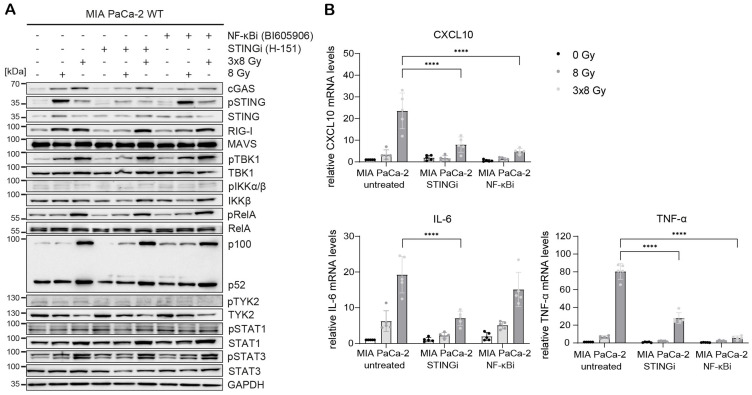
Methods: Human PDAC cell lines (PANC-1, MIA PaCa-2, BxPC-3) were treated with X-rays and protons. Immunogenic cell death was measured based on HMGB1 release. Cytosolic dsDNA and dsRNA were analysed by immunofluorescence microscopy. Cell cycle progression, MHC-I and PD-L1 expression were determined by flow cytometry. Galectin-1 and IFNB1 were measured by ELISA. The expression levels and the phosphorylation status of the cGAS/STING and RIG-I/MAVS signalling pathways were analysed by western blotting, the expression of IFNB1 and proinflammatory cytokines was determined by RT-qPCR and genome-wide by RNA-seq. CRISPR-Cas9 knock-outs and inhibitors were used to elucidate the relevance of STING, MAVS and NF-κB for radiation-induced IFNB1 activation.

Results: We demonstrate that a clinically relevant X-ray hypofractionation regimen (3x8 Gy) induces immunogenic cell death and activates IFNB1 and proinflammatory cytokines. Fractionated radiation induces G2/M arrest and accumulation of cytosolic DNA in PDAC cells, which partly originates from mitochondria. RNA-seq analysis shows a global upregulation of type I interferon response and NF-κB signalling in PDAC cells following 3x8 Gy. Radiation-induced immunogenic response is regulated by STING, MAVS and NF-κB. In addition to immunostimulation, radiation also induces immunosuppressive galectin-1. No significant changes in MHC-I or PD-L1 expression were observed. Moreover, PDAC cell lines show similar radiation-induced immune effects when exposed to single-dose protons or photons.

Conclusion: Our findings provide a rationale for combinatorial radiation-immunomodulatory treatment approaches in PDAC using conventional photon-based or proton beam radiotherapy.

Keywords: MAVS; NF-κB; STING; interferon; pancreatic cancer; protons; radiation.


Conflict of interest statement

The authors declare that the research was conducted in the absence of any commercial or financial relationships that could be construed as a potential conflict of interest.

Figures

Figure 1
Figure 1
Radiation induces immunogenic cell death in human PDAC cell lines. (A) Cell survival curves of PANC-1, MIA PaCa-2 and BxPC-3 after reference X-ray (black circles) or proton irradiation (red triangles) (N=4-5). (B) Soluble HMGB1 was measured by ELISA in the supernatant 72 h post radiation (N=5). Data points represent mean values ± standard deviation. *≤ 0.05; **≤ 0.01; ***≤ 0.005, ****≤0.0001
Figure 2
Figure 2
Radiation induces cytosolic dsDNA accumulation in human PDAC cell lines. (A, B) Cytosolic dsDNA content in PANC-1, MIA PaCa-2 and BxPC-3 cells 24 h post radiation with different doses of X-rays (black circles) or protons (red triangles). 5 cells were analysed per experiment, 4 independent experiments were performed. (A) Quantification and (B) representative images are shown. Scale bar = 20 µm. (C) Flow cytometry analysis of cell cycle distribution in untreated cells and 24 h after treatment with 3x8 Gy or 24 Gy X-rays (N = 4). (D) Percentage of micronuclei-positive PANC-1, MIA PaCa-2 and BxPC-3 cells exposed to different doses of X-rays. A cytokinesis block was induced with cytochalasin B 48 h after radiation for 24 h. Micronuclei-positive binucleated cells were scored. A minimum of 50 cells were analysed per experiment (N=3). (E) Colocalization analyses of dsDNA and TFAM in untreated cells and 24 h after treatment with 3x8 Gy X-rays using Airyscan high resolution imaging (N=3). Quantification and representative images are shown. Scale bar = 20 µm. Data points represent mean values ± standard deviation. *≤ 0.05; **≤ 0.01; ***≤ 0.005, ****≤0.0001.
Figure 3
Figure 3
Hypofractionated irradiation (3x8 Gy) activates IFNB1 in human PDAC cells. Activation of the cGAS/STING pathway 24 h after X-ray (black circles) or proton (red triangles) irradiation was analysed by western blotting and RT-qPCR in (A-C) PANC-1, (D-F) MIA PaCa-2 and (G-I) BxPC-3. (A, D, G) Representative western blots and (B, E, H) quantification based on five replicates are shown. GAPDH was used as a loading control. (C, F, I) Relative expression of IFNB1 and the interferon-stimulated gene (ISG) CXCL10 compared to 0 Gy was determined by RT-qPCR (N=5). No qPCR Ct values could be determined for MIA PaCa-2 IFNB1. Data points represent mean values ± standard deviation. *≤ 0.05; **≤ 0.01; ***≤ 0.005, ****≤0.0001.
Figure 4
Figure 4
Comparative expression levels and activation of cGAS/STING, RIG-I/MAVS, NF-κB and STAT proteins in untreated and irradiated PDAC cells. PANC-1, MIA PaCa-2 and BxPC-3 cells were untreated or irradiated with 8 Gy or 3x8 Gy X-rays and harvested 72 h after radiation. (A) Representative western blots and (B) RT-qPCR analysis of IFNB1, CXCL10 and IL-6 compared to 0 Gy. Data points represent mean values of 5 independent experiments ± standard deviation. *≤ 0.05; **≤ 0.01.
Figure 5
Figure 5
Type I interferon response and NF-κB signalling are globally upregulated in human PDAC cells after fractionated X-ray dose. (A-C) RNA-seq analysis in (A) PANC-1, (B) MIA PaCa-2 and (C) BxPC-3 cells 24 h after 3x8 Gy of X-rays compared to untreated. (D-F) RNA-seq analysis in (D) PANC-1, (E) MIA PaCa-2 and (F) BxPC-3 (A) PANC-1, (B) MIA PaCa-2 and (C) BxPC-3 cells 72 h after 3x8 Gy of X-rays compared to untreated. Genes with fold-change>2, p<0.05 are marked in red as upregulated or blue as downregulated (N=3). (G) GO analysis of upregulated genes shows enrichment of genes involved in type I interferon response and NF-κB signalling. Normalized enrichment scores (NESs) for Hallmark gene sets (MSigDB) between indicated irradiation treatments and controls in PANC-1, MIA PaCa-2 and BxPC-3 cells. Shown are pathways significant in at least 4 comparisons with significance level p < 0.05 adjusted for multiple testing. (H) Log2 fold changes in gene expression between indicated irradiation and control samples of chosen genes.
Figure 6
Figure 6
STING, MAVS and NF-κB drive radiation-induced immunogenic signalling in PANC-1. PANC-1 WT, STING KO and MAVS KO cells were untreated (0 Gy) or irradiated with 8 Gy or 3x8 Gy X-rays and harvested 72 h after radiation. WT cells were also treated with STINGi (10 µM H-151) or NF-κBi (10 µM BI-605906) without or with radiation. Cells were pretreated with inhibitors for 1 h before irradiation and kept until harvesting. (A) Representative western blot analysis of cGAS/STING, RIG-I/MAVS, IFN-1 and NF-κB signalling. (B) Relative mRNA levels of IFNB1, CXCL10, IL-6 and TNF-α compared to untreated WT cells. (C) IFNB1 was measured in the supernatant of PANC-1 cells by ELISA. Data points represent mean values of 5 independent experiments ± standard deviation. *≤ 0.05; **≤ 0.01; ***≤ 0.005.
Figure 7
Figure 7
STING, MAVS and NF-κB drive radiation-induced immunogenic signalling in BxPC-3. BxPC-3 WT, STING KO and MAVS KO cells were untreated (0 Gy) or irradiated with 8 Gy or 3x8 Gy X-rays and harvested 72 h after radiation. WT cells were also treated with STINGi (10 µM H-151) or NF-κBi (10 µM BI-605906) without or with radiation. Cells were pretreated with inhibitors for 1 h before irradiation and kept until harvesting. (A) Representative western blot analysis of cGAS/STING, RIG-I/MAVS, IFN-1 and NF-κB signalling. (B) Relative mRNA levels of IFNB1, CXCL10, IL-6 and TNF-α compared to untreated WT cells. (C) IFNB1 was measured in the supernatant of BxPC-3 cells by ELISA. Data points represent mean values of 4-5 independent experiments ± standard deviation. *≤ 0.05; **≤ 0.01; ***≤ 0.005, ****≤0.0001.
Figure 8
Figure 8
STING and NF-κB drive radiation-induced immunogenic signalling in MIA PaCa-2. MIA PaCa-2 cells were either untreated (0 Gy) or irradiated with 8 Gy or 3x8 Gy X-rays and harvested 72 h after radiation without or with STINGi (10 µM H-151) or NF-κBi (10 µM BI-605906). (A) Representative western blot analysis of cGAS/STING, RIG-I/MAVS, IFN-1 and NF-κB signalling. (B) Relative mRNA levels of CXCL10, IL-6 and TNF-α compared to untreated WT cells. Data points represent mean values of 5 independent experiments ± standard deviation. ****≤0.0001.
Figure 9
Figure 9
The effects of radiation on Galectin-1, MHC-I and PD-L1 in PDAC cells. (A) Galectin-1 was measured by ELISA in the supernatant of PDAC cells exposed to different doses of X-ray (black circles) or proton (red triangles) radiation 24 h post radiation and also 72 h after 3x8 Gy (N=5). (B) MHC-I expression was analysed by flow cytometry (N=3). (C,D) PD-L1 levels were measured by (C) RT-qPCR (N=5) and (D) FACS (N=3). Data points represent mean values of 3 to 5 independent experiments ± standard deviation. *≤ 0.05; **≤ 0.01; ***≤ 0.005, ****≤0.0001.
Figure 10
Figure 10
Radiation-induced immunogenic signalling in PDAC cell lines. (A) Damaged mitochondria release dsDNA and dsRNA, which activate cGAS/STING and RIG-I/MAVS signalling pathways respectively. Activation of these pathways results in the translocation of transcription factors IRF3 and NF-κB into the nucleus, where they induce the expression of IFNB1 and proinflammatory cytokines such as CXCL10, IL-6 and TNF-α. Activation of the non-canonical NF-κB pathway dampens the induction of IFNB1. Canonical NF-κB signalling can also be induced by the ATM kinase, which is activated by double-strand DNA breaks (DSBs) induced by genotoxic stress. ATM phosphorylates NEMO and promotes its monoubiquitination, which is required for translocation into the cytoplasm where it activates the IKK complex. ATM also induces K63-linked ubiquitination of STING through the p53-IFI16-TRAF6 complex, which activates canonical NF-κB. (B) STING, MAVS and NF-κB are responsible for radiation-induced expression of IFNB1 and proinflammatory cytokines in PDAC cell lines. MAVS is the dominant pathway in PANC-1, STING in BxPC-3, while non-canonical NF-κB is induced in MIA PaCa-2 and dampens IFNB1 expression. NF-κB signalling is more pronounced in PANC-1 and MIA PaCa-2, which harbour oncogenic KRAS mutations G12D and G12C respectively.

References

    1. National Cancer Institue . Cancer Stat Facts: Pancreatic Cancer . Available online at: https://seer.cancer.gov/statfacts/html/pancreas.html. (Accessed April 04, 2022)
    1. Kolbeinsson HM, Chandana S, Wright GP, Chung M. Pancreatic cancer: A review of current treatment and novel therapies. J Invest Surg. (2023) 36:2129884. doi: 10.1080/08941939.2022.2129884 - DOI - PubMed
    1. Mukherji R, Debnath D, Hartley ML, Noel MS. The role of immunotherapy in pancreatic cancer. Curr Oncol. (2022) 29:6864–92. doi: 10.3390/curroncol29100541 - DOI - PMC - PubMed
    1. Henriksen A, Dyhl-Polk A, Chen I, Nielsen D. Checkpoint inhibitors in pancreatic cancer. Cancer Treat Rev. (2019) 78:17–30. doi: 10.1016/j.ctrv.2019.06.005 - DOI - PubMed
    1. Karamitopoulou E. Tumour microenvironment of pancreatic cancer: immune landscape is dictated by molecular and histopathological features. Br J Cancer. (2019) 121:5–14. doi: 10.1038/s41416-019-0479-5 - DOI - PMC - PubMed

MeSH terms