Review
. 2024 Sep 20:15:1476904.
doi: 10.3389/fimmu.2024.1476904. eCollection 2024.

CD8+ T cell exhaustion and its regulatory mechanisms in the tumor microenvironment: key to the success of immunotherapy

Affiliations
Review

CD8+ T cell exhaustion and its regulatory mechanisms in the tumor microenvironment: key to the success of immunotherapy

Biao Zhang et al. Front Immunol. .

Abstract

A steady dysfunctional state caused by chronic antigen stimulation in the tumor microenvironment (TME) is known as CD8+ T cell exhaustion. Exhausted-like CD8+ T cells (CD8+ Tex) displayed decreased effector and proliferative capabilities, elevated co-inhibitory receptor generation, decreased cytotoxicity, and changes in metabolism and transcription. TME induces T cell exhaustion through long-term antigen stimulation, upregulation of immune checkpoints, recruitment of immunosuppressive cells, and secretion of immunosuppressive cytokines. CD8+ Tex may be both the reflection of cancer progression and the reason for poor cancer control. The successful outcome of the current cancer immunotherapies, which include immune checkpoint blockade and adoptive cell treatment, depends on CD8+ Tex. In this review, we are interested in the intercellular signaling network of immune cells interacting with CD8+ Tex. These findings provide a unique and detailed perspective, which is helpful in changing this completely unpopular state of hypofunction and intensifying the effect of immunotherapy.

Keywords: CD8 + T cell; T cell exhaustion; adoptive T cell treatment; immune checkpoint blockade; tumor microenvironment.


Conflict of interest statement

The authors declare that the research was conducted in the absence of any commercial or financial relationships that could be construed as a potential conflict of interest.

Figures
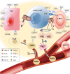
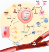
Figure 1
Figure 1
How do tumor cells instigate immune cells into the TME and make them accomplices? T cells in the TME, through their T cell receptor (TCR), recognize the antigen presented by MHC on the APC (including tumor cells) and secrete effector cytokines to exert antitumor immunity. IL-6, which is released by tumor cells in the TME, not only restrains the antitumor immunity of effector T cells but also recruits MDSCs into the TME. Hypoxia in the TME makes HIF-1 α highly expressed. With the mediation of hypoxia-inducible factor-1α (HIF-1α) and IL-6, MDSCs induce monocyte myeloid-derived suppressor cells (m-MDSCs) to differentiate into TAMs for obstructing T cell activation and effector response. In addition, IL-10 produced by MDSCs attracts CD4+ T cells to enter the TME and develop into Tregs with TGF-β. The interaction between IL-35 released by CD177+ Tregs and IL-10 released by PD-1+ Tregs inhibits CD8+ T cells from eliminating tumor cells and mediates T-cell exhaustion.
Figure 2
Figure 2
How can good helpers of CD8+ T cells inhibit Tex and assist anti-tumor immunity in the TME? Immature DCs can sense chemokines secreted by tumor cells to develop into APCs and enter the TME to recognize tumor antigens. Tumor antigens are expressed by APC, which further stimulates CD8+ T cells to develop into Teff and memory T cells (Tmem). Plasmacytoid pre-dendritic cells (pDCs) secrete IFN-α/β, which can improve Teff activity and encourage the maturation of APCs into conventional dendritic cell 1(cDC1) to produce IL-2. IL-2 produced by cDC1 will bind to the IL-2R on Teffs to mediate the STAT5/TOX pathway to inhibit excessive antigen-stimulated Teffs toward exhaustion. In addition, IL-2 can also encourage CD8+ T cell growth. However, excessive IFN-α/β production by pDCs would promote Teff toward CD8+ Tex.

References

    1. Ritter AT, Asano Y, Stinchcombe JC, Dieckmann NM, Chen BC, Gawden-Bone C, et al. . Actin depletion initiates events leading to granule secretion at the immunological synapse. Immunity. (2015) 42:864–76. doi: 10.1016/j.immuni.2015.04.013 - DOI - PMC - PubMed
    1. Dolina JS, Van Braeckel-Budimir N, Thomas GD, Salek-Ardakani S. Cd8(+) T cell exhaustion in cancer. Front Immunol. (2021) 12:715234. doi: 10.3389/fimmu.2021.715234 - DOI - PMC - PubMed
    1. Moskophidis D, Lechner F, Pircher H, Zinkernagel RM. Virus persistence in acutely infected immunocompetent mice by exhaustion of antiviral cytotoxic effector T cells. Nature. (1993) 362:758–61. doi: 10.1038/362758a0 - DOI - PubMed
    1. McLane LM, Abdel-Hakeem MS, Wherry EJ. Cd8 T cell exhaustion during chronic viral infection and cancer. Annu Rev Immunol. (2019) 37:457–95. doi: 10.1146/annurev-immunol-041015-055318 - DOI - PubMed
    1. Beltra JC, Manne S, Abdel-Hakeem MS, Kurachi M, Giles JR, Chen Z, et al. . Developmental relationships of four exhausted Cd8(+) T cell subsets reveals underlying transcriptional and epigenetic landscape control mechanisms. Immunity. (2020) 52:825–41.e8. doi: 10.1016/j.immuni.2020.04.014 - DOI - PMC - PubMed

Substances