[Preprint]. 2024 Sep 7:2024.09.07.611779.
doi: 10.1101/2024.09.07.611779.

Mechano-osmotic signals control chromatin state and fate transitions in pluripotent stem cells

Affiliations

Mechano-osmotic signals control chromatin state and fate transitions in pluripotent stem cells

Kaitlin P McCreery et al. bioRxiv. .

Update in

  • Mechano-osmotic signals control chromatin state and fate transitions in pluripotent stem cells.
    McCreery KP, Stubb A, Stephens R, Fursova NA, Cook A, Kruse K, Michelbach A, Biggs LC, Keikhosravi A, Nykänen S, Pulkkanen R, Hydén-Granskog C, Zou J, Lackmann JW, Niessen CM, Vuoristo S, Miroshnikova YA, Wickström SA. McCreery KP, et al. Nat Cell Biol. 2025 Oct;27(10):1757-1770. doi: 10.1038/s41556-025-01767-x. Epub 2025 Sep 29. Nat Cell Biol. 2025. PMID: 41023488 Free PMC article.

Abstract

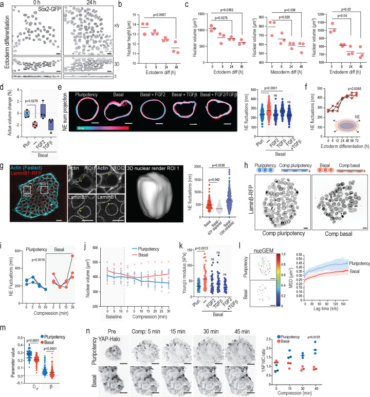
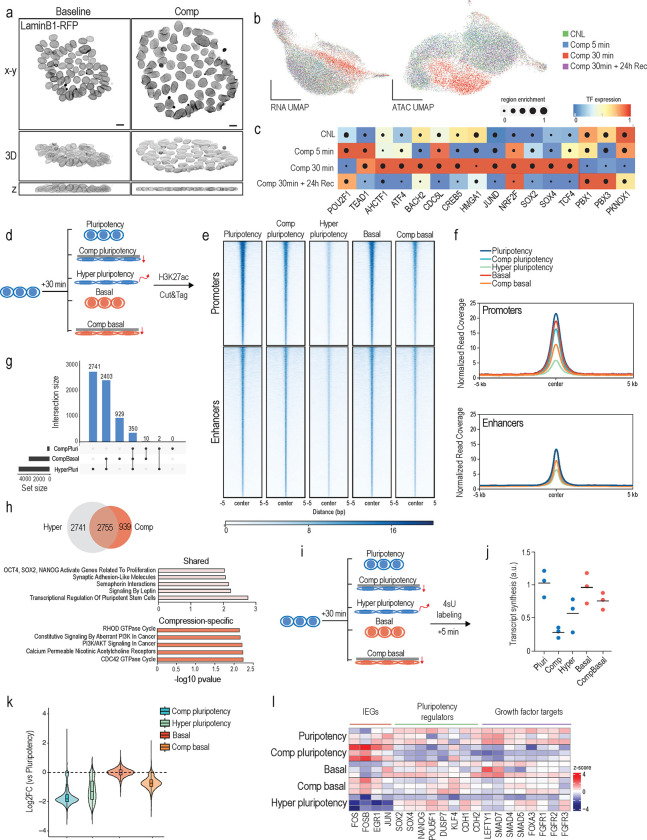
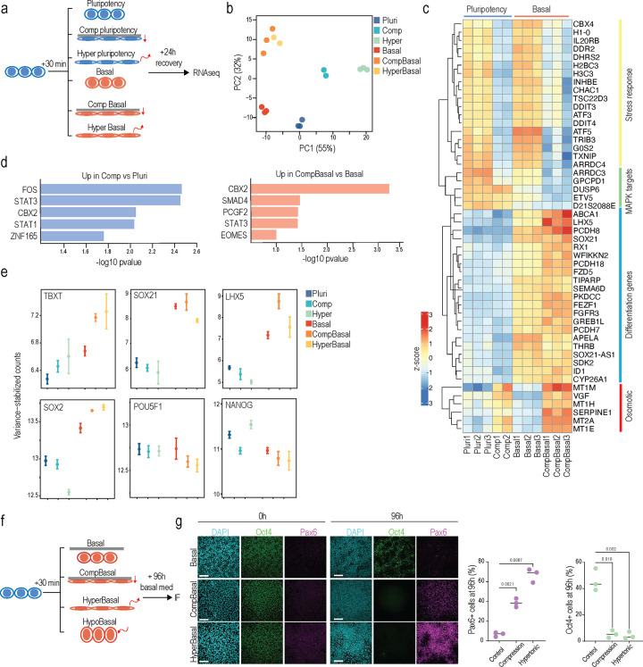
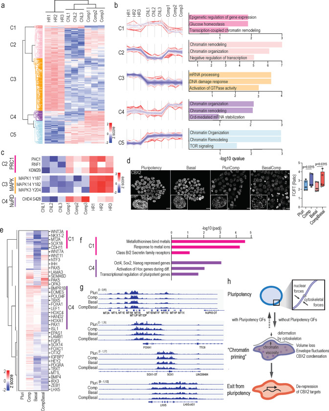
Acquisition of specific cell shapes and morphologies is a central component of cell fate transitions. Although signaling circuits and gene regulatory networks that regulate pluripotent stem cell differentiation have been intensely studied, how these networks are integrated in space and time with morphological transitions and mechanical deformations to control state transitions remains a fundamental open question. Here, we focus on two distinct models of pluripotency, primed pluripotent stem cells and pre-implantation inner cell mass cells of human embryos to discover that cell fate transitions associate with rapid changes in nuclear shape and volume which collectively alter the nuclear mechanophenotype. Mechanistic studies in human induced pluripotent stem cells further reveal that these phenotypical changes and the associated active fluctuations of the nuclear envelope arise from growth factor signaling-controlled changes in chromatin mechanics and cytoskeletal confinement. These collective mechano-osmotic changes trigger global transcriptional repression and a condensation-prone environment that primes chromatin for a cell fate transition by attenuating repression of differentiation genes. However, while this mechano-osmotic chromatin priming has the potential to accelerate fate transitions and differentiation, sustained biochemical signals are required for robust induction of specific lineages. Our findings uncover a critical mechanochemical feedback mechanism that integrates nuclear mechanics, shape and volume with biochemical signaling and chromatin state to control cell fate transition dynamics.


Conflict of interest statement

Declaration of interests The authors declare no conflict of interest.

Figures

Figure 1:
Figure 1:. Exit from primed pluripotency is associated with mechano-osmotic remodeling of the nucleus
(a) Representative top views (x-y), 3D reconstructions and cross sections (z), of Sox2-GFP-tagged hiPSCs undergoing ectodermal differentiation for the indicated time points (scale bars 15 μm). (b) Quantification of nuclear height from hiPSCs undergoing ectodermal differentiation for the indicated time points (n= 3 independent experiments with >600 nuclei/condition/experiment; ANOVA/Dunnett’s). (c) Quantification of nuclear volume from hiPSCs undergoing tri-lineage differentiation for the indicated time points (n= 3 independent experiments with >600 nuclei/condition/experiment; ANOVA/Dunnett’s). (d) Quantification of change in nuclear volume upon exposure to culture medium/growth factors indicated (n=4 independent experiments with >60 nuclei/condition/experiment; Kruskal-Wallis/Dunn’s). (e) Representative projections of nuclear envelope movements as a function of time upon pluripotency factor removal and adding back specific growth factors. Note that removal of pluripotency factors triggers immediate fluctuations that can be rescued by adding back TGF-β and FGF2 (scale bars 5μm; n= 3 independent experiments with >140 nuclei/experiment; ANOVA/Kruskal-Wallis). (f) Quantification of rapid nuclear envelope fluctuations in iPSCs undergoing ectodermal differentiation for time points indicated (n= 3 independent experiments with >120 nuclei/condition/experiment; Kruskal-Wallis/Dunn’s). (g) Representative snapshots of live imaging movies of Lamin-B-RFP tagged iPSC in basal medium, stained with FastAct to label Actin. Note perinuclear actin rings surrounding nuclei and actin-rich bleb like structures with corresponding nuclear deformation (scale bars 20 and 10 μm; images representative of 5 movies). (h) Quantification of nuclear fluctuations from cells in basal medium with or without ATP depletion. Note attenuation of fluctuations upon ATP-depletion (n>150 nuclei/condition pooled across 3 independent experiments; ANOVA/Kruskal Wallis). (i, j) Schematic of experimental outline, representative images (i), and quantification (j) of nuclear envelope fluctuations in cells compressed in pluripotency or basal medium for time points indicated. Note decreased fluctuations pluripotency condition and an increase in basal medium (scale bars 10 μm; n> 150 nuclei/condition pooled across 3 independent experiments; ANOVA/ Kruskal-Wallis). (k) Quantification of nuclear volume evolution over time from of Sox-GFP-tagged hiPSCs live imaged directly after a media change into pluripotency or basal medium, followed by compression. Line represents median volume and individual dots are average colony volumes at indicated timepoints. (n= 10 colonies/condition pooled across 6independent experiments). (l) AFM force indentation experiments of iPS cell nuclei within 20 min of media switch. Note increased elastic modulus of cells in basal media conditions, restored by adding FGF2 (n > 70 nuclei/condition pooled across 5 independent experiments; ANOVA/Kruskal-Wallis). (m) Representative tracks of nucGEM particles. Colors represent average rate of diffusion per tracked particle (scale bars 5 μm). (n, o) Quantification of mean squared displacement versus lag time per nucGEM particle (h) and nucGEM diffusion and diffusivity exponent b (n=3 independent experiments with >140 nuclei/experiment; ANOVA/ Kruskal-Wallis). (p) Representative snapshots of live imaging and quantification of HALO-tagged endogenous YAP localization in cells compressed to 5 μm height in pluripotency or basal medium. Note YAP nuclear entry in pluripotency condition but not in basal medium upon compression (scale bars 30 μm; n= 3 independent experiments with >100 nuclei/experiment; ANOVA/Friedman).
Figure 2:
Figure 2:. Nuclear flattening primes chromatin for spontaneous differentiation
(a) Representative top views, side views and 3D reconstructions of LaminB1-RFP-tagged hiPSCs subjected to compression (Scale bars 10 μm; images representative of 6 independent experiments). (b) Uniform manifold approximation and projection (UMAP) of scRNA- and scATAC-seq from hiPSCs subjected to compression for timepoints indicated (c) Heatmap of predicted regulons enriched in compressed cells from SCENIC+ analyses of the multiome data. (d) Schematic of experimental outline for genome-wide mapping of H3K27ac changes. (e, f) Heat map (e) and metaplot (f) analysis of mean H3K27ac levels at active promoters and predicted active enhancer regions. Note reduction in H3K27ac enrichment at promoters across all conditions and at enhancers in cells compressed in basal medium or exposed to hypertonic shock. (g) UpSet plot showing an overlap of enhancers decommissioned in compression and hypertonic shock conditions. (h) Venn diagram and Reactome pathway enrichment of compression-specific and shared decommissioned enhancers as defined in (g). (i) Schematic of experimental outline for quantification of the nascent transcriptome. (j) Quantification of RNA synthesis across conditions. Note reduced synthesis across all conditions compared to pluripotency medium condition. (k) Quantification of total changes in nascent RNA production across conditions relative to the pluripotency medium condition. (l) Heatmap of altered nascent RNA levels of relevant transcripts from TTseq. Note increased levels of immediate early genes specifically in cells compressed in pluripotency medium while key pluripotency and growth factor regulators are repressed.
Figure 3:
Figure 3:. Mechano-osmotic signals control kinetics of lineage commitment
(a, b) Schematic of experimental outline (a) and PCA plot (b) of bulk RNA sequencing in cells subjected to compression or hypertonic shock and recovery in the indicated media conditions. Note divergence of transcriptome of hypertonic shock and compression in pluripotency medium and convergence in basal medium. (c) Heatmap of top variable genes from bulk RNA seq in conditions indicated. Note increase in differentiation gene expression in cells compressed in basal medium. (d) Transcription factor binding enrichment analysis from genes upregulated in the bulk RNAseq for the indicated conditions. (e) Representative examples of gene expression changes across the conditions. Note increased expression of differentiation genes in basal compressed and hypertonic shock conditions. (f, g) Schematic of experimental outline (f), representative images and quantification (g) of iPSCs immunostained for Oct4 and Pax6 after compression or hypertonic shock. Note increased differentiation in compressed cells or cells exposed to hypertonic shock (n= 3 independent experiments with >650 nuclei/experiment; ANOVA/Kruskal-Wallis).
Figure 4:
Figure 4:. Osmotic pressure controls CBX2 condensation to gate gene repression
(a) Heatmap and Euclidian distance dendrogram of differentially abundant phosphosites quantified by mass spectrometry in cells subjected to compression (comp) or hypertonic (HR) stress. (b) Distance-based clustering of phosphosites and GO-term analyses show changes specific or common to the specific stresses. (c) Example heatmaps of differentially abundant phosphoproteins from (b). (d) Representative images and quantification of CBX2 condensation in hiPSCs (Scale bars 20 μm; images representative of n= 5 independent experiments with >20 nuclei/condition/experiment; RM-ANOVA/Holm-Sidak). (e) Heatmap and Euclidian distance dendrogram of differential CBX2 occupancy quantified by CUT&Run in cells subjected to removal of pluripotency factors (basal), compression (comp) or compression in basal medium, normalized to pluripotency condition. (f) Reactome analysis of genes in clusters 1 and 4 implicate metal-binding genes with reduced CBX2 occupancy in both basal medium and basal medium compression condition whereas pluripotency genes show reduced CBX2 in compression in basal medium. (g) Representative tracks of genes with altered CBX2. (h) Model of how intranuclear and cytoskeletal forces influence iPSC exit from pluripotency. Under conditions with pluripotency growth factors (GFs), nuclear mechanics are maintained and differentiation is prevented under volumetric stress, restoring pluripotency gene expression. In the absence of pluripotency GFs, osmotic stress leads to nuclear envelope fluctuations and CBX2 condensation, priming chromatin for a cell state transition. This ultimately causes de-repression of CBX2 target genes, facilitating exit from pluripotency.

References

    1. Kalkan T. & Smith A. Mapping the route from naive pluripotency to lineage specification. Philos Trans R Soc Lond B Biol Sci 369 (2014). - PMC - PubMed
    1. Dixon J.R. et al. Chromatin architecture reorganization during stem cell differentiation. Nature 518, 331–336 (2015). - PMC - PubMed
    1. Pelham-Webb B., Murphy D. & Apostolou E. Dynamic 3D Chromatin Reorganization during Establishment and Maintenance of Pluripotency. Stem Cell Reports 15, 1176–1195 (2020). - PMC - PubMed
    1. Tyser R.C.V. et al. Single-cell transcriptomic characterization of a gastrulating human embryo. Nature 600, 285–289 (2021). - PMC - PubMed
    1. Lovicu F.J., McAvoy J.W. & de Iongh R.U. Understanding the role of growth factors in embryonic development: insights from the lens. Philos Trans R Soc Lond B Biol Sci 366, 1204–1218 (2011). - PMC - PubMed

Publication types

LinkOut - more resources