[Preprint]. 2024 Sep 16:rs.3.rs-5058118.
doi: 10.21203/rs.3.rs-5058118/v1.

A Broad Spectrum Lasso Peptide Antibiotic Targeting the Bacterial Ribosome

Affiliations

A Broad Spectrum Lasso Peptide Antibiotic Targeting the Bacterial Ribosome

Gerard Wright et al. Res Sq. .

Update in

  • A broad-spectrum lasso peptide antibiotic targeting the bacterial ribosome.
    Jangra M, Travin DY, Aleksandrova EV, Kaur M, Darwish L, Koteva K, Klepacki D, Wang W, Tiffany M, Sokaribo A, Chen X, Deng Z, Tao M, Coombes BK, Vázquez-Laslop N, Polikanov YS, Mankin AS, Wright GD. Jangra M, et al. Nature. 2025 Apr;640(8060):1022-1030. doi: 10.1038/s41586-025-08723-7. Epub 2025 Mar 26. Nature. 2025. PMID: 40140562 Free PMC article.

Abstract

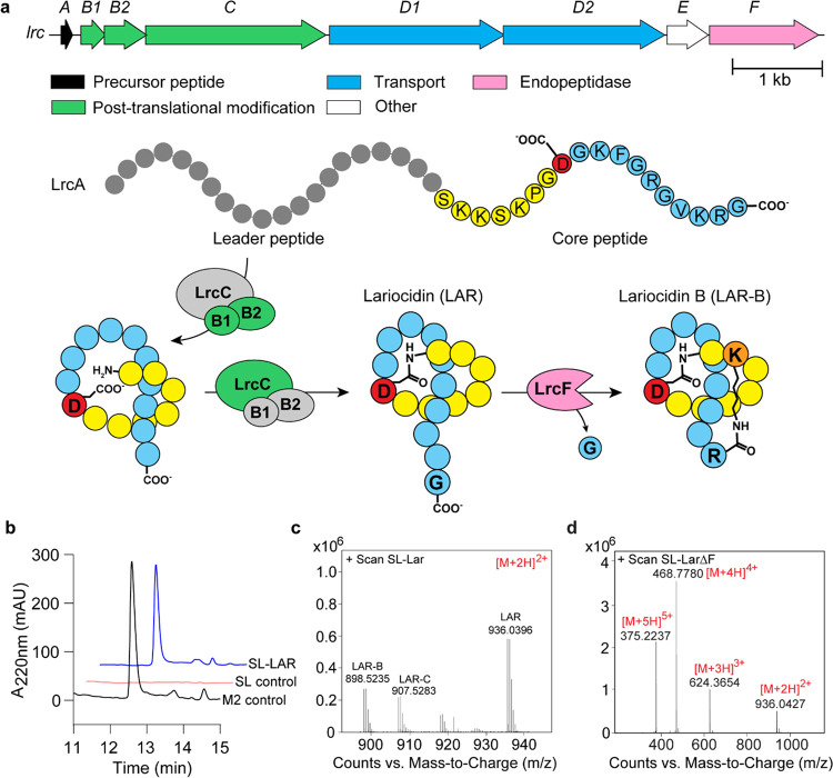
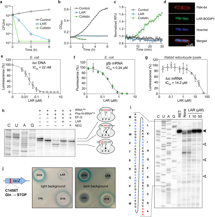
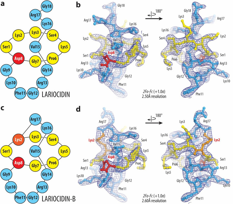
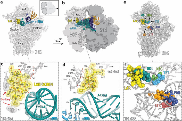
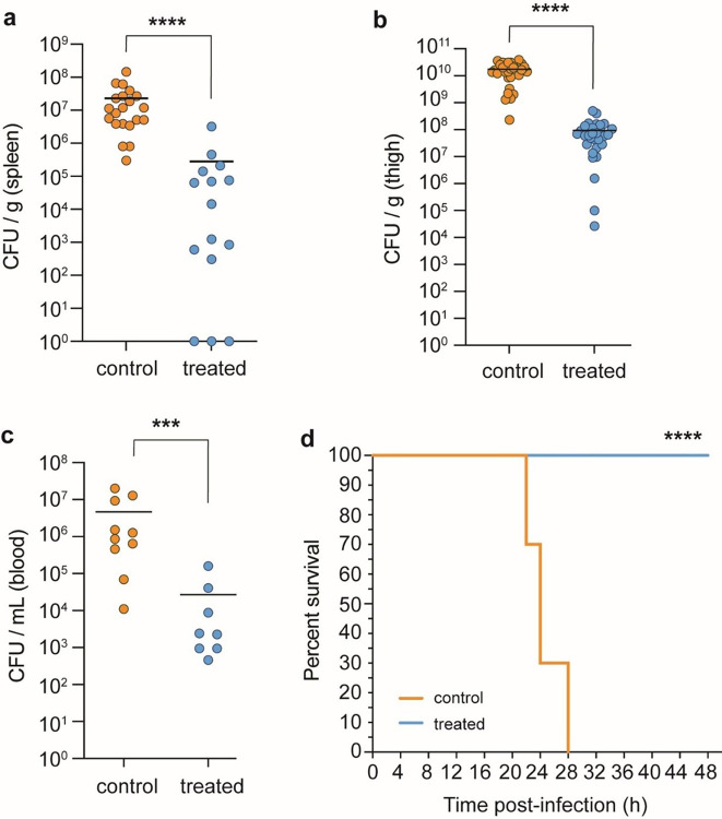
Lasso peptides, biologically active molecules with a distinct structurally constrained knotted fold, are natural products belonging to the class of ribosomally-synthesized and posttranslationally modified peptides (RiPPs). Lasso peptides act upon several bacterial targets, but none have been reported to inhibit the ribosome, one of the main antibiotic targets in the bacterial cell. Here, we report the identification and characterization of the lasso peptide antibiotic, lariocidin (LAR), and its internally cyclized derivative, lariocidin B (LAR-B), produced by Paenabacillussp. M2, with broad-spectrum activity against many bacterial pathogens. We show that lariocidins inhibit bacterial growth by binding to the ribosome and interfering with protein synthesis. Structural, genetic, and biochemical data show that lariocidins bind at a unique site in the small ribosomal subunit, where they interact with the 16S rRNA and aminoacyl-tRNA, inhibiting translocation and inducing miscoding. LAR is unaffected by common resistance mechanisms, has a low propensity for generating spontaneous resistance, shows no human cell toxicity, and has potent in vivo activity in a mouse model of Acinetobacter baumannii infection. Our finding of the first ribosome-targeting lasso peptides uncovers new routes toward discovering alternative protein synthesis inhibitors and offers a new chemical scaffold for developing much-needed antibacterial drugs.

Keywords: Lasso peptide; RiPP; antibiotic; decoding; drug resistance; inhibitor; ribosome; tRNA; translocation.


Conflict of interest statement

COMPETING INTERESTS STATEMENT The authors declare no competing interests.

Figures

Figure 1
Figure 1. Lariocidin and its biosynthetic gene cluster.
(a) Top, gene composition of the lrc BGC. Bottom, posttranslational modification of the LrcA precursor peptide leads to the production of LAR-A and LAR-B. (b) Heterologous expression of lrc BGC in Streptomyces lividans and analysis of LAR in cell-free supernatant. The panel shows chromatographic analysis of LAR produced in the heterologous host. SL-Lar S. lividans pIJ10257-lrc; M2 control is LAR purified from the native producer Paenibacillus M2; SL-control is S. lividans with the empty vector (without lrc BGC). (c) LC-MS analysis of LAR purified from the heterologous host. All masses shown are ions corresponding to [M+2H]2+. (d) LC-MS analysis of LAR produced by heterologous host from the lrc operon with the lrcF gene deleted, showing exclusive production of LAR (or LAR-A), but not LAR-B and LAR-C variants.
Figure 2
Figure 2. LAR exhibits bactericidal activity and targets bacterial protein synthesis.
(a) Reduction in viable cell counts (colony forming units, CFU) of E. coli cultures treated with LAR at 10xMIC (40 μg/ml) or the cell membrane-targeting lytic antibiotic colistin at 10xMIC (5 μg/ml). (b) Effect of LAR or colistin on the density of exponential E. coli cell cultures. In a and b, data are presented as mean ±SD (standard deviation) of three technical replicates and are representatives of two biological replicates with similar results. (c) A propidium iodide accumulation assay was used to assess the effect of LAR on cell permeabilization. Colistin was used as a positive control. The y-axis represents relative fluorescence units (RFU) normalized by the initial fluorescence at time 0. Plot points represent the mean of three biological replicates, with error bars representing SD. (d) LAR-BODIPY accumulates in the cytoplasm of E. coli cells. Green; LAR-BODIPY fluorescence; red: membrane stain Fm-4–64; blue: DNA stain Hoechst 33342. (e, f) Effect of LAR on E. coli protein synthesis in the cell-free transcription-translation system programmed with firefly luciferase-encoding plasmid (e) or GFP mRNA (f). (g) Inhibition of protein biosynthesis in rabbit reticulocyte lysate programmed with luc mRNA by LAR in varying concentrations. In e–g, data points represent the mean of three experiments with error bars indicating SD. (h) LAR inhibits ribosome translocation. The pretranslocation complex is assembled from the E. coli ribosome, short mRNA with the AUG and UUC codons, deacylated tRNAiMet in the P-site, and N-acetyl-Phe-tRNAPhe in the A-site. The addition of elongation factor G (EF-G) and GTP promotes translocation, which is detected by extension of the primer annealed to the mRNA 3’ end. Adding LAR or a control antibiotic negamycin (NEG), a known inhibitor of translocation, interferes with the movement of mRNA/tRNA complex through the ribosome. (i) Toeprinting analysis of ribosomes stalled on a model mRNA template in the presence of increasing concentrations of LAR. The inhibitor of translation initiation retapamulin (RET) was used as a control. ‘None’ – no antibiotic control. The bands representing ribosomes stalled at the start codon are indicated by a black arrowhead, and those of ribosomes stalled at internal mRNA codons are indicated by open arrowheads. (j) LAR induces miscoding as evidenced by the ability of E. coli cells harboring the lacZ gene with a premature stop-codon to produce functional β-galactosidase when exposed to subinhibitory concentrations of LAR (visualized as a blue halo around the zone of cell growth inhibition on indicator plates). The known miscoding-inducing antibiotics gentamicin (GEN) and streptomycin (STR) were used as positive controls; chloramphenicol (CHL) served as a negative control. The plate was imaged with light and dark backgrounds to reveal the blue halo reflecting β-galactosidase expression (left image) and zones of inhibition of cell growth around the drops of applied antibiotics (right image).
Figure 3
Figure 3. Structures and electron density maps of ribosome-boundLAR-A and LAR-B.
(a, c) Schematic diagrams of the lasso peptides LAR-A (a) and LAR-B (c) highlighting their N-terminal residues 1–7 (yellow), branching point at residue 8 (red), and C-terminal residues 9–18 (blue). Lys2 residues of LAR-B forming the second isopeptide bond is colored orange. (b, d) 2Fo-FcFourier electron density maps of LAR (b) and LAR-B (d) in complex with the T. thermophilus 70S ribosome (blue mesh). The refined models of lariocidins are displayed in their respective electron density maps after the refinements contoured at 1.0σ. Color scheme as in panels (a) and (c), respectively.
Figure 4
Figure 4. Structure of LAR in complex with the T. thermophilus 70S ribosome.
(a, b) Overview of the LAR binding site (yellow) in the bacterial ribosome, viewed from two different perspectives. The 30S subunit is shown in light grey, the 50S subunit is dark grey, the mRNA is cyan, and the A-, P-, and E-site tRNAs are colored teal, blue, and orange, respectively. In (a), the 30S subunit is viewed from the inter-subunit side, as indicated by the inset (the 50S subunit and parts of the tRNAs are removed for clarity). The view in panel b is a cross-cut section through the nascent peptide exit tunnel. (c, d) Close-up views of the LAR’s interactions with the small ribosomal subunit. The E. colinumbering of the 16S rRNA nucleotides is used. H-bonds between LAR, rRNA, and A-site tRNA are indicated with dashed lines. In (d), the mRNA nucleotides are numbered relative to the first nucleotide of the P-site codon. (e, f) Superposition of structures of antibiotics binding in the vicinity of the decoding center on the small ribosomal subunit. Overview (e) and close-up view (f) of the ribosome-bound LAR (yellow) relative to the binding sites of other antibiotics targeting the A site of the small ribosomal subunit: odilorhabdin (ODL, teal), tetracycline (TET, blue), negamycin (NEG, green), streptomycin (STR, light red), paromomycin (PAR, dark blue), viomycin (VIO, orange).
Figure 5
Figure 5. Therapeutic efficacy of Lar in a mouse neutropenic thigh infection model.
(a-c) Reduction in bacterial burden (A. baumannii C0286) after 24 hr post administration of LAR as measured by colony forming units (cfu) per gram of tissue or per mL of blood. (a)Bacterial burden in the spleen. control (n = 21) and treated (n = 15) groups. (b)Thigh bacterial burden in control (n = 32) and treated (n = 30) groups. (c)Blood bacterial burden in control (n = 10) and treated (n = 8) groups. Data points are from individual animals and horizontal lines represent the group means. Significance was determined with a two-tailed Mann-Whitney test (*P≤0.05; **P<0.01; ***P<0.001;****P<0.0001). (d) Kaplan-Meier test showing group survival across select time points throughout A. baumannii thigh infection in vehicle control and LAR-treated mice. ****P <0.0001, Log-rank (Mantel-Cox) test.

References

    1. Antimicrobial Resistance, C. Global burden of bacterial antimicrobial resistance in 2019: a systematic analysis. Lancet 399, 629–655 (2022). 10.1016/S0140-6736(21)02724-0 - DOI - PMC - PubMed
    1. Liu Y., Ding S. Y., Shen J. Z. & Zhu K. Nonribosomal antibacterial peptides that target multidrug-resistant bacteria. Nat Prod Rep 36, 573–592 (2019). 10.1039/c8np00031j - DOI - PubMed
    1. Sieber S. A. & Marahiel M. A. Molecular mechanisms underlying nonribosomal peptide synthesis: Approaches to new antibiotics. Chem Rev 105, 715–738 (2005). 10.1021/cr0301191 - DOI - PubMed
    1. Ongpipattanakul C. et al. Mechanism of Action of Ribosomally Synthesized and Post-Translationally Modified Peptides. Chem Rev 122, 14722–14814 (2022). 10.1021/acs.chemrev.2c00210 - DOI - PMC - PubMed
    1. Arnison P. G. et al. Ribosomally synthesized and post-translationally modified peptide natural products: overview and recommendations for a universal nomenclature. Nat Prod Rep 30, 108–160 (2013). 10.1039/c2np20085f - DOI - PMC - PubMed

Publication types

LinkOut - more resources