

A case of spontaneous direct vertebral artery - External vertebral venous plexus fistula in the upper cervical portion
- PMID: 39373003
- PMCID: PMC11450823
- DOI: 10.25259/SNI_736_2024
A case of spontaneous direct vertebral artery - External vertebral venous plexus fistula in the upper cervical portion
Abstract
Background: Spontaneous direct vertebral artery-external vertebral venous plexus (VA-EVVP) fistula is a rare disease that presents in patients with neurofibromatosis type 1 (NF-1) or trauma.
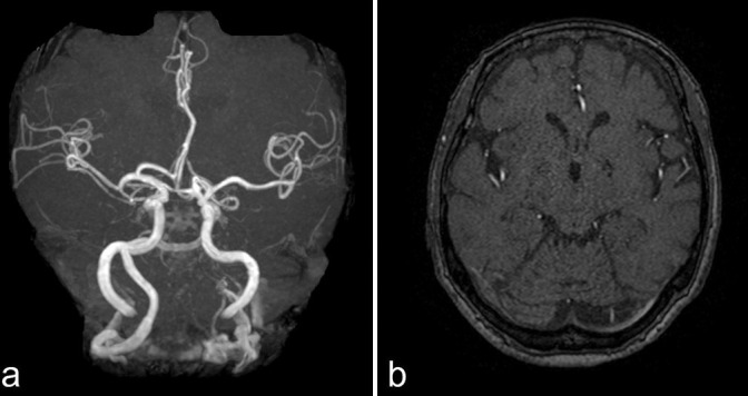
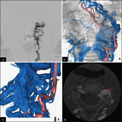
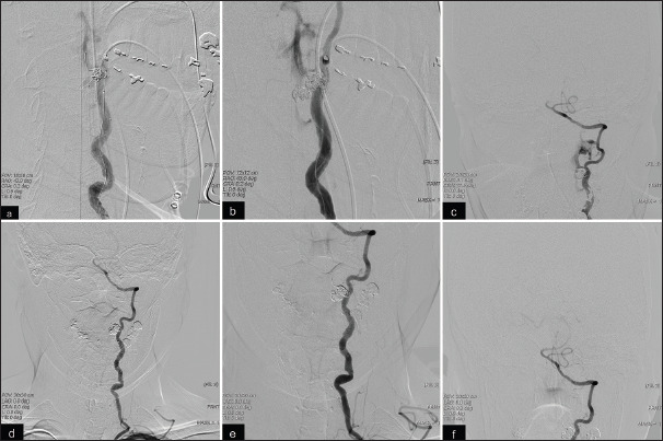
Case description: An 82-year-old female patient with no neurological deficits or trauma presented to our hospital with right hemianopsia. Head magnetic resonance imaging (MRI) revealed left occipital cerebral infarction and magnetic resonance angiography demonstrated high signal intensity in the left transverse sinus (TS). The attending doctor diagnosed an old infarction on the left occipital lobe and dural arteriovenous fistula (AVF) in the TS. After 3 years after the first diagnosis, her new attending doctor re-checked the MRI and performed digital subtraction angiography (DSA). The DSA examination revealed a single-hole AVF between the vertebral artery and external vertebral plexus at the C2 level, which was diagnosed as upper cervical VA-EVVP. The patient presented with tinnitus due to a high-flow VA-EVVP fistula, so we performed coil embolization of the fistula under general anesthesia using a double-catheter technique and achieved subtotal embolization, which diminished the intracranial reflux. The 6-month follow-up DSA image revealed complete obliteration of the AVF.
Conclusion: We report a rare case of upper cervical VA-EVVP fistula in a patient with no history of trauma and relevant medical conditions. Coil embolization of the fistula was performed using a combination of balloon-assisted and double-catheter techniques. Although the patient showed residual shunt flow after the intervention, follow-up DSA revealed complete obliteration. These findings should provide novel insights for the treatment strategy against VA-EVVP fistula.
Keywords: Arteriovenous fistula; Coil embolization; Spontaneous; direct vertebral artery-external vertebral venous plexus fistula.
Copyright: © 2024 Surgical Neurology International.
Conflict of interest statement
There are no conflicts of interest.
Figures
References
-
- Bahar S, Chiras J, Carpena JP, Meder JF, Bories J. Spontaneous vertebro-vertebral arterio-venous fistula associated with fibro-muscular dysplasia. Report of two cases Neuroradiology. 1984;26:45–9. - PubMed
-
- Chen C, Wu Y, Zhao K, Duan G, Liu J, Huang Q. Endovascular treatment of vertebro-vertebral arteriovenous fistula in neurofibromatosis type I: A report of two cases and literature review with a focus on endovascular treatment. Clin Neurol Neurosurg. 2021;207:106806. - PubMed
-
- Deans WR, Bloch S, Leibrock L, Berman BM, Skultety FM. Arteriovenous fistula in patients with neurofibromatosis. Radiology. 1982;144:103–7. - PubMed
-
- Gao P, Chen Y, Zhang H, Zhang P, Ling F. Vertebral arteriovenous fistulae (AVF) in neurofibromatosis type 1: A report of two cases. Turk Neurosurg. 2013;23:289–93. - PubMed
Publication types
LinkOut - more resources
Full Text Sources
Research Materials
Miscellaneous