. 2024 Nov 5;12(11):e0426923.
doi: 10.1128/spectrum.04269-23. Epub 2024 Oct 7.

The development of single-domain VHH nanobodies that target the Candida albicans cell surface

Affiliations

The development of single-domain VHH nanobodies that target the Candida albicans cell surface

Giuseppe Buda De Cesare et al. Microbiol Spectr. .

Abstract

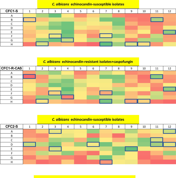
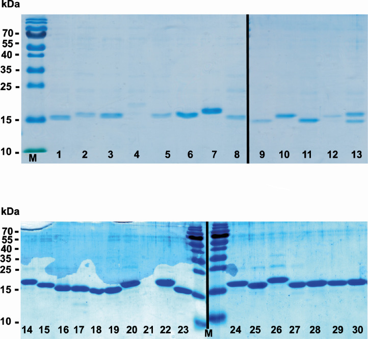
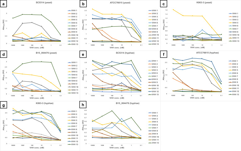
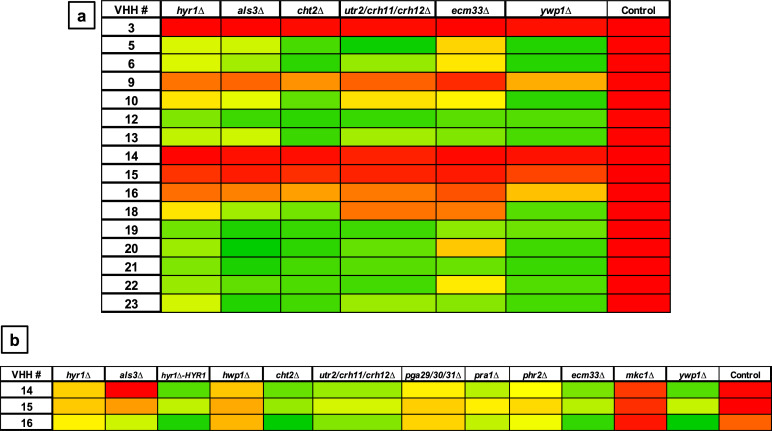
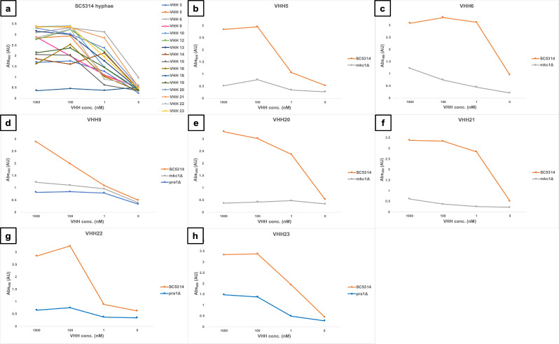
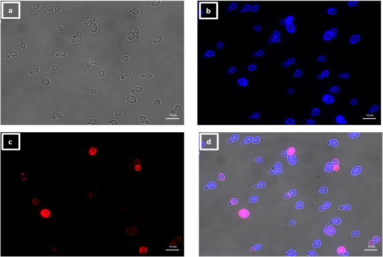
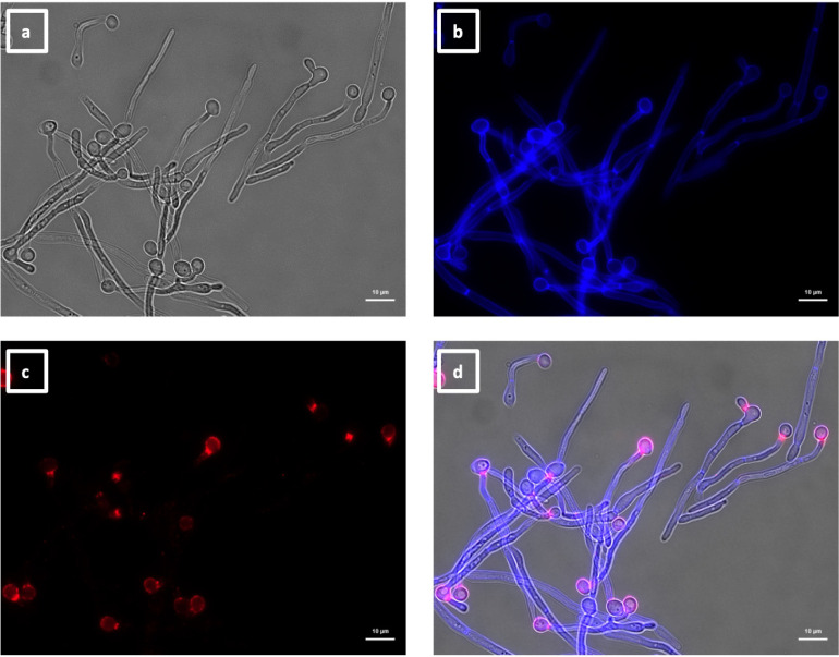
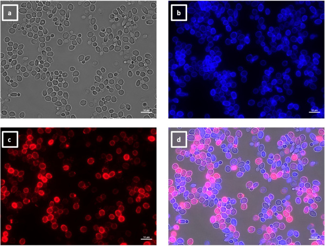
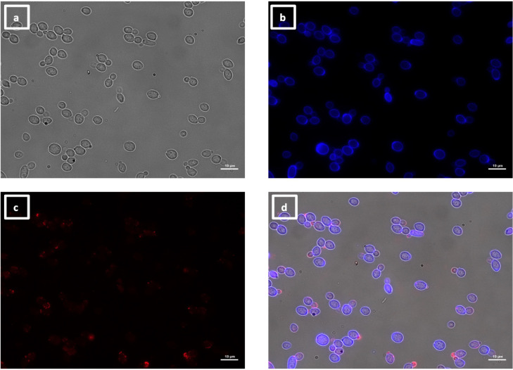
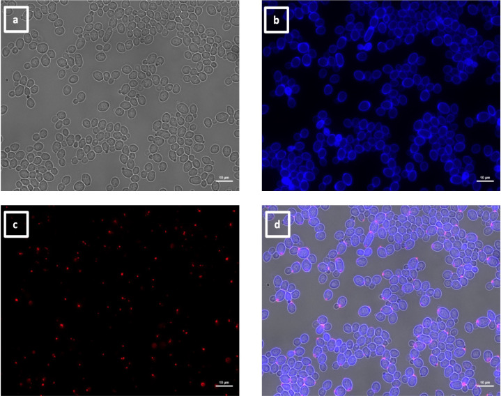
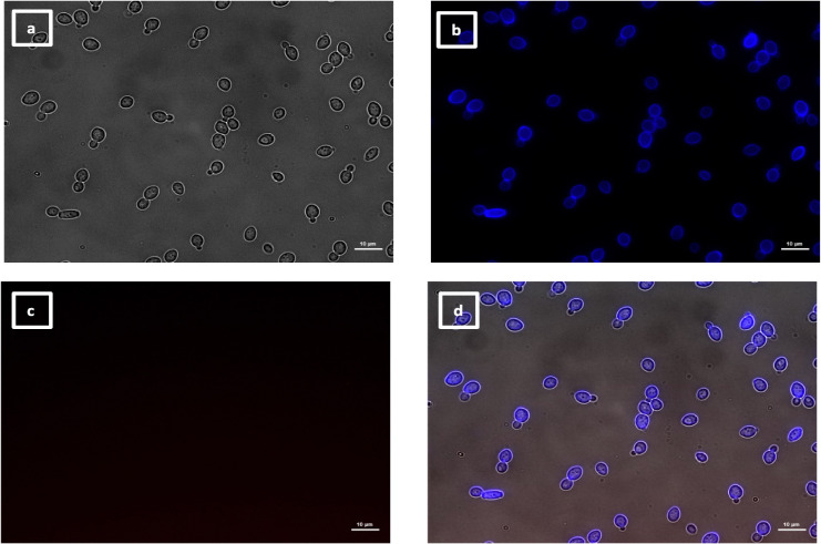
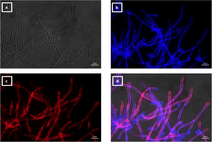
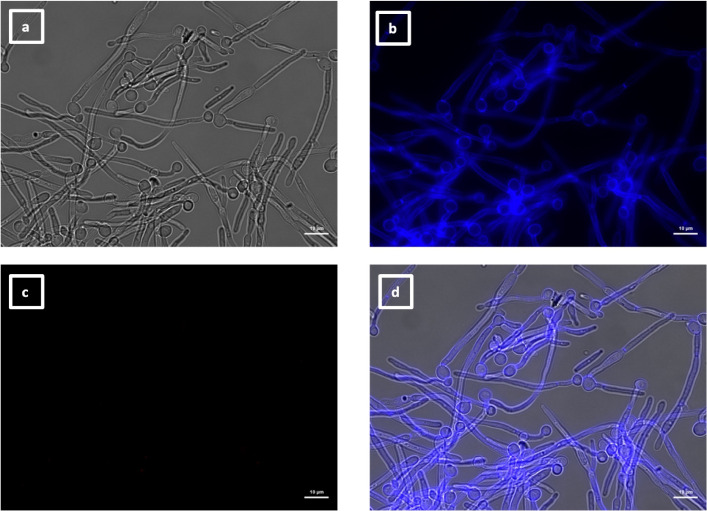
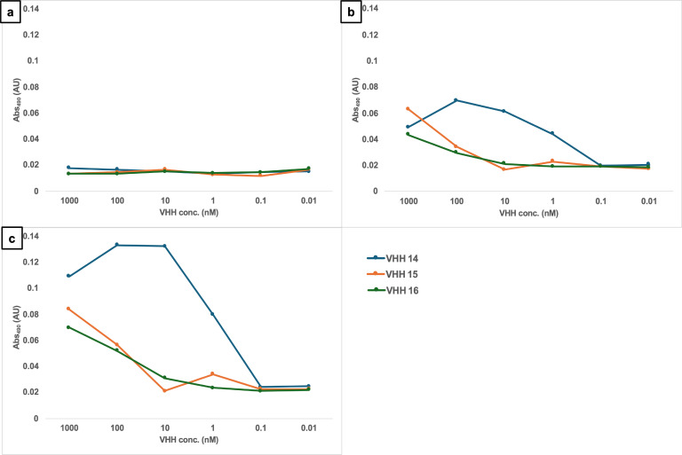
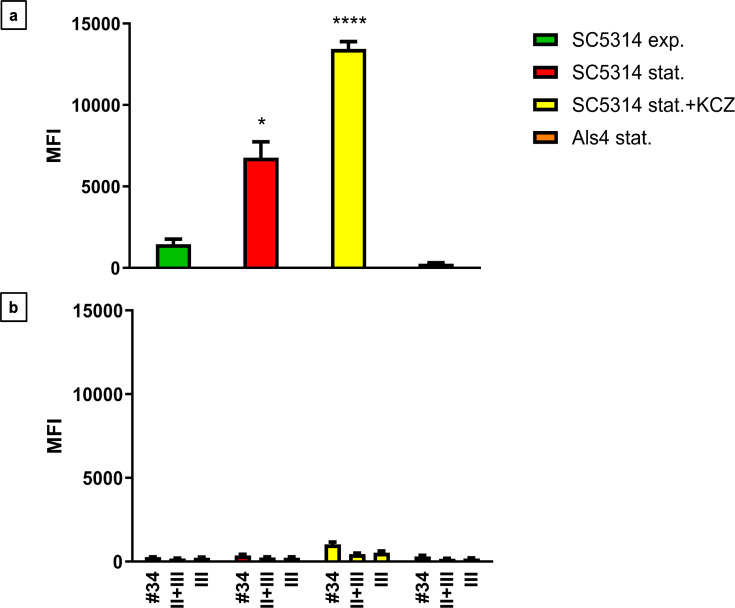
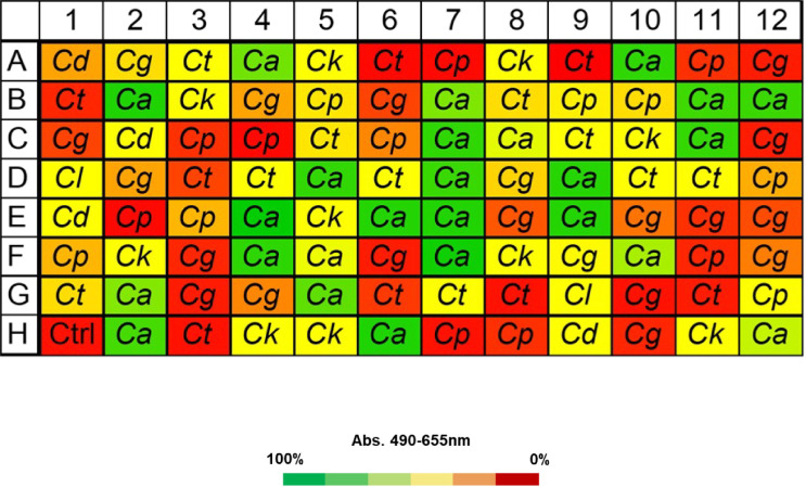
Candida albicans causes life-threatening invasive infections that are hard to diagnose and treat, with drug resistance leading to treatment failure. The goal of this study was to develop VHH (single variable domain on a heavy chain) nanobodies to detect drug-resistant infections. Llamas were immunized with a mixture of heat killed and fixed C. albicans cells of different morphologies. Llama lymphocyte RNA was used to generate phage display libraries that were tested for binding to C. albicans cells or cell wall fractions, and single antibody domains were isolated. The libraries were panned against echinocandin-resistant C. albicans isolates and counter-selected against echinocandin-susceptible isolates with the aim of isolating binding domains specific for antigens on drug-resistant cells. Thirty diverse VHH nanobodies were selected, and binding characteristics were assessed via dose-response ELISA. Binding was tested against a variety of C. albicans isolates and other Candida species, indicating that the VHHs were specific for C. albicans. The VHH nanobodies were sorted into four distinct groups based on their binding patterns. Two of the groups bound preferentially to the yeast cell poles and hyphae, respectively. Nanobody binding to C. albicans deletion mutants was tested by fluorescence microscopy and ELISA to identify the antigen targets. VHH19 nanobody, belonging to the largest group, recognized the Als4 adhesin. VHH14 antibody in the hyphae-specific group recognized Als3. None of the isolated VHH nanobodies was selective for drug-resistant clinical isolates. Our data indicate that this approach can generate valuable single-domain antibodies specific to C. albicans proteins.IMPORTANCEThe human fungal pathogen Candida albicans causes a range of diseases from superficial mucosal infections such as oral and vaginal thrush to life-threatening, systemic infections. Accurate and rapid diagnosis of these infections remains challenging, and currently, there are no rapid ways to diagnose drug-resistant infections without performing drug susceptibility testing from blood culture, which can take several days. In this proof-of-concept study, we have generated a diverse set of single domain VHH antibodies (nanobodies) from llamas that recognize and bind specifically to C. albicans cell surface. The nanobodies were classified into four groups based on their binding patterns, for example, cell poles or hyphae. Specific nanobodies were verified as recognizing the important adhesin Als4 or the hyphae associated invasin Als3, respectively. The data validate the approach that small VHH antibody domains hold future promise for diagnostic applications and as probes to study the fungal cell surface.

Keywords: Candida albicans; antibodies; fungal cell wall.


Conflict of interest statement

E.D. and T.C.V. are affiliated with QVQ company.

Figures

Fig 1
Fig 1
Whole cell ELISA of C. albicans isolates with periplasmic extracts containing the VHHs fused with the phage. Absorbance of ELISA performed with periplasmic extracts containing clones from libraries created after the second round of selection: clones from library 7 tested on echinocandin-susceptible (CFC1-S) and -resistant isolates treated with caspofungin (CFC1-R + CAS); clones from library 11 tested on echinocandin-susceptible (CFC2-S) and -resistant isolates treated with caspofungin (CFC2-R + CAS). The clones highlighted in blue were selected for further characterization based on differential binding between drug-resistant and drug-sensitive isolates with or without caspofungin treatment. The absorbance was calculated at 490 nm and subtracted from the background signal at 655 nm. The color code for the absorbance was set on the signal intensity of the negative controls (empty wells in H6 and H12; isotype control in well H5; and phage with empty pUR8100 plasmid in well H11). Zero percent indicates the signal intensity of the negative controls (red), and 100%, the highest signal in the plate (green). See Fig. S2 of supplementary materials for more information on the layout of the master plate.
Fig 2
Fig 2
SDS-PAGE of VHH antibodies stained with Coomassie blue. The numbers in each lane correspond to the VHH number displayed in Table 2. M: PageRulerTM prestained protein ladder.
Fig 3
Fig 3
Dose-response ELISA of selected VHHs against echinocandin-resistant (K063-3 and B15_004476) and -susceptible (SC5314 and ATCC76615) isolates of C. albicans. Serial dilutions of the VHHs (from 10 µM to 0.1 nM) were tested against: (a) SC5314 yeast; (b) ATCC76615 yeast; (C) K063-3 yeast; (d) B15_004476 yeast; (e) SC5314 hyphae; (f) ATCC76615 hyphae; (g) K063-3 hyphae; and (h) B15_004476 hyphae.
Fig 4
Fig 4
Whole-cell ELISAs with cell wall mutants and selected VHHs. The antibodies were tested against formaldehyde-inactivated cells, and the absorbance was measured at 490 nm (subtracting the background 655 nm) for yeast cells (a) and hyphae (b). Control is SC5314. The color code for the absorbance was set on the background signal given by the control (no cells). Zero percent indicates the signal intensity of the negative controls (red), and 100%, the highest signal in the plate (green). The measurements were the average of two technical replicates.
Fig 5
Fig 5
Dose-response ELISA of selected VHHs against filamentous forms of wild-type and cell wall mutants of C. albicans. Serial dilutions of the VHHs (1 µM, 100 µM, and 1 nM) were tested against (a) SC5314. Binding of the mkc1Δ mutant was tested with (b) VHH5, (c) VHH6, (d) VHH9, (e) VHH20, and (f) VHH21. Binding to the pra1Δ mutant was assessed for (d) VHH9, (g) VHH22, and (h) VHH23.
Fig 6
Fig 6
Immunolabeling of C. albicans yeast with VHH19. Yeast cells of C. albicans strain SC5314 were grown in YPD medium overnight at 30°C and fixed with formaldehyde. The samples were stained with calcofluor white and labeled with VHH19, and a rabbit anti-VHH polyclonal antibody and a goat anti-Rabbit IgG–Alexa Fluor 647–conjugated antibody for detection. Panels indicate: (a) DIC, (b) calcofluor white, (c) Alexa Fluor 647, and (d) merged view. Cells were imaged using a Zeiss Axio Imager M2 microscope.
Fig 7
Fig 7
Immunolabeling of C. albicans hyphae with VHH19. Hyphae of C. albicans strain SC5314 were grown in RPMI-1640 medium at 37°C for 6 h and fixed with formaldehyde. The samples were stained with calcofluor white and labeled with VHH19, and a rabbit anti-VHH polyclonal antibody and a goat anti-Rabbit IgG–Alexa Fluor 647–conjugated antibody for detection. Panels indicate: (a) DIC, (b) calcofluor white, (c) Alexa Fluor 647, and (d) merged view. Cells were imaged using a Zeiss Axio Imager M2 microscope.
Fig 8
Fig 8
Immunolabeling of C. albicans als4Δ yeast with VHH19. Yeast cells of C. albicans strain als4Δ were grown in YPD medium overnight at 30°C and fixed with formaldehyde. The samples were labeled with calcofluor white and VHH19, and a rabbit anti-VHH polyclonal antibody and a goat anti-Rabbit IgG–Alexa Fluor 647–conjugated antibody for detection. Panels indicate: (a) DIC, (b) calcofluor white, (c) Alexa Fluor 647, and (d) merged view. Cells were imaged using a Zeiss Axio Imager M2 microscope.
Fig 9
Fig 9
Immunolabeling of ketoconazole-treated C. albicans yeast with VHH19. Yeast cells of C. albicans strain SC5314 were grown in YPD medium with ketoconazole for 6 h at 30°C and fixed with formaldehyde. The samples were stained with calcofluor white and labeled with VHH19, and a rabbit anti-VHH polyclonal antibody and a goat anti-Rabbit IgG–Alexa Fluor 647–conjugated antibody for detection. Panels indicate: (a) DIC, (b) calcofluor white, (c) Alexa Fluor 647, and (d) merged view. Cells were imaged using a Zeiss Axio Imager M2 microscope.
Fig 10
Fig 10
Immunolabeling of C. albicans yeast with VHH9. Yeast cells of C. albicans strain SC5314 were grown in YPD medium overnight at 30°C and fixed with formaldehyde. The samples were stained with calcofluor white and labeled with VHH9, and a rabbit anti-VHH polyclonal antibody and a goat anti-Rabbit IgG–Alexa Fluor 647–conjugated antibody for detection. Panels indicate: (a) DIC, (b) calcofluor white, (c) Alexa Fluor 647, and (d) merged view. Cells were imaged using a Zeiss Axio Imager M2 microscope.
Fig 11
Fig 11
Immunolabeling of C. albicans pseudohyphae with VHH9. Pseudohyphae of C. albicans strain SC5314 were grown in YPD medium overnight at 30°C, starved for 24 h, transferred into RPMI-1640 medium at 30°C for 6 h, and then fixed with formaldehyde. The samples were stained with calcofluor white and labeled with VHH9, and a rabbit anti-VHH polyclonal antibody and a goat anti-Rabbit IgG–Alexa Fluor 647–conjugated antibody for detection. Panels indicate: (a) DIC, (b) calcofluor white, (c) Alexa Fluor 647, and (d) merged view. Cells were imaged using a Zeiss Axio Imager M2 microscope.
Fig 12
Fig 12
Immunolabeling of C. albicans yeast with VHH2. Yeast cells of C. albicans strain SC5314 were grown in YPD medium overnight at 30°C and fixed with formaldehyde. The samples were stained with calcofluor white and labeled with VHH2, and a rabbit anti-VHH polyclonal antibody and a goat anti-Rabbit IgG–Alexa Fluor 647–conjugated antibody for detection. Panels indicate: (a) DIC; (b) calcofluor white; (c) Alexa Fluor 647; and (d) merged view. Cells were imaged using a Zeiss Axio Imager M2 microscope.
Fig 13
Fig 13
Immunolabeling of C. albicans yeast with VHH14. Yeast cells of C. albicans strain SC5314 were grown in YPD medium overnight at 30°C and fixed with formaldehyde. The samples were stained with calcofluor white and labeled with VHH14, and a rabbit anti-VHH polyclonal antibody and a goat anti-Rabbit IgG–Alexa Fluor 647–conjugated antibody for detection. Panels indicate: (a) DIC; (b) calcofluor white; (c) Alexa Fluor 647; and (d) merged view. Cells were imaged using a Zeiss Axio Imager M2 microscope.
Fig 14
Fig 14
Immunolabeling of C. albicans hyphae with VHH14. Hyphae of C. albicans strain SC5314 were grown in RPMI-1640 medium at 37°C for 6 h and fixed with formaldehyde. The samples were stained with calcofluor white and and labeled with VHH14, and a rabbit anti-VHH polyclonal antibody and a goat anti-Rabbit IgG–Alexa Fluor 647–conjugated antibody for detection. Panels indicate: (a) DIC; (b) calcofluor white; (c) Alexa Fluor 647; and (d) merged view. Cells were imaged using a Zeiss Axio Imager M2 microscope.
Fig 15
Fig 15
Immunolabeling of C. albicans als3Δ hyphae with VHH14. Hyphae of C. albicans als3Δ were grown in RPMI-1640 medium at 37°C for 6 h and fixed with formaldehyde. The samples were stained with calcofluor white and and labeled with VHH14, and a rabbit anti-VHH polyclonal antibody and a goat anti-Rabbit IgG–Alexa Fluor 647–conjugated antibody for detection. Panels indicate: (a) DIC; (b) calcofluor white; (c) Alexa Fluor 647; and (d) merged view. Cells were imaged using a Zeiss Axio Imager M2 microscope.
Fig 16
Fig 16
Dose-response ELISA of hyphae-specific VHHs against C. albicans hyphae. Serial dilutions of the VHH14 (blue), VHH15 (orange), and VHH16 (green) were tested against C. albicans SC5314 (a) yeast, (b) germ tubes, and (c) hyphae.
Fig 17
Fig 17
Flow cytometry of C. albicans yeast cells labeled with VHH19. The MFI was measured for C. albicans als4Δ mutant in stationery phase (Als4 stat.), SC5314 strain in exponential (SC5314 exp.), stationary phase (SC5314 stat.), or SC5314 treated with ketoconazole (SC5314 stat.+ KCZ) immunolabeled with (a) VHH19 and (b) isotype control (#34), no VHH control (II + III), and detection antibody-only control (III). The values represented three independent measurements, and the statistical significance was calculated with one-way ANOVA (n = 350,000 events analyzed, *P < 0.05, ****P < 0.00005).
Fig 18
Fig 18
Whole-cell ELISA experiment to show species specificity of VHH19. VHH19 was tested for binding against C. albicans (Ca) and other Candida species: C. dubliniensis (Cd), C. tropicalis (Ct), C. parapsilosis (Cp), C. glabrata (Cg), C. krusei (Ck), and C. lusitaniae (Cl). Each well of the 96-well plates was coated with fixed fungal cells from each of the indicated species, and ELISA was performed with VHH19. According to absorbance values, VHH19 only recognized C. albicans isolates, indicated by the green squares. The absorbance was measured at 490 nm and subtracted from the background signal at 655 nm. The color code for the absorbance was set on the signal intensity of the empty well negative control (Ctrl): 0% indicates the signal intensity of the negative control (red) and 100%, the highest signal in the plate (green).

References

    1. Odds FC. 2010. Molecular phylogenetics and epidemiology of Candida albicans. Future Microbiol 5:67–79. doi:10.2217/fmb.09.113 - DOI - PubMed
    1. Odds FC. 1987. Candida infections: an overview. Crit Rev Microbiol 15:1–5. doi:10.3109/10408418709104444 - DOI - PubMed
    1. Perlroth J, Choi B, Spellberg B. 2007. Nosocomial fungal infections: epidemiology, diagnosis, and treatment. Med Mycol 45:321–346. doi:10.1080/13693780701218689 - DOI - PubMed
    1. Fisher MC, Alastruey-Izquierdo A, Berman J, Bicanic T, Bignell EM, Bowyer P, Bromley M, Brüggemann R, Garber G, Cornely OA, Gurr SJ, Harrison TS, Kuijper E, Rhodes J, Sheppard DC, Warris A, White PL, Xu J, Zwaan B, Verweij PE. 2022. Tackling the emerging threat of antifungal resistance to human health. Nat Rev Microbiol 20:557–571. doi:10.1038/s41579-022-00720-1 - DOI - PMC - PubMed
    1. Schmalreck AF, Lackner M, Becker K, Fegeler W, Czaika V, Ulmer H, Lass-Flörl C. 2014. Phylogenetic relationships matter: antifungal susceptibility among clinically relevant yeasts. Antimicrob Agents Chemother 58:1575–1585. doi:10.1128/AAC.01799-13 - DOI - PMC - PubMed

LinkOut - more resources