. 2024 Dec 2;156(12):e202413604.
doi: 10.1085/jgp.202413604. Epub 2024 Oct 7.

Functional role of myosin-binding protein H in thick filaments of developing vertebrate fast-twitch skeletal muscle

Affiliations

Functional role of myosin-binding protein H in thick filaments of developing vertebrate fast-twitch skeletal muscle

Andrew F Mead et al. J Gen Physiol. .

Erratum in

Abstract

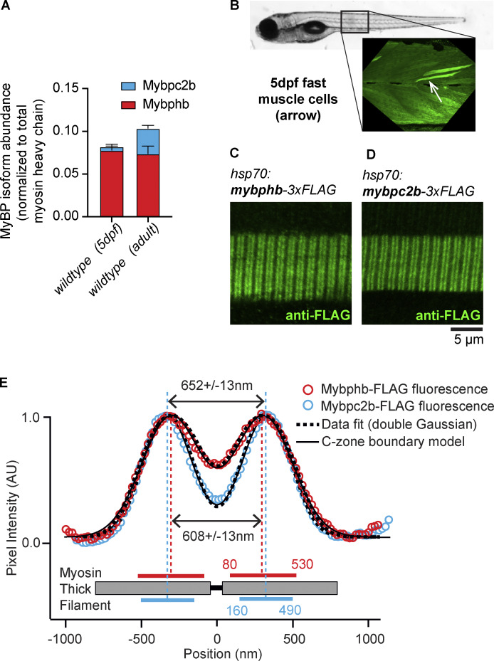
Myosin-binding protein H (MyBP-H) is a component of the vertebrate skeletal muscle sarcomere with sequence and domain homology to myosin-binding protein C (MyBP-C). Whereas skeletal muscle isoforms of MyBP-C (fMyBP-C, sMyBP-C) modulate muscle contractility via interactions with actin thin filaments and myosin motors within the muscle sarcomere "C-zone," MyBP-H has no known function. This is in part due to MyBP-H having limited expression in adult fast-twitch muscle and no known involvement in muscle disease. Quantitative proteomics reported here reveal that MyBP-H is highly expressed in prenatal rat fast-twitch muscles and larval zebrafish, suggesting a conserved role in muscle development and prompting studies to define its function. We take advantage of the genetic control of the zebrafish model and a combination of structural, functional, and biophysical techniques to interrogate the role of MyBP-H. Transgenic, FLAG-tagged MyBP-H or fMyBP-C both localize to the C-zones in larval myofibers, whereas genetic depletion of endogenous MyBP-H or fMyBP-C leads to increased accumulation of the other, suggesting competition for C-zone binding sites. Does MyBP-H modulate contractility in the C-zone? Globular domains critical to MyBP-C's modulatory functions are absent from MyBP-H, suggesting that MyBP-H may be functionally silent. However, our results suggest an active role. In vitro motility experiments indicate MyBP-H shares MyBP-C's capacity as a molecular "brake." These results provide new insights and raise questions about the role of the C-zone during muscle development.


Conflict of interest statement

Disclosures: D.M. Warshaw reported grants from Edgewise Therapeutics outside the submitted work. No other disclosures were reported.

Figures

Figure 1.
Figure 1.
The sarcomere “C-zone” is home to the MyBP-C/H family of regulatory proteins. (A) A schematic of the vertebrate skeletal muscle sarcomere, consisting of interdigitating myosin thick- and actin thin-filaments showing the “C-zone” (black) and expanded view of half-thick filament illustrating a mixture of MyBP-C/H molecules within the C-zone with their N- and C-termini identified. (B) Domain structure and homology among MyBP-C and MyBP-H isoforms showing conservation of C-terminal, myosin thick filament anchoring domains. (C) Normalized abundance of individual myosin heavy chain isoforms (with gene names in parentheses) in embryonic hindlimb buds (n = 3 replicates of 6–10 pooled limb buds each) and prenatal and adult rat tibialis anterior (TA) muscle samples (n = 3 per timepoint) relative to total myosin heavy chain abundance at each timepoint. (D) Abundance of individual myosin binding protein (MyBP) isoforms in embryonic hindlimb bud (none detected) and prenatal and adult rat TA muscle samples, relative to total muscle myosin heavy chain. Data are presented as means ± 1 SD. (E) A midpoint-rooted Maximum Likelihood consensus tree of the MyBP family based on an alignment of three C-terminal globular domains (294 amino acids) conserved among all family members from four species. The percentage of trees, from 100 bootstrapped replicates, in which the associated sequences clustered together is shown next to the branches. Initial tree(s) for the heuristic search were obtained automatically by applying Neighbor-Join and BioNJ algorithms to a matrix of pairwise distances estimated using the JTT model and then selecting the topology with superior log likelihood value. The tree is drawn to scale with branch lengths equal to the number of substitutions per site. Sequences cluster into four distinct clades, corresponding to the four paralogous MyBP gene family members. Orthologous sequences from each species are represented within each clade, indicating divergence and specialization early in vertebrate evolution. The grouping of mammalian MyBP-H and MyBP-HL, as well as zebrafish fMyBP-C (a/b) and MyBP-H (a/b) is indicative of more recent, lineage-specific gene duplication events (nodes with black dots).
Figure S1.
Figure S1.
Genetically modified zebrafish. (A) Transient mosaic expression of FLAG-tagged MyBP was accomplished by transposase-mediated random integration of exogenous MyBP cDNA. We used the modular “tol2kit” approach to assemble the transgene constructs as described in Materials and methods. Briefly, pTol2-Hsp70I:mybphb-3XFLAG and pTol2-hsp70I:mybpc2b-3XFLAG plasmids were generated by cloning chemically synthesized transgene cDNA, together with hsp70I promoter and SV40 late poly-A sequences, into a backbone vector containing tol2 ITR sequence and a cardiac-specific eGFP reporter cassette. (B) The mybphb null allele (Figs. 3, 4, and 5) was generated by creating double-strand breaks in exons 1 and 4 using spCas9 protein precomplexed to guide RNAs to those exons (see Table S5), followed by cell-mediated repair and excision of the intervening ∼12 kb of genomic DNA. Genotyping was accomplished by PCR using primers a, b, c, as shown (see Table S5). Precise characterization of the mutant allele was accomplished by PCR followed by Sanger sequencing. (C) The SA10810 mutation in mybpc2b (Fig. 6) consists of a single base substitution in exon 7 creating a premature STOP codon. Genotyping was accomplished by PCR and bidirectional Sanger sequencing of the mutation site (see Table S5). Source data are available for this figure: SourceData FS1.
Figure S2.
Figure S2.
Immunofluorescent imaging and LCMS of stable Tg:hsp70:mybpc2b-FLAG transgenic zebrafish larvae (F3 generation) and sibling wildtype controls after heat shock treatment (see Materials and methods). (A, inset) Confocal image of heat-shocked and anti-FLAG immunostained Tg:hsp70:mybpc2b-FLAG 5 dpf larval tail shows widespread “doublet” staining pattern. (B) LCMS of 5 dpf heat shocked Tg(hsp70:mybpc2b-FLAG) larval tails shows no significant increase in accumulation of Mypbc2b peptides versus wildtype.
Figure 2.
Figure 2.
Quantification and distribution of MyBP-C and MyBP-H isoforms in fast-twitch zebrafish myotomal muscle. (A) Quantitative LCMS of 5 dpf larval tails (n = 3) and adult, fast-twitch muscle samples (n = 3) reveal two MyBP protein isoforms present at levels above the threshold for quantification: Mybphb (MyBP-H) and Mybpc2b (fMyBP-C) and presented as the relative molar ratio to total myosin heavy chain. (B) Mosaic fluorescence from immunostained FLAG peptide in 5 dpf larval tail injected with hsp70:mypbhb-3xFLAG construct 24 h after heat shock. Fast-twitch myotomal muscle cells (arrow) have a typical trapezoidal shape and off-axis orientation. (C and D) In fast-twitch larval muscle cells expressing transgenic hsp70:mypbhb-3xFLAG (C) or hsp70:mypbc2b-3xFLAG (D), anti-FLAG antibodies label two bands in each sarcomere, creating a fluorescence doublet pattern along the fiber. (E) For each construct, the aligned and integrated intensity of these two bands (open circles) (see Materials and methods) are well fit with two Gaussian peaks (dashed curves) with separations of 608 ± 13 nm (hsp70i:mypbhb-3xFLAG) and 652 ± 13 nm (hsp70:mypbc2b-3xFLAG). Theoretical intensity profiles generated by an analytical model (solid curves) are fit to the experimental data by assuming that the antibody fluorescence is equally distributed across regions of the thick filament, bounded at points 80 and 530 nm (Mybphb-3XFLAG) or 160 and 490 nm (Mybpc2b-3XFLAG) from the sarcomere center. These regions correspond well to the dimensions and location of the mammalian skeletal muscle C-zone. Data are presented as means ± 1 SD.
Figure 3.
Figure 3.
Effect of a mybphb deletion allele on MyBP isoform accumulation and morphology in 5 dpf larval tails. (A) Quantitative LCMS of tails from heterozygous (mybphb+/−) and homozygous (mybphb−/−) larvae show reduction and ablation of Mybphb, respectively, compared with wildtype. Statistics were calculated using a two-tailed Student’s t test. Magenta or cyan * denote P < 0.05 between bars of the corresponding colors. In each case, Mybpc2b accumulation increases slightly, but does not fully compensate for the loss of Mybphb. (B–D)Mybphb−/− larvae display no gross morphological phenotype or change in relative or absolute fast-twitch or slow-twitch muscle cross-section area. (E–H) TEM images of longitudinal (E and G) and transverse (F and H) ∼80 nm sections of 5 dpf fast-twitch muscle cells revealed no deviations from normal muscle ultrastructure in the absence of Mybphb. Statistics were calculated using a two-tailed Student’s t test. ns denotes P > 0.05. Data are presented as Means ± 1 SD.
Figure S3.
Figure S3.
TEM images of longitudinal sections of fast myotomal muscle from three individual wildtype (left) and three individual mybphb −/− (right) 5 dpf zebrafish larvae.
Figure S4.
Figure S4.
TEM images of transverse sections of fast myotomal muscle from three individual wildtype (left) and three individual mybphb −/− (right) 5 dpf zebrafish larvae.
Figure 4.
Figure 4.
Small-angle x-ray scattering from live 5 dpf wildtype and mybphb −/− tails. (A–C) The distances between planes in the paracrystalline lattice formed by myofibrillar actin and myosin filaments are measured by the spacing of equatorial x-ray scattering reflections. (A) TEM of zebrafish fast-twitch muscle cross section and schematic showing the distribution of actin and myosin filaments, and major spatial planes created by the array of actin, thin and myosin, thick filaments. (B) Representative x-ray scatter showing 1,0 and 1,1 equatorial reflections caused by planes in A. The circumferential spread of the reflections around the origin is caused by the inhomogeneity of muscle fiber angles within the tail. (C) Representative plot of background-subtracted radial intensity over the angle phi showing position and relative intensity of 1,0 and 1,1 reflections before and after stretch. (D) Spacing of d1,0 plane in wildtype (n = 7) and mybphb−/− (n = 8) tails at rest length (L0), after being stretched to rest length + 10% (L10), and during a 300 Hz, electrically stimulated tetanic contraction at L10 (Tetanus). Note: we previously showed that a tetanic contraction at L10 results in a 10% shortening of the muscle length back to its rest length of ∼L0 due to tissue compliance at the ends of the preparation (Mead et al., 2020). (E) An increase in the ratio of 1,1 to 1,0 reflection intensities (I1,1/I1,0) can indicate a shift of mass from thick to thin filaments (i.e., myosin heads moving towards or attaching to the thin filament). No such shift was seen in either group with stretch or tetanic stimulation. Data are presented as means ± 1 SD.
Figure 5.
Figure 5.
Myotomal muscles from mybphb −/− larvae function normally. (A and B) 10 dpf larvae react to a mild mechanical stimulus with a brief burst of high-speed swimming. No difference was observed between wildtype (n = 48) and mybphb−/− (n = 27) sibling larvae in average velocity during the escape maneuver (A) or total distance traveled (B). (C–H) In vitro mechanical function of 5 dpf larval tails (Wildtype, n = 3; mybphb−/−, n = 3). Mybphb−/− larvae developed normal forces in response to a single 0.4 ms electrical stimulus (twitch, D), and to a 100 ms, 300 Hz train of stimuli (tetanus, E). (F) Twitch full width at half maximum (FWHM), which depends on rates of activation and relaxation, was also unaffected. (G and H) To measure the force/velocity relationship, tails were allowed to shorten at a series of fixed velocities during tetanic stimulation. Active force at the end of each ramp (a) was normalized to isometric force after recovery (b) and plotted against muscle velocity in H. No difference in force/velocity was seen over the range of velocities possible within the limits of the instrumentation. Significance was determined using a two-tailed Student’s t test. ns denotes P > 0.05. Data are presented as Means ± 1 SD.
Figure 6.
Figure 6.
Mybphb acts as a molecular brake in the C-zone. (A) Abundance of MyBP isoforms in fast-twitch muscle samples from adult wildtype and mybpc2b−/− zebrafish relative to total myosin heavy chain. A homozygous null mutation in mybpc2b (SA10810) ablates Mybpc2b and increases the abundance of Mybphb, maintaining a consistent molar ratio of MyBPs to myosin heavy chains observed in the wiltype zebrafish. (B–D) The sliding of fluorescently labeled actin filaments along native myosin thick filaments isolated from wildtype and mybpc2b−/− adult zebrafish fast muscle. (B) An illustration of wildtype and mutant thick filaments with C-zones colored to represent MyBP isoform content as in A. The native thick filament assay observes the movement of a fluorescently labeled actin filament being propelled over the thick filament surface by the myosin heads emanating from the thick filament. Actin filament displacement trajectories have an initial fast phase associated with the D-zone, which is devoid of MyBP, followed by a potential slower phase as it moves over the MyBP within the C-zone. (C) Actin filament displacement versus time data for 13 different actin filaments moving over different wildtype fast-twitch muscle thick filaments demonstrating dual-phase trajectories as illustrated in B with the fast velocity phase (grey data) and slow velocity phase (green data) identified. (D) The fast (grey) and slow (green) velocity phases for actin filament motion over wildtype thick filaments containing a mix of Mybphb and Mybpc2b (74 thick filaments from n = 4 animals) as in A and mybpc2b−/− thick filaments containing exclusively Mybphb (116 thick filaments from n = 3 animals). Statistics were calculated using a two-tailed Student’s t test. ns denotes P > 0.05. Data are presented as means ± 1 SD.

Update of

References

    1. Ackermann, M.A., and Kontrogianni-Konstantopoulos A.. 2013. Myosin binding protein-C slow: A multifaceted family of proteins with a complex expression profile in fast and slow twitch skeletal muscles. Front. Physiol. 4:391. 10.3389/fphys.2013.00391 - DOI - PMC - PubMed
    1. Ackermann, M.A., and Kontrogianni-Konstantopoulos A.. 2011. Myosin binding protein-C: A regulator of actomyosin interaction in striated muscle. J. Biomed. Biotechnol. 2011:636403. 10.1155/2011/636403 - DOI - PMC - PubMed
    1. Ackermann, M.A., Patel P.D., Valenti J., Takagi Y., Homsher E., Sellers J.R., and Kontrogianni-Konstantopoulos A.. 2013. Loss of actomyosin regulation in distal arthrogryposis myopathy due to mutant myosin binding protein-C slow. FASEB J. 27:3217–3228. 10.1096/fj.13-228882 - DOI - PMC - PubMed
    1. Adekeye, T. E., Teets E.M., Tomak E.A., Waterman S.L., Sprague K.A., White A., Coffin M.L., Varga S.M., Easterbrooks T.E., Shepherd S.J., et al. 2024. Fast-twitch myofibrils grow in proportion to Mylpf dosage in the zebrafish embryo. bioRxiv. 10.1101/2024.09.18.613721 - DOI
    1. Bähler, M., Eppenberger H.M., and Wallimann T.. 1985. Novel thick filament protein of chicken pectoralis muscle: The 86 kd protein. I. Purification and characterization. J. Mol. Biol. 186:381–391. 10.1016/0022-2836(85)90112-3 - DOI - PubMed

MeSH terms

LinkOut - more resources