. 2025 Mar;43(2):61-73.
doi: 10.1007/s00774-024-01555-x. Epub 2024 Oct 7.

Disulfiram ameliorates bone loss in ovariectomized mice by suppressing osteoclastogenesis

Affiliations

Disulfiram ameliorates bone loss in ovariectomized mice by suppressing osteoclastogenesis

Tatsuyuki Fukui et al. J Bone Miner Metab. 2025 Mar.

Abstract

Introduction: Disulfiram (DSF), known as an anti-alcoholism drug, has been reported to suppress osteoclast differentiation in vitro; however, it remains uncertain whether DSF is effective in preventing osteoclastogenesis in vivo. This study aimed to investigate the effect of DSF administration in osteoporotic mice and its contribution to osteoclastogenesis in vivo.

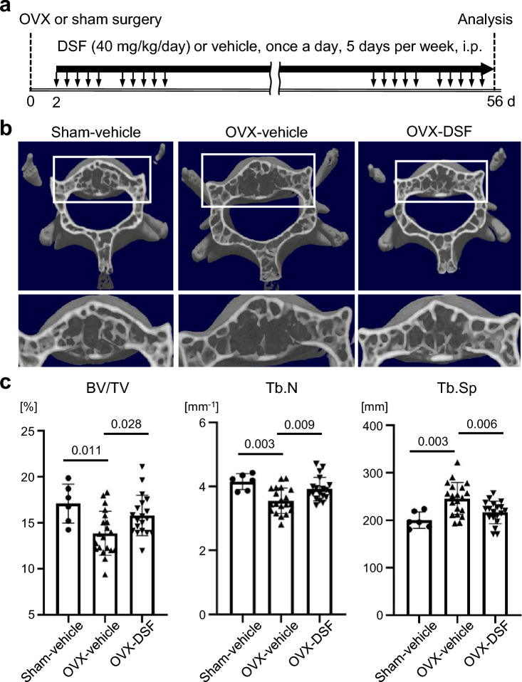
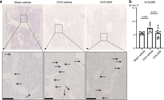
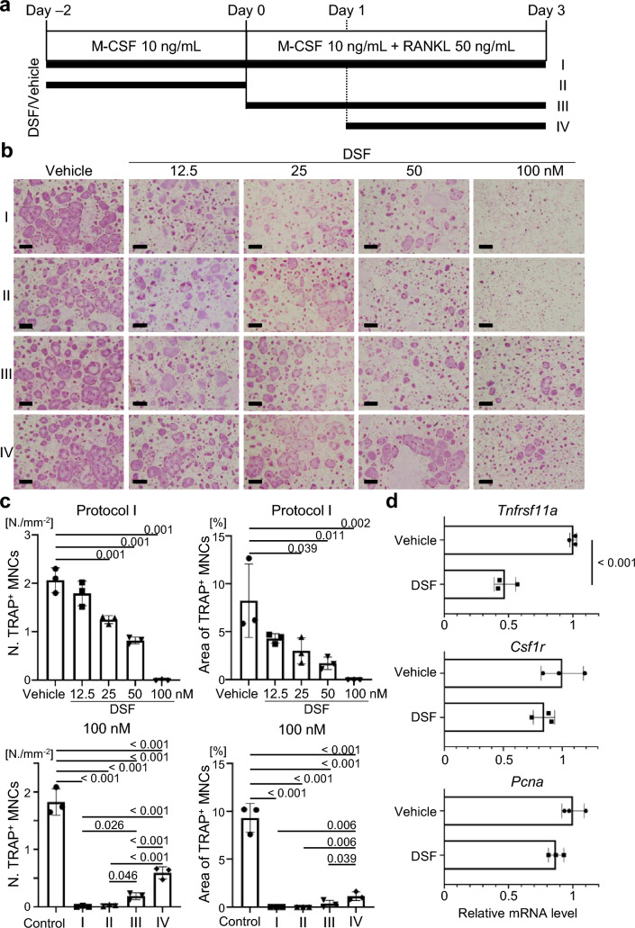
Materials and methods: The bone phenotype of ovariectomized mice, both treated and untreated with DSF, was examined using microcomputed tomography analysis. Osteoclastic and osteoblastic parameters were assessed through bone morphometric analysis. The direct effect of DSF on osteoblastogenesis in vitro was evaluated via a primary osteoblast culture experiment. The expression of genes related to DSF targets (Nup85, Ccr2, and Ccr5) in osteoclast-lineage cells was examined using scRNA-seq analysis and flow cytometry analysis using the bone marrow cells from ovariectomized mice. The impact of DSF on osteoclast-lineage cells was assessed using primary cultures of osteoclasts.

Results: DSF administration ameliorated ovariectomy-induced bone loss and mitigated the increase of osteoclasts without affecting osteoblastogenesis. The scRNA-seq data revealed that osteoclast precursor cells expressed Nup85, Ccr2, and Ccr5. CCR2 and CCR5-positive cells in osteoclast precursor cells within bone marrow increased following ovariectomy, and this increase was canceled by DSF administration. Finally, we found that DSF had a significant inhibitory effect on osteoclastogenesis in the early stage by suppressing Tnfrsf11a expression.

Conclusion: This study demonstrates that DSF could be a candidate for osteoporosis therapies because it suppresses osteoclastogenesis from an early stage in vivo.

Keywords: DSF; Osteoclast precursor; Osteoclastogenesis; Osteoporosis; scRNA-seq analysis.


Conflict of interest statement

Declarations. Conflict of interest: The Department of Osteoimmunology is an endowment department, supported with unrestricted grants from AYUMI Pharmaceutical Corporation, ELEOM, Kondo Cotton Spinning, JCR Pharmaceuticals, MIKIHOUSE, MITSUI FUDOSAN, Meiji, Noevir, TAKENAKA, TENNENBUTSU IKAGAKU KENKYU ZAIDAN and Yakult. Ethical approval: This article does not contain any studies with human participants performed by any of the authors. Informed consent: For this type of study, formal consent is not required.

Figures

Fig. 1
Fig. 1
DSF administration improves the OVX-induced bone loss. a Schematic illustration of the time schedule for OVX and administration of DSF or vehicle. b Representative μCT images of the lumbar vertebrae of sham-operated mice treated with vehicle and OVX-operated mice treated with vehicle or DSF. c Vertebral architectures. BV/TV, bone volume per total volume; Tb.N, trabecular number; Tb.Sp, trabecular separation. Data are presented as the mean (SD), and p-values were determined using Tukey’s multiple comparisons test. Significant p-values are shown
Fig. 2
Fig. 2
DSF administration ameliorates the increased osteoclast number in OVX mice. a Representative images of TRAP staining of the vertebral tissue section. Arrows indicate osteoclasts. Scale bars, 100 μm. b Quantitative analysis of TRAP-positive osteoclast numbers. Data are presented as the mean (SD), and p-values were determined using Tukey’s multiple comparisons test. Significant p-values are shown. N.Oc/BS, osteoclast number per bone surface
Fig. 3
Fig. 3
DSF administration has no obvious effect on osteoblast function and differentiation either in vivo or in vitro. a Histomorphometric analysis of the lumbar vertebrae. b Representative images in histological sections of the lumbar vertebrae of toluidine blue staining (left) and calcein double labeling for determining bone formation (right). Osteoids are indicated between red arrows, and orange arrowheads indicate osteoblasts. Black bars, 20 μm. White bars, 5 μm. c (upper) Representative images of alkaline phosphatase (ALP) staining and alizarin red S staining of murine cranial cells incubated in osteogenic media. (lower) Quantitative analysis of the stain intensity for ALP staining and Alizarin Red S staining. Data are presented as the mean (SD), and p-values were determined using Tukey’s multiple comparisons test. Significant p-values are shown. Ob.S/BS, osteoblast surface per bone surface; BFR/BS, bone formation rate per bone surface; DM, differentiation medium
Fig. 4
Fig. 4
Osteoclast precursor cells (OCPs) express Ccr2, Ccr5, and FROUNT based on scRNA-seq analysis. a Uniform manifold approximation and projection (UMAP) visualization of 11 clusters in the osteoclast culture system. b Separated UMAPs at Days 0, 1, and 3. c Dot plot showing the expression of osteoclast marker genes in the identified clusters. d Dot plot showing the expression of Ccr2, Ccr5, and FROUNT in the identified clusters. e Expression of Ccr2, Ccr5, and FROUNT genes in the UMAP visualization
Fig. 5
Fig. 5
DSF administration suppresses the increase in the percentages of CCR2- or CCR5- positive cells in the OCPs in OVX mice. a, b Percentage of CCR2- (a, lower right box) or CCR5- positive cells (a, upper box) in Lin CD11b−/lo CD115+ CD117+ OCPs. The data are representative of more than two independent experiments. Data are presented as the mean (SD), and p-values were determined using Tukey’s multiple comparisons test. Significant p-values are shown
Fig. 6
Fig. 6
The inhibitory effect of DSF on osteoclastogenesis is observed from an early stage in vitro. a Schematic diagram of the time schedule for the exposure of cultured mouse osteoclasts to DSF or vehicle. b Representative images of TRAP staining of cultured murine osteoclasts. Scale bars, 100 μm. c Numbers of TRAP-positive multinucleated (i.e., more than three nuclei) osteoclasts in areas (N./mm2). d Relative mRNA expression levels of Tnfrsf11a, Csf1r, and Pcna genes at Day 0. Data are presented as the mean (SD), and p-values were determined using Tukey’s multiple comparisons test or Student’s t-test. Significant p-values are shown

References

    1. Hernlund E, Svedbom A, Ivergård M, Compston J, Cooper C, Stenmark J, McCloskey EV, Jönsson B, Kanis JA (2013) Osteoporosis in the European Union: medical management, epidemiology and economic burden. A report prepared in collaboration with the International Osteoporosis Foundation (IOF) and the European Federation of Pharmaceutical Industry Associations (EFPIA). Arch Osteoporos. 10.1007/s11657-013-0136-1 - PMC - PubMed
    1. Walker MD, Shane E (2023) Postmenopausal osteoporosis. N Engl J Med 389:1979–1991. 10.1056/NEJMcp2307353 - PubMed
    1. Riggs BL, Parfitt AM (2005) Drugs used to treat osteoporosis: the critical need for a uniform nomenclature based on their action on bone remodeling. J Bone Miner Res 20:177–184. 10.1359/jbmr.041114 - PubMed
    1. Brown JP, Prince RL, Deal C, Recker RR, Kiel DP, de Gregorio LH, Hadji P, Hofbauer LC, Alvaro-Gracia JM, Wang H, Austin M, Wagman RB, Newmark R, Libanati C, San Martin J, Bone HG (2009) Comparison of the effect of denosumab and alendronate on bmd and biochemical markers of bone turnover in postmenopausal women with low bone mass: a randomized, blinded, phase 3 trial. J Bone Miner Res 24:153–161. 10.1359/jbmr.0809010 - PubMed
    1. Ann DG (1994) Bisphosphonates : structure-activity relationships and therapeutic implications. Medicina 57:61–64

LinkOut - more resources