

doi: 10.1073/pnas.2407246121.
Epub 2024 Oct 7.
The perivascular space is a conduit for cerebrospinal fluid flow in humans: A proof-of-principle report
Affiliations
- PMID: 39374384
- PMCID: PMC11494350
- DOI: 10.1073/pnas.2407246121
Item in Clipboard
The perivascular space is a conduit for cerebrospinal fluid flow in humans: A proof-of-principle report
Proc Natl Acad Sci U S A.
.
Abstract
The glymphatic pathway was defined in rodents as a network of perivascular spaces (PVSs) that facilitates organized distribution of cerebrospinal fluid (CSF) into the brain parenchyma. To date, perivascular CSF and cerebral interstitial fluid exchange has not been shown in humans. Using intrathecal gadolinium contrast-enhanced MRI, we show that contrast-enhanced CSF moves through the PVS into the parenchyma, supporting the existence of a glymphatic pathway in humans.
Keywords: MRI; cerebrospinal fluid; glymphatic; perivascular space.
Conflict of interest statement
Competing interests statement:The authors declare no competing interest.
Figures
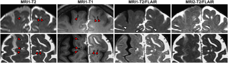
Visualization of perivascular spaces by intrathecal contrast-enhanced brain MRI. Intrathecal contrast-enhanced brain MRI in coronal (A–D) and axial planes (E–H). T2 (A and E), T1 (B and F), and T2/FLAIR (C and G) sequences from timepoint 1, and T2/FLAIR from timepoint 2 (D and H) are shown. Some MV-PVSs decrease in signal intensity while others increase between timepoints 1 and 2. Red arrowheads: MV-PVS on T1 and T2 sequences. White arrow: Postcontrast, nonenhancing MV-PVS. White arrowheads: Enhancing MV-PVSs (D and H).
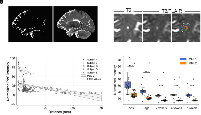
Contrast-enhanced CSF and parenchymal signal relative to the perivascular space. (A) Example of the contrast-containing subarachnoid mask (Left) derived from enhancing subarachnoid spaces on T2/FLAIR (Right). (B) Scatterplot of normalized PVS intensity by distance between all MV-PVSs and the contrast-containing subarachnoid mask. Shaded region = 95% CI. (C) Cross-section of an enhancing MV-PVS shown on T2 (Left), T2/FLAIR (Center), and T2/FLAIR with regions of interest expanding away from the PVS (Right). Red-PVS; orange-PVS edge; green-2 voxels from PVS edge; blue-4 voxels from PVS edge; white-7 voxels from PVS edge. (D) Boxplot of normalized signal intensity by ROI and MRI timepoint for MV-PVS enhancing at timepoint 1.
References
-
- Rennels M. L., Gregory T. F., Blaumanis O. R., Fujimoto K., Grady P. A., Evidence for a ‘Paravascular’ fluid circulation in the mammalian central nervous system, provided by the rapid distribution of tracer protein throughout the brain from the subarachnoid space. Brain Res. 326, 47–63 (1985). - PubMed
MeSH terms
Substances
Grants and funding
LinkOut - more resources
Full Text Sources
Medical
Research Materials
