. 2024 Nov;300(11):107860.
doi: 10.1016/j.jbc.2024.107860. Epub 2024 Oct 5.

Upregulation of nuclear protein Hemgn by transcriptional repressor Gfi1 through repressing PU.1 contributes to the anti-apoptotic activity of Gfi1

Affiliations

Upregulation of nuclear protein Hemgn by transcriptional repressor Gfi1 through repressing PU.1 contributes to the anti-apoptotic activity of Gfi1

Binod G C et al. J Biol Chem. 2024 Nov.

Abstract

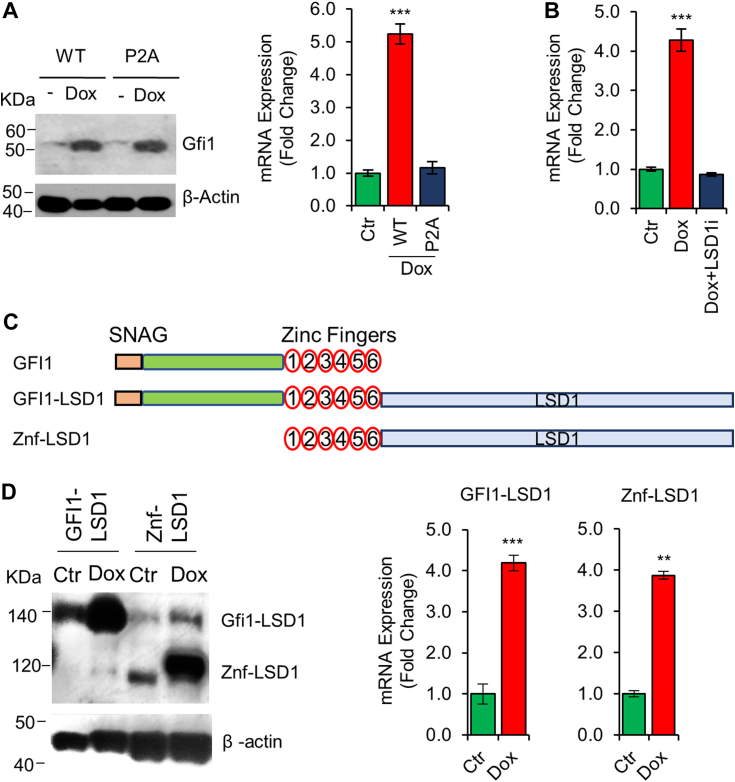
Gfi1 is a transcriptional repressor that plays a critical role in hematopoiesis. The repressive activity of Gfi1 is mediated mainly by its SNAG domain that interacts with and thereby recruits the histone demethylase LSD1 to its target genes. An important function of Gfi1 is to protect hematopoietic cells against stress-induced apoptosis, which has been attributed to its participation in the posttranscriptional modifications of p53 protein, leading to suppression of p53 activity. In this study, we show that Gfi1 upregulated the expression of Hemgn, a nuclear protein, through a 16-bp promoter region spanning from +47 to +63 bp relative to the transcription start site (TSS), which was dependent on its interaction with LSD1. We further demonstrate that Gfi1, Ikaros, and PU.1 are bound to this 16-bp region. However, while Ikaros activated Hemgn and collaborated with Gfi1 to augment Hemgn expression, it was not required for Gfi1-mediated Hemgn upregulation. In contrast, PU.1 repressed Hemgn and inhibited Hemgn upregulation by Gfi1. Notably, PU.1 knockdown and deficiency, while augmenting Hemgn expression, abolished Hemgn upregulation by Gfi1. PU.1 (Spi-1) is repressed by Gfi1. We show here that PU.1 repression by Gfi1 preceded and correlated well with Hemgn upregulation. Thus, our data strongly suggests that Gfi1 upregulates Hemgn by repressing PU.1. In addition, we demonstrate that Hemgn upregulation contributed to the anti-apoptotic activity of Gfi1 in a p53-independent manner.

Keywords: Gfi1; Hemgn; Ikaros; PU.1; apoptosis; cell death; histone demethylase; transcription; transcription repressor.


Conflict of interest statement

Conflict of interest The authors declare that they have no conflicts of interest with the contents of this article.

Figures

Figure 1
Figure 1
Gfi1 upregulates Hemgn expression.A and B, cells as indicated were left untreated (Ctr) or treated with Dox (1 μg/ml) for 24 h, followed by evaluation of Hemgn mRNA and protein expression by qRT-PCR (A) and Western blot analysis (B). C, Hemgn mRNA levels were analyzed in Lin- BM cells from Gfi1+/+ and Gfi1−/− mice. D, BaF/Gfi1 (left panel) and Ramos/Gfi1 (right panel) cells were left untreated (Ctr) or treated with Dox for 6 h and then incubated with or without Doxo for 16 h prior to evaluation of Hemgn mRNA levels. Data are presented as mean ± SD (N = 3). Statistically significant differences in Hemgn expression levels: ∗∗p < 0.01; ∗∗∗p < 0.001.
Figure 2
Figure 2
LSD1 is required for Gfi1-mediated Hemgn upregulation.A, BaF/Gfi1 and BaF/Gfi1P2A cells were left untreated or treated with Dox (1 μg/ml) for 24 h and examined for expression of Gfi1 proteins (left panel) and Hemgn mRNA (right panel). B, BaF/Gfi1 cells were treated with Dox overnight and then with LSD1i (1 μM) for 24 h prior to evaluation of Hemgn mRNA levels. C, schematic diagrams of Gfi1-LSD1 and Znf-LSD1 fusion proteins. D, BaF3 cells expressing the Dox-inducible fusion proteins as indicated were left untreated or treated with Dox for 24 h and then examined for expression of the fusion proteins using the anti-Flag antibody (left panel) and Hemgn mRNA (right panel). Data are shown as mean ± SD (n = 3). Statistical significances: ∗∗p < 0.01; ∗∗∗p < 0.001.
Figure 3
Figure 3
The 16-bp region spanning from +47 to +63 bp of Hemgn is essential for Gfi1-mediated Hemgn upregulation.A, schematic diagrams of the various mouse Hemgn promoter fragments (upper panel) and the different deletion mutants derived from fragment VI (middle panel), and the nucleotide sequence of Hemgn promoter spanning from +33 to +63 bp and the mutations introduced in this region of fragment II. B, the reporter constructs containing the different promoter fragments were transfected into BaF/Gfi1 cells, followed by treatment with Dox (1 μg/ml) for 24 h. Promoter activation, shown as fold changes as compared to untreated cells, was determined. Data are shown as mean ± SD (n = 3). ∗∗p < 0.01; ∗∗∗p < 0.001.
Figure 4
Figure 4
Gfi1 and Ikaros bind to the Hemgn core promoter.A, cells as indicated were cultured with and without Dox for 24 h. ChIP assays were performed using antibodies against Gfi1 and Ikaros or IgG as a control, followed by PCR to amplify the Hemgn core promoter and the 3-kb upstream region. B, expression of Gfi1 and Ikaros in BaF/Ik and BaF/Gfi1/Ik cells. C, BaF3/Gfi1/Ik cells were transfected with the reporter plasmid containing fragment I or fragment VII (upper panel). The green and red arrows denote the forward and reverse primers, respectively, used to amplify the proximal (P1) and distal (P2) plasmid sequences. ChIP experiments were conducted as in A using cells transfected with fragment I (middle panel) and fragment VII (bottom panel). Data are shown as mean ± SD (n = 3). ∗∗p < 0.01; ∗∗∗p < 0.001.
Figure 5
Figure 5
PU.1 binds to the Hemgn core promoter. BaF/Gfi1/PUER cells were transfected with the reporter plasmid containing fragment I or fragment VII, and cultured without or with Dox (1 μg/ml) and 4-HT (100 nM) for 24 h. ChIP experiments were carried out using antibodies against Gfi1, PU.1 or IgG. A, qPCR was conducted to amplify the Hemgn core promoter and the 3 kb upstream region in cells transfected with fragment VII (upper panel). Expression of PUER along with endogenous PU.1 and Gfi1 was examined (lower panel). B, the P1 and P2 plasmid sequences were amplified using cells transfected with fragment I (upper panel) or fragment VII (lower panel). Data are shown as mean ± SD (n = 3). ∗∗p < 0.01; ∗∗∗p < 0.001.
Figure 6
Figure 6
Ikaros collaborates with Gfi1 to activate Hemgn promoter.A, BaF/Ik and BaF/Gfi1/Ik cells were transfected with reporter plasmids containing fragment I or VII and then cultured without (Ctr) or with Dox (1 μg/ml) for 24 h prior to examination of luciferase activity. B, Hemgn mRNA levels were examined in BaF/Gfi1, BaF/Ik and BaF/Gfi1/Ik cells cultured in the absence (Ctr) or presence of Dox for 24 h. C, Hemgn mRNA levels were examined in JE131/Gfi1, JE131/Ik and JE131/Gfi1/Ik cells untreated or treated with Dox for 24 h (left panel). Data are shown as mean ± SD (n = 3). ∗∗p < 0.01; ∗∗∗p < 0.001. Expression of Gfi1 and Ikaros proteins was confirmed by Western blot analysis (right panel).
Figure 7
Figure 7
PU.1 represses Hemgn and inhibits Gfi1-mediated activation of Hemgn.A, BaF/Gfi1/PUER cells were transfected with reporter plasmids containing fragment I or VII and cultured without or with Dox (1 μg/ml), 4HT (100 μM) or both for 24 h prior to evaluation of luciferase activity. B, BaF/Gfi1/PUER, 32D and Lin- BM cells transduced with PUER retroviral construct were cultured without or with 4HT, Dox or both as indicated for 24 h prior to examination of Hemgn mRNA levels by qRT-PCR. Data are shown as mean ± SD (n = 3). ∗∗p < 0.01; ∗∗∗p < 0.001.
Figure 8
Figure 8
PU.1 is required for Gfi1-mediated upregulation of Hemgn.A, Hemgn mRNA (left panel) and protein (right panel) levels were examined in PUER/Gfi1 cells untreated or treated with Dox (1 μg/ml) for 24 h. B, BaF/Gfi1 cells were transduced with empty or two different PU.1 shRNA-expressing lentiviruses (498 and 501) and examined for Hemgn expression following Dox induction for 24 h (upper panel). PU.1 knockdown was confirmed by Western blot analysis (lower panel). C, BaF/Gfi1 cells were treated with Dox for the indicated times and examined for expression of Gfi1 and PU.1 proteins (upper panel) and Hemgn mRNA (middle panel). Lower panel: Graphical presentation of Gfi1, PU.1, and Hemgn expression. The levels of Gfi1 and PU.1 proteins were based on the densities of bands determined using Image J software. Data are shown as mean ± SD (n = 3). ∗∗p < 0.01; ∗∗∗p < 0.001.
Figure 9
Figure 9
Knockdown of Hemgn diminishes the protective effect of Gfi1 on DNA damage-induced apoptosis. BaF/Gfi1 and Ramos/Gfi1 cells were transduced with empty or Hemgn shRNA-expressing lentiviral constructs, followed by treatment with Dox (1 μg/ml) for 6 h prior to treatment with Doxo (200 ng/ml for BaF/Gfi1 cells and 2 mg/ml for Ramos/Gfi1 cells) for 24 h. A, the apoptosis of BaF/Gfi1 cells was examined by Annexin V assay. Shown is a representative flow cytometry experiment. The numbers denote percentages of live cells. B, Data from 3 independent flow cytometry experiments are presented. C, live Ramos/Gfi1 cells were quantitated using MTS assay. The numbers in (B) and (C) indicate increases in the viability of Doxo-treated cells cultured in the presence versus absence of Dox, and are shown as mean ± SD (n = 3). ∗∗p < 0.01; ∗∗∗p < 0.001. D, Hemgn expression was examined by Western blot analysis.
Figure 10
Figure 10
Restoration of Hemgn expression in Gfi1−/−BM cells partially rescues the hypersensitivity to DNA damage. Lin cells were isolated from WT and Gfi1−/− mice. Gfi1−/− cells were transduced with the empty or Hemgn-expressing retrovirus and sorted based on GFP. Cells were then treated with Doxo (50 ng/ml) for 24 h and examined for apoptosis by flow cytometry based on annexin V staining. A, shown is a representative experiment of flow cytometry. B, data from 3 independent flow cytometry experiments are presented. Data are shown as mean ± SD (n = 3). ∗∗p < 0.01; ∗∗∗p < 0.001. C, expression of Hemgn protein in the different cell populations was examined by Western blot analysis.

References

    1. Grimes H.L., Chan T.O., Zweidler-McKay P.A., Tong B., Tsichlis P.N. The Gfi-1 proto-oncoprotein contains a novel transcriptional repressor domain, SNAG, and inhibits G1 arrest induced by interleukin-2 withdrawal. Mol. Cell Biol. 1996;16:6263–6272. - PMC - PubMed
    1. Zeng H., Yücel R., Kosan C., Klein-Hitpass L., Möröy T. Transcription factor Gfi1 regulates self-renewal and engraftment of hematopoietic stem cells. EMBO J. 2004;23:4116–4125. - PMC - PubMed
    1. Hock H., Hamblen M.J., Rooke H.M., Schindler J.W., Saleque S., Fujiwara Y., et al. Gfi-1 restricts proliferation and preserves functional integrity of haematopoietic stem cells. Nature. 2004;431:1002–1007. - PubMed
    1. Khandanpour C., Kosan C., Gaudreau M.C., Dührsen U., Hébert J., Zeng H., et al. Growth factor independence 1 protects hematopoietic stem cells against apoptosis but also prevents the development of a myeloproliferative-like disease. Stem Cells. 2011;29:376–385. - PubMed
    1. Spooner C.J., Cheng J.X., Pujadas E., Laslo P., Singh H. A recurrent network involving the transcription factors PU.1 and Gfi1 orchestrates innate and adaptive immune cell fates. Immunity. 2009;31:576–586. - PMC - PubMed

Publication types

MeSH terms

LinkOut - more resources