. 2024 Oct 23;11(10):ENEURO.0271-23.2024.
doi: 10.1523/ENEURO.0271-23.2024. Print 2024 Oct.

Role of Membrane Estrogen Receptor Alpha on the Positive Feedback of Estrogens on Kisspeptin and GnRH Neurons

Affiliations

Role of Membrane Estrogen Receptor Alpha on the Positive Feedback of Estrogens on Kisspeptin and GnRH Neurons

Mélanie C Faure et al. eNeuro. .

Abstract

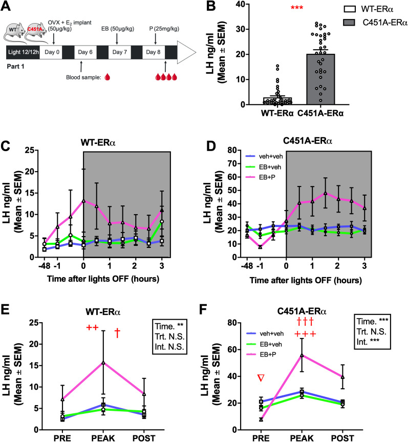
Estrogens act through nuclear and membrane-initiated signaling. Estrogen receptor alpha (ERα) is critical for reproduction, but the relative contribution of its nuclear and membrane signaling to the central regulation of reproduction is unclear. To address this question, two complementary approaches were used: estetrol (E4) a natural estrogen acting as an agonist of nuclear ERs, but as an antagonist of their membrane fraction, and the C451A-ERα mouse lacking mERα. E4 dose- dependently blocks ovulation in female rats, but the central mechanism underlying this effect is unknown. To determine whether E4 acts centrally to control ovulation, its effect was tested on the positive feedback of estradiol (E2) on neural circuits underlying luteinizing hormone (LH) secretion. In ovariectomized females chronically exposed to a low dose of E2, estradiol benzoate (EB) alone or combined with progesterone (P) induced an increase in the number of kisspeptin (Kp) and gonadotropin-releasing hormone (GnRH) neurons coexpressing Fos, a marker of neuronal activation. E4 blocked these effects of EB, but not when combined to P. These results indicate that E4 blocked the central induction of the positive feedback in the absence of P, suggesting an antagonistic effect of E4 on mERα in the brain as shown in peripheral tissues. In parallel, as opposed to wild-type females, C451A-ERα females did not show the activation of Kp and GnRH neurons in response to EB unless they are treated with P. Together these effects support a role for membrane-initiated estrogen signaling in the activation of the circuit mediating the LH surge.

Keywords: GnRH neurons; kisspeptin neurons; LH surge; estetrol; mERα; preoptic area.


Conflict of interest statement

The authors declare no competing financial interests.

Figures

Figure 1.
Figure 1.
Profiles of LH changes induced by estradiol benzoate (EB) alone or in combination with progesterone (P) in ovariectomized WT-ERα (white) or C451A-ERα (gray mice). A, Protocol used to induce a positive feedback: females were ovariectomized (OVX), chronically treated with estradiol (E2) from day 0 to day 8, injected with estradiol benzoate on day 7, and injected with progesterone or its vehicle (sesame oil) on day 8. B, On day 6, C451A-ERα females (n = 32) showed higher baseline LH levels than WT-ERα females (n = 31; Mann–Whitney test). C, D, Profiles of LH levels measured every 30 min starting 1 h before lights off following treatment on day 8 in WT-ERα and C451A-ERα females, respectively. C, LH profiles obtained in WT-ERα mice (OVX + E2 + veh + veh n = 12, OVX + E2 + EB + veh n = 11, OVX + E2 + EB + P n = 14). D, LH profiles obtained in C451A-ERα mice (OVX + E2 + veh + Veh n = 10, OVX + E2 + EB + veh n = 11 OVX + E2 + EB + P n = 12). E, Regardless of treatment, WT-ERα females showed an increased LH concentration at one time point (peak) between 0 and 2.5 h after lights off compared with prior (day 6, pre) and during 3 h after lights off (post; two-way ANOVA; Tukey’s post hoc test following significant time effect: ++p < 0.01 vs “pre”; p < 0.05 vs “post”). F, EB + P induced an increased LH concentration in C451A-ERα females within 0 and 2.5 h after lights off (peak) compared with prior (day 6, pre) and during 3 h after lights off (post; two-way ANOVA; Tukey’s post hoc test, following significant interaction: +++p = 0.001 vs “pre” within same treatment; †††p = 0.001 vs “post“ within same treatment; p < 0.05 EB + P “pre” vs “post” within same treatment. Symbols in the statistical boxes: *, **, ***, p < 0.05, 0.01, 0.001; N.S., nonsignificant).
Figure 2.
Figure 2.
Effect of mERα absence on the positive feedback of estrogens on LH concentration and the activation of the associated neurocircuits. A, Protocol used to induce positive feedback: following a first round of injections to induce the positive feedback (Fig. 1), the E2 implant was replaced by a new one on Day 30, and females were treated again with veh + veh, EB + veh, or EB + P on Days 38 and 39. Blood and brains were collected 30–60 min after lights off. B, In WT-ERα females (white), EB + P, but not EB + veh, induced a significant rise in LH (Kruskal–Wallis test: **p < 0.01 vs veh + veh), while in C451A-ERα females (gray), EB + veh induced a significant reduction in LH (Kruskal–Wallis test: *p < 0.05 vs veh + veh; Mann–Whitney tests: ##, ### < 0.01, 0.001 vs WT-ERα within same treatment). C, WT-ERα females displayed more kisspeptin (Kp) neurons in RP3 V (AVPv + PeN) than C451A-ERα females (two-way ANOVA). D, A higher percentage of Kp neurons coexpressed Fos following EB and EB + P than veh + veh in WT-ERα, while only EB + P induced such activation in C451A-ERα (two-way ANOVA; * and ***, p < 0.05 and 0.001 vs veh + veh same genotype; $$$p < 0.0001 vs EB + veh same genotype; ###p < 0.001 vs same treatment in WT-ERα). E, GnRH neurons counted in POA were slightly more abundant in C451A-ERα females than in WT-ERα females (two-way ANOVA). F, A higher percentage of GnRH neurons coexpressed Fos following EB and EB + P than veh + veh in WT-ERα, while only EB + P induced such activation in C451A-ERα (two-way ANOVA; ***, p < 0.001 vs veh + veh same genotype; $$$p < 0.0001 vs EB + veh same genotype; ##p < 0.01 vs same treatment in WT-ERα). Sample size: B, C. WT-ERα: veh + veh, n = 14, veh + EB, n = 14, EB + P, n = 13, C451A-ERα: veh + veh, n = 11, veh + EB, n = 11, EB + P, n = 13. CF. WT-ERα: veh + veh, n = 13, veh + EB, n = 14, EB + P, n = 13, C451A-ERα: veh + veh, n = 12, veh + EB, n = 11, EB + P, n = 13. Symbols in the statistical boxes: *, **, ***, p < 0.05, 0.01, 0.001; N.S., nonsignificant.
Figure 3.
Figure 3.
Representative photomicrographs of Kp-IR neurons (in blue) and their coexpression of the neuronal activity marker Fos (in orange) as a function of the treatment and genotype. Black arrows point at double-labeled neurons, while white arrows point at single-labeled neurons.
Figure 4.
Figure 4.
Representative photomicrographs of GnRH-IR neurons (in blue) and their coexpression of the neuronal activity marker Fos (in orange) as a function of the treatment and genotype. Black arrows point at double-labeled neurons, while white arrows point at single-labeled neurons.
Figure 5.
Figure 5.
Effect of estetrol on the LH surge induced by estradiol and the neurocircuits underlying this response. A, Protocol used to induce a positive feedback. WT mice were ovariectomized (OVX) on day 0, treated with subcutaneous estradiol (E2) implant from day 0 to day 9, and injected on day 8 with estradiol benzoate (EB) alone or combined with estetrol (E4, 200 µg, s.c.) or their vehicle (sesame oil) and on day 9 with progesterone (P) or its vehicle (sesame oil). Blood samples were collected prior to treatment on day 8 and within 1 h of lights off on day 9, when brains were also collected for immunohistological analyses. B, LH levels did not differ between groups (n = 9) on day 8. C, Females treated with EB alone (n = 9) or EB + E4 (n = 9) did not show a LH surge compared with veh + veh (n = 9) unless they were treated with P (EB + P, n = 9, and EB + E4 + P, n = 9). D, F, The number of kisspeptin (Kp) neurons in RP3V (AVPv + PeN, D) or GnRH neurons in POA (F) did not differ across treatments (Kp: veh + veh, n = 7, EB, n = 9, EB + P, n = 7, EB + E4, n = 6, EB + E4 + P, n = 7; GnRH: veh + veh, n = 9, EB, n = 8, EB + P, n = 9, EB + E4, n = 8, EB + E4 + P, n = 8). E, G, The percentages of Kp (E) and GnRH (G) neurons coexpressing Fos were higher in females treated with EB, EB + P, and EB + E4 + P than females treated with veh + P and EB + E4 (same sample sizes as in D and F). All data were analyzed by one-way ANOVA followed by Tukey’s post hoc test when significant: *, **, and *** p < 0.05, 0.01, and 0.001 versus veh + P; #, p < 0.05 versus EB; Δ, ΔΔ, and ΔΔΔ, p < 0.05, 0.01, and 0.001 versus EB + E4.

References

    1. Abot A, et al. (2014) The uterine and vascular actions of estetrol delineate a distinctive profile of estrogen receptor alpha modulation, uncoupling nuclear and membrane activation. EMBO Mol Med 6:1328–1346. 10.15252/emmm.201404112 - DOI - PMC - PubMed
    1. Abraham IM, Han S-K, Todman MG, Korach KS, Herbison AE (2003) Estrogen receptor Beta mediates rapid estrogen actions on gonadotropin-releasing hormone neurons in vivo. J Neurosci 23:5771–5777. 10.1523/JNEUROSCI.23-13-05771.2003 - DOI - PMC - PubMed
    1. Acconcia F, Fiocchetti M, Busonero C, Fernandez VS, Montalesi E, Cipolletti M, Pallottini V, Marino M (2021) The extra-nuclear interactome of the estrogen receptors: implications for physiological functions. Mol Cell Endocrinol 538:111452. 10.1016/j.mce.2021.111452 - DOI - PubMed
    1. Adlanmerini M, et al. (2014) Mutation of the palmitoylation site of estrogen receptor alpha in vivo reveals tissue-specific roles for membrane versus nuclear actions. Proc Natl Acad Sci U S A 111:E283–290. 10.1073/pnas.1322057111 - DOI - PMC - PubMed
    1. Alvisi RD, Diniz GB, Da-Silva JM, Bittencourt JC, Felicio LF (2016) Suckling-induced Fos activation and melanin-concentrating hormone immunoreactivity during late lactation. Life Sci 148:241–246. 10.1016/j.lfs.2016.02.038 - DOI - PubMed

LinkOut - more resources