. 2024 Oct 7;10(4):e004664.
doi: 10.1136/rmdopen-2024-004664.

Methotrexate treatment hampers induction of vaccine-specific CD4 T cell responses in patients with IMID

Collaborators, Affiliations

Methotrexate treatment hampers induction of vaccine-specific CD4 T cell responses in patients with IMID

Laura Y L Kummer et al. RMD Open. .

Abstract

Objectives: Methotrexate (MTX) is one of the most commonly used medications to treat rheumatoid arthritis (RA). However, the effect of MTX treatment on cellular immune responses remains incompletely understood. This raises concerns about the vulnerability of these patients to emerging infections and following vaccination.

Methods: In the current study, we investigated the impact of MTX treatment in patients with immune-mediated inflammatory disease on B and CD4 T cell SARS-CoV-2 vaccination responses. Eighteen patients with RA and two patients with psoriatic arthritis on MTX monotherapy were included, as well as 10 patients with RA without immunosuppressive treatment, and 29 healthy controls. CD4 T and B cell responses were analysed 7 days and 3-6 months after two SARS-CoV-2 messenger RNA vaccinations. High-dimensional flow cytometry analysis was used to analyse fresh whole blood, an activation-induced marker assay to measure antigen-specific CD4 T cells, and spike probes to study antigen-specific B cells.

Results: Seven days following two SARS-CoV-2 vaccinations, total B and T cell counts were similar between MTX-treated patients and controls. In addition, spike-specific B cell frequencies were unaffected. Remarkably, the frequency of antigen-specific CD4 T cells was reduced in patients using MTX and correlated strongly with anti-RBD IgG antibodies. These results suggest that decreased CD4 T cell activity may result in slower vaccination antibody responses in MTX-treated patients.

Conclusion: Taken together, MTX treatment reduces vaccine-induced CD4 T cell activation, which correlates with lower antibody responses.

Trial registration number: NL8900.

Keywords: B-lymphocytes; T-lymphocytes; autoimmune diseases; methotrexate.


Conflict of interest statement

Competing interests: FE, GW, SMvH and TWK report (governmental) grants from ZonMw to study immune response after SARS-Cov-2 vaccination in auto-immune diseases. FE also reports grants from Prinses Beatrix Spierfonds, CSL Behring, Kedrion, Terumo BCT, Grifols, Takeda Pharmaceutical and GBS-CIDP Foundation; consulting fees from UCB Pharma and CSl Behring; honoraria from Grifols. All other authors report no disclosures relevant to the manuscript.

Figures
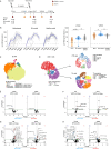
Figure 1
Figure 1. SARS-CoV-2 antibody responses and fresh whole blood analysis. (A) Study design with timing of SARS-CoV-2 mRNA-1273 vaccination and peripheral blood collection. (B) Dynamics of anti-RBD antibody titres over time, with comparisons between MTX and RA control, as well as HC versus RA control at V1D28 (MTX n=19, RA control n=10, HC n=26) and at V2D28 (MTX n=20, RA control n=9, HC n=27). Horizontal bars represent the median. (C–D) Fit-SNE map and cluster identification of B cell subsets, and (D) CD4 T cell subsets in whole blood using FlowSOM analysis, including data from both MTX-treated patients (n=18) and HCs (n=24) at baseline and V2D7. (E–F) Volcano plot displaying differentially expressed B cell and CD4 T cell populations in counts per µL, with a log2 fold change >0.5 and p value <0.5 (E) between HC and MTX and (F) between V1pre and V2D7. Numbers in the top left and right corner of volcano plots indicate the number of differentially expressed populations. (G–H) Volcano plot illustrating percentage of CD4 T cells expressing PD1, CTLA4, TIM3, TIGIT, CD137, CD40L or CD38/HLA-DR, with a log2 fold change >0.5 and p value <0.5 (G) between V1pre and V2D7 and (H) between MTX and HC. Statistical significance was determined using the Wilcoxon ranked sum test for unpaired comparisons and the Wilcoxon signed rank test for paired comparisons. Bonferroni-Holm correction for multiple comparisons was used to adjust for multiple testing. HC, healthy control; MTX, methotrexate; ns, not significant; PBMC, peripheral blood mononuclear cell; RA, rheumatoid arthritis; RBD, receptor binding domain.
Figure 2
Figure 2. Spike-specific B cell vaccination responses are not affected by MTX treatment. (A) Percentage of spike-specific and RBD-specific B cells of total CD19+B cells at V2D7 (MTX n=16, RA control n=10, HC n=10) and V2M3-6 (MTX n=12, RA control n=6, HC n=9). Horizontal bars represent the median. (B) Stacked bar graph illustrating the mean proportion of the 16 spike-specific B cell clusters as a percentage of total CD19+ B cells. (C) Heatmap displaying the differential cluster expression between MTX versus RA control and RA control versus HCs at V2D7 and V2M3-6. (D) Kinetics of significantly differentially expressed B cell clusters (as observed in 1C) over time. (E) Correlation analysis of %spike-specific B cells at V2D7 vs anti-RBD antibody titre at V2D7 or V2D28, in both MTX-treated patients and controls. (F) Correlation analysis of spike-specific IgG+ clusters at V2D7 with anti-RBD antibody titre at V2D7 in MTX-treated patients. Statistical significance was determined using the Wilcoxon ranked sum test for unpaired comparisons with Bonferroni-Holm correction for multiple comparisons. Correlations were calculated using Spearman’s rank correlation. ASC, antibody secreting cell; HC, healthy control; MTX, methotrexate; ns, not significant; RA, rheumatoid arthritis; RBD, receptor binding domain.
Figure 3
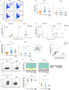
Figure 3. MTX reduces spike-induced CD40L+CD137+ CD4 T cells post-SARS-CoV-2 vaccination. (A) Representative flow cytometry plots of spike-induced and DMSO-induced (background) CD40L+CD137+ CD4 T cells at V2D7 (MTX n=16, RA control n=10, HC n=10) and at V2M3-6 (MTX n=12, RA control n=6, HC n=9). (B) Percentage of spike-induced CD40L+CD137+ CD4 T cells per group. Background was subtracted. Horizontal bars represent the median. (C–D) Correlation analysis of spike-induced CD40L+CD137+ CD4 T cells at V2D7 with (C) anti-RBD antibody titre at V2D28 or (D) spike-specific B cells at V2D7, in both MTX-treated patients and controls. (E) Percentage of spike-induced CD4 T cells expressing two or three of the following cytokines: interleukin-2, tumour necrosis factor-α and interferon-γ. (F) Correlation analysis of spike-induced CD40L+CD137+ CD4 T cells and spike-induced double or triple cytokine-producing CD4 T cells, at V2D7. (G) Representative flow cytometry plots of CD27 and CD45RA expression on spike-induced CD40L+CD137+ CD4 T cells. (H) Phenotypic distribution of central memory (CD27+CD45RA−), effector memory (CD27−CD45RA−), naïve (CD27+CD45RA+) and TEMRA (CD27−CD45RA+) spike-induced CD40L+CD137+ CD4 T cells. (I) Representative flow cytometry plots of expression of PD1 and ICOS on spike-induced CD40L+CD137+ CD4 T cells. (J) Percentage of PD1 and ICOS expressing spike-induced CD40L+CD137+ CD4 T cells per time point. Statistical significance was determined using Wilcoxon ranked sum test for unpaired comparisons with Bonferroni-Holm correction for multiple comparisons. Correlations were calculated using Spearman’s rank correlation. ICOS, inducible T-cell costimulator; MTX, methotrexate; ns, not significant; PD1, programmed cell death 1; RA, rheumatoid arthritis; RBD, receptor binding domain.
Figure 4
Figure 4. A third SARS-CoV-2 vaccination shows a trend of increased spike-specific B and CD4 T cell responses. (A) Schematic overview of third SARS-CoV-2 mRNA-1273 vaccination, provided 109–113 days post-second vaccination to MTX-treated patients (n=8). (B) Anti-RBD antibody titre dynamics in MTX-treated patients after second and third SARS-CoV-2 vaccination. (C) Percentage of spike-induced CD40L+CD137+ CD4 T cells, (D) spike-induced double or triple cytokine-producing CD4 T cells, (E) spike-specific B cells and (F) RBD-specific B cells 7 days post-second vaccination (V2D7), at the day of third vaccination (V3pre) and 7 days post-third vaccination (V3D7). Statistical significance was determined using Wilcoxon signed-rank test with Bonferroni-Holm correction for multiple comparisons. HC, healthy control; MTX, methotrexate; ns, not significant; RA, rheumatoid arthritis; RBD, receptor binding domain.

References

    1. Freeman ML, Clagett BM, Moisi D, et al. Methotrexate Inhibits T Cell Proliferation but Not Inflammatory Cytokine Expression to Modulate Immunity in People Living With HIV. Front Immunol. 2022;13:924718. doi: 10.3389/fimmu.2022.924718. - DOI - PMC - PubMed
    1. Cronstein BN, Aune TM. Methotrexate and its mechanisms of action in inflammatory arthritis. Nat Rev Rheumatol. 2020;16:145–54. doi: 10.1038/s41584-020-0373-9. - DOI - PubMed
    1. Kapetanovic MC, Roseman C, Jönsson G, et al. Antibody response is reduced following vaccination with 7-valent conjugate pneumococcal vaccine in adult methotrexate-treated patients with established arthritis, but not those treated with tumor necrosis factor inhibitors. Arthritis Rheum. 2011;63:3723–32. doi: 10.1002/art.30580. - DOI - PubMed
    1. Kapetanovic MC, Nagel J, Nordström I, et al. Methotrexate reduces vaccine-specific immunoglobulin levels but not numbers of circulating antibody-producing B cells in rheumatoid arthritis after vaccination with a conjugate pneumococcal vaccine. Vaccine (Auckl) 2017;35:903–8. doi: 10.1016/j.vaccine.2016.12.068. - DOI - PubMed
    1. Kobie JJ, Zheng B, Bryk P, et al. Decreased influenza-specific B cell responses in rheumatoid arthritis patients treated with anti-tumor necrosis factor. Arthritis Res Ther. 2011;13:R209. doi: 10.1186/ar3542. - DOI - PMC - PubMed