

Quantitative susceptibility mapping at 7 T in COVID-19: brainstem effects and outcome associations
- PMID: 39375207
- PMCID: PMC7616766
- DOI: 10.1093/brain/awae215
Quantitative susceptibility mapping at 7 T in COVID-19: brainstem effects and outcome associations
Erratum in
-
Correction to: Quantitative susceptibility mapping at 7 T in COVID-19: brainstem effects and outcome associations.Brain. 2025 Jan 7;148(1):e3. doi: 10.1093/brain/awae332. Brain. 2025. PMID: 39468699 Free PMC article. No abstract available.
Abstract
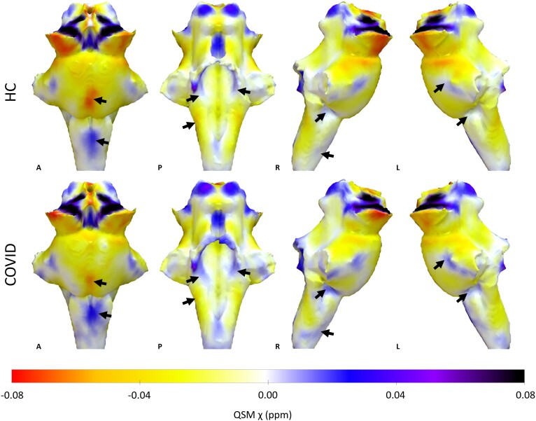
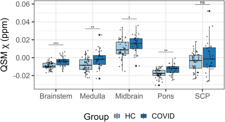
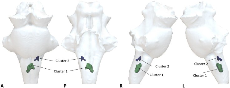
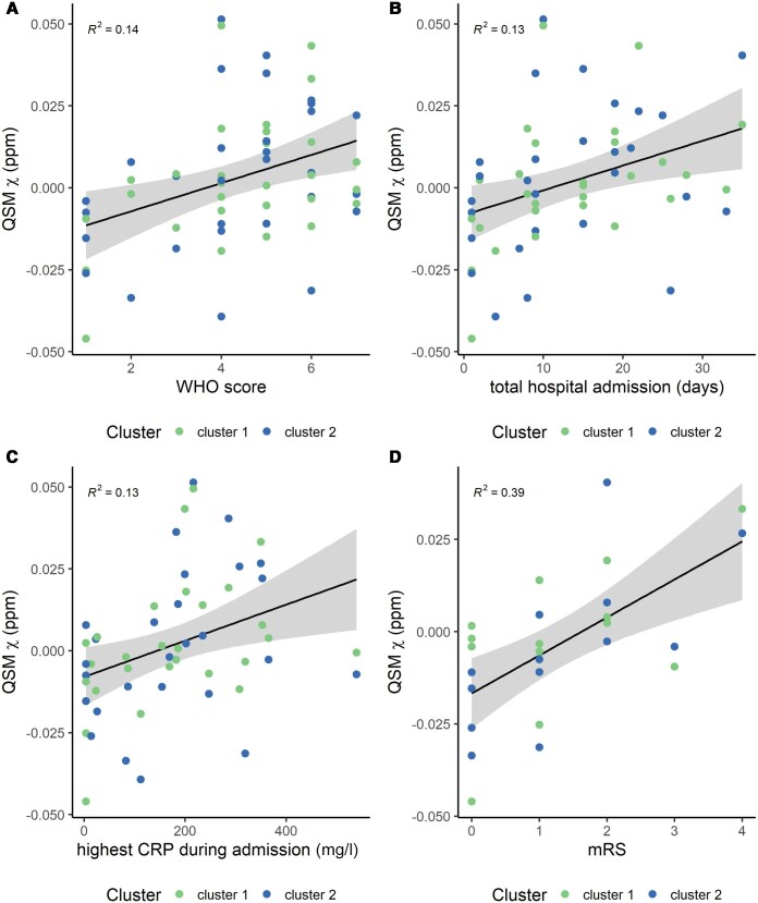
Post-mortem studies have shown that patients dying from severe acute respiratory syndrome coronavirus (SARS-CoV-2) infection frequently have pathological changes in their CNS, particularly in the brainstem. Many of these changes are proposed to result from para-infectious and/or post-infection immune responses. Clinical symptoms such as fatigue, breathlessness, and chest pain are frequently reported in post-hospitalized coronavirus disease 2019 (COVID-19) patients. We propose that these symptoms are in part due to damage to key neuromodulatory brainstem nuclei. While brainstem involvement has been demonstrated in the acute phase of the illness, the evidence of long-term brainstem change on MRI is inconclusive. We therefore used ultra-high field (7 T) quantitative susceptibility mapping (QSM) to test the hypothesis that brainstem abnormalities persist in post-COVID patients and that these are associated with persistence of key symptoms. We used 7 T QSM data from 30 patients, scanned 93-548 days after hospital admission for COVID-19 and compared them to 51 age-matched controls without prior history of COVID-19 infection. We correlated the patients' QSM signals with disease severity (duration of hospital admission and COVID-19 severity scale), inflammatory response during the acute illness (C-reactive protein, D-dimer and platelet levels), functional recovery (modified Rankin scale), depression (Patient Health Questionnaire-9) and anxiety (Generalized Anxiety Disorder-7). In COVID-19 survivors, the MR susceptibility increased in the medulla, pons and midbrain regions of the brainstem. Specifically, there was increased susceptibility in the inferior medullary reticular formation and the raphe pallidus and obscurus. In these regions, patients with higher tissue susceptibility had worse acute disease severity, higher acute inflammatory markers, and significantly worse functional recovery. This study contributes to understanding the long-term effects of COVID-19 and recovery. Using non-invasive ultra-high field 7 T MRI, we show evidence of brainstem pathophysiological changes associated with inflammatory processes in post-hospitalized COVID-19 survivors.
Keywords: 7T MRI; brainstem; coronavirus disease of 2019; inflammation; quantitative susceptibility mapping.
© The Author(s) 2024. Published by Oxford University Press on behalf of the Guarantors of Brain.
Conflict of interest statement
K.P. is named as co-inventor on a provisional UK patent application titled ‘Use of cerebral nitric oxide donors in the assessment of the extent of brain dysfunction following injury'. K.P. is named as co-inventors on a provisional UK patent titled ‘Discordant sensory stimulus in VR based exercise’ UK Patent office application: 2204698.1 filing date 31/3/2022.
Figures
Comment in
-
Exploring the long-term neurological consequences of COVID-19.Nat Rev Neurol. 2025 Feb;21(2):65. doi: 10.1038/s41582-025-01056-z. Nat Rev Neurol. 2025. PMID: 39820146 No abstract available.
References
-
- Conklin J, Frosch MP, Mukerji S, et al. Cerebral microvascular injury in severe COVID-19. medRxiv. [Preprint] https://doi:10.1101/2020.07.21.20159376 - DOI - PMC - PubMed
MeSH terms
Grants and funding
- NIHR203316/NIHR Oxford Health Biomedical Research Centre
- NIHR
- BHF Centre of Research Excellence, Oxford
- 0009118/University of Oxford Medical Sciences Division COVID-19 Research Response Fund
- NIHR203312/NIHR Cambridge Biomedical Research Centre
- WT_/Wellcome Trust/United Kingdom
- BRC-1215-20008/NIHR Oxford Biomedical Research Centre
- Academy of Medical Sciences/The Health Foundation Clinician Scientist Fellowship
- Cambridge Centre for Parkinson-Plus
- 103838/WT_/Wellcome Trust/United Kingdom
- MC_UU_00030/14/MRC_/Medical Research Council/United Kingdom
LinkOut - more resources
Full Text Sources
Medical
Research Materials
Miscellaneous
