. 2024 Oct 7;15(10):732.
doi: 10.1038/s41419-024-07108-4.

HNRNPC mediates lymphatic metastasis of cervical cancer through m6A-dependent alternative splicing of FOXM1

Affiliations

HNRNPC mediates lymphatic metastasis of cervical cancer through m6A-dependent alternative splicing of FOXM1

Yun-Yun Liu et al. Cell Death Dis. .

Abstract

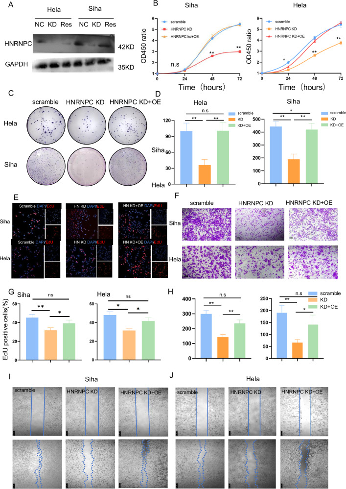
Cervical cancer (CCa) patients with lymph node (LN) metastasis face poor prognoses and have limited treatment options. Aberrant N6-methyladenosine (m6A) modification of RNAs are known to promote tumor metastasis, but their role in CCa remains unclear. Our study reveals that HNRNPC, an alternative splicing (AS) factor and m6A reader, increases tumor-related variants through m6A-dependent manner, thereby promoting lymphatic metastasis in CCa. We found that HNRNPC overexpression correlates with lymphatic metastasis and poorer prognoses in CCa patients. Functionally, knocking down HNRNPC markedly inhibited the migration and invasion of several CCa cell lines, while supplementing HNRNPC restored the malignant phenotypes of these cells. Mechanistically, HNRNPC regulates exon skipping of FOXM1 by binding to its m6A-modified motif. Mutating the m6A site on FOXM1 weakened the interaction between HNRNPC and FOXM1 pre-RNA, leading to a reduction in the metastasis-related FOXM1-S variant. In conclusion, our findings demonstrate that m6A-dependent alternative splicing mediated by HNRNPC is essential for lymphatic metastasis in CCa, potentially providing novel clinical markers and therapeutic strategies for patients with advanced CCa.


Conflict of interest statement

The authors declare no competing interests.

Figures

Fig. 1
Fig. 1. HNRNPC overexpression in cervical cancer: clinical and prognostic implications.
A TCGA data reveals elevated HNRNPC expression in tumors across various cancer types compared to normal tissues. B Significantly increased HNRNPC expression observed in TCGA cervical cancers. CE GEO database analysis confirms high HNRNPC expression in CCa tissues compared to normal tissues (GSE7803, GSE6791, GSE63514). F PCR analysis shows significant upregulation of HNRNPC in CCa tumors compared to adjacent tissues in a local cohort (n = 10). G Local cohort PCR data indicates higher HNRNPC levels in lymphatic metastasis CCa compared to patients without lymph node metastasis and normal cervix. H TCGA data analysis links high HNRNPC RNA expressions to shorter overall survival (n = 292, P value = 0.048). I HNRNPC expression patterns in cervical adenocarcinoma and squamous cell carcinoma from two independent gynecology departments. J Kaplan–Meier curves demonstrate high HNRNPC expression as an unfavorable prognostic factor for overall survival in both local cohorts (cohort1: n = 91, cohort2: n = 96) with P values of 0.007 and 0.008, respectively. *P < 0.05, **P < 0.01, ***P < 0.001. LN lymph node, TCGA The Cancer Genome Atlas, AC adenocarcinoma, SC squamous cell carcinoma.
Fig. 2
Fig. 2. Impact of HNRNPC Downregulation on CCa Cell Proliferation, Invasion, and Migration In Vitro.
A Western blot confirms successful downregulation of HNRNPC expression in H229 and MS751 cells using siRNAs; siRNA sequence-1 exhibits higher efficacy. B CCK8 assay demonstrates reduced proliferative capacity of H229 and MS751 cells upon HNRNPC downregulation (OD value = (ODexperiment-ODblank)/((OD0h-ODblank), n = 4)). C, D Clone formation assay reveals decreased cell proliferation following HNRNPC downregulation (n = 3). E, G EdU assay illustrates diminished proliferation of H229 and MS751 cells after HNRNPC silencing (n = 3). F, H Transwell assay shows decreased invasiveness of CCa cells upon HNRNPC downregulation (n = 3). I, J Wound healing assay under serum-free culture conditions indicates reduced migration ability of H229 and MS751 cells 48 h after HNRNPC knocking down. *P < 0.05, **P < 0.01, n.s: P > 0.05. Si1 siRNA sequence-1, si2 siRNA sequence-2.
Fig. 3
Fig. 3. HNRNPC drives proliferation, invasion, and migration of CCa cells in vitro.
A Western blot confirms successful rescue of HNRNPC expression in HNRNPC-silenced Hela and Siha cells through HNRNPC cDNA. B CCK8 assay reveals increased proliferation of Hela and Siha cells induced by HNRNPC (OD value = (ODexperiment-ODblank)/((OD0h-ODblank), n = 4)). C, D Clone formation assay demonstrates enhanced cell proliferation upon HNRNPC rescue (n = 3). E, G EdU assay shows increased proliferation of Hela and Siha cells after HNRNPC rescue (n = 3). F, H Transwell assay indicates elevated invasiveness of CCa cells following HNRNPC rescue (n = 3). I, J Wound healing assay under serum-free culture conditions shows heightened migration ability of Hela and Siha cells with HNRNPC rescue. *P < 0.05, **P < 0.01, n.s:P > 0.05. NC negative control, KD knock down of HNRNPC, RES rescue of HNRNPC, OE over-expression of HNRNPC.
Fig. 4
Fig. 4. HNRNPC facilitates CCa metastasis to regional lymph nodes in vivo through the footpad-popliteal lymphatic metastasis model.
A Diagrammatic illustration of footpad injection. B Anatomical atlas displaying footpad tumor and popliteal lymphatic metastasis (green arrow denotes the primary tumor on the footpad of Balb/c nude mice, black arrow indicates metastatic regional lymph nodes on the popliteal). C Fluorescence imaging of footpad tumor and popliteal lymphatic metastasis (white arrow signifies the primary tumor on the footpad, yellow arrow indicates metastatic regional lymph nodes on the popliteal). D Histogram demonstrating reduced in vivo lymph node metastasis rates in HNRNPC knock-down cells (n = 5). E, F Immunohistochemistry (IHC) of pan-CK staining in popliteal lymph nodes of mice injected with Hela scramble cells. G, H Hematoxylin and eosin (HE) staining of popliteal lymph nodes in mice injected with Hela scramble cells. I, J IHC of pan-CK staining in popliteal lymph nodes of mice injected with HNRNPC knocked-down Hela cells. K, L HE staining of popliteal lymph nodes in mice injected with HNRNPC knocked-down Hela cells. NC negative control, KD knock down of HNRNPC, LN negative lymph node, LN+ positive lymph node, HE hematoxylin-eosin staining.
Fig. 5
Fig. 5. HNRNPC mediates alternative splicing of multiple molecules, including the key transcriptional factor FOXM1.
A Volcano plots representing differentially expressed genes after HNRNPC silencing in Hela and Siha cells. B GSEA enrichment plots for Hela and Siha cells. C Histograms depicting differentially expressed alternative splicing events in Hela and Siha cells. D Volcano plots illustrating exon skipping events. E Venn diagram depicting shared alternative splicing events in Hela and Siha cells. F Schematic representation of skipped exons (SE) in FOXM1, showing consistent exon retention after HNRNPC knockdown in both cell lines. G PCR validation confirms retention of the FOXM1-L variant after HNRNPC silencing, which is reduced upon HNRNPC overexpression. *P < 0.05, **P < 0.01. KD knock down of HNRNPC, OE over expression of HNRNPC, TF transcription factor, DEG differently expressed gene, SE skipped exon, AS alternative splicing, RI retained intron, MTE mutually exclusive exons, A5SS alternative 5’ splice site, A3SS alternative 3’ splice site.
Fig. 6
Fig. 6. HNRNPC-Mediated Increase in FOXM1-S Variant Plays a Crucial Role in CCa Metastasis.
A RNA levels of FOXM1-L and -S variants in cervical cancer tissues. B RNA levels of FOXM1-L and -S variants in paired samples of cervical cancer and adjacent tissues. C Regression analysis and fitting curve depicting the relationship between HNRNPC and FOXM1-S expressions in cervical cancer samples. D, E Transwell assay results indicating that the FOXM1-S variant rescues cell invasion ability, unlike the FOXM1-L variant. F Western blot results demonstrating elevated MMP proteins and EMT pathway molecules after complementing FOXM1-S rather than FOXM1-L. G RIP-PCR comparing different areas of wild-type and m6A sites mutated FOXM1. *P < 0.05, **P < 0.01, n.s P > 0.05. RIP RNA binding protein immunoprecipitation, wt wild type, mt m6A sites mutation, WTAP KD knocking down WTAP.
Fig. 7
Fig. 7. Hypothesized mechanism diagram: HNRNPC-mediated m6A-dependent binding to FOXM1 promotes lymph node metastasis in CCa through alternative splicing.
In cervical cancer cells, when the VIII exon of FOXM1 undergoes m6A modification, the nuclear-located splicing factor HNRNPC recognizes and binds to the FOXM1 pre-RNA. This interaction promotes the skipping of the VIIa exon of FOXM1, leading to an elevation in the FOXM1-S variant. FOXM1-S variants are then translated into transcriptionally active FOXM1 proteins, promoting the expression of MMP and EMT-related genes, ultimately enhancing lymph node metastasis.

References

    1. Siegel RL, Miller KD, Wagle NS, Jemal A. Cancer statistics, 2023. CA Cancer J Clin. 2023;73:17–48. - PubMed
    1. Abu-Rustum NR, Yashar CM, Arend R, Barber E, Bradley K, Brooks R, et al. NCCN Guidelines(R) insights: cervical cancer, version 1.2024. J Natl Compr Cancer Netw. 2023;21:1224–33. - PubMed
    1. Matsuo K, Machida H, Mandelbaum RS, Konishi I, Mikami M. Validation of the 2018 FIGO cervical cancer staging system. Gynecol Oncol. 2019;152:87–93. - PMC - PubMed
    1. Brown M, Assen FP, Leithner A, Abe J, Schachner H, Asfour G, et al. Lymph node blood vessels provide exit routes for metastatic tumor cell dissemination in mice. Science. 2018;359:1408–11. - PubMed
    1. Zhong S, Guo Q, Chen X, Luo X, Long Y, Chong T, et al. The inhibition of YTHDF3/m(6)A/LRP6 reprograms fatty acid metabolism and suppresses lymph node metastasis in cervical cancer. Int J Biol Sci. 2024;20:916–36. - PMC - PubMed

MeSH terms