. 2024 Oct 7;15(1):8673.
doi: 10.1038/s41467-024-52701-y.

Structure of the Nipah virus polymerase phosphoprotein complex

Affiliations

Structure of the Nipah virus polymerase phosphoprotein complex

Ge Yang et al. Nat Commun. .

Abstract

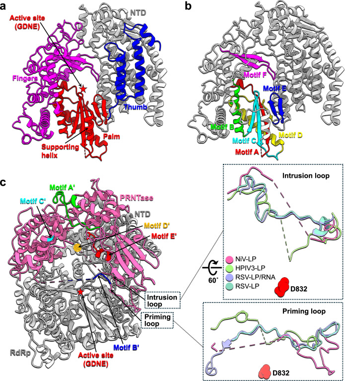
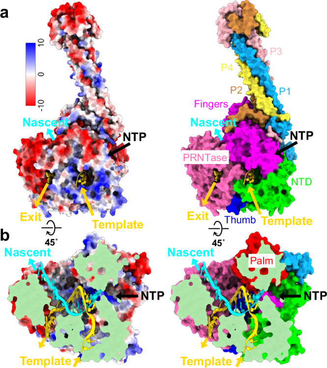
The Nipah virus (NiV), a member of the Paramyxoviridae family, is notorious for its high fatality rate in humans. The RNA polymerase machinery of NiV, comprising the large protein L and the phosphoprotein P, is essential for viral replication. This study presents the 2.9-Å cryo-electron microscopy structure of the NiV L-P complex, shedding light on its assembly and functionality. The structure not only demonstrates the molecular details of the conserved N-terminal domain, RNA-dependent RNA polymerase (RdRp), and GDP polyribonucleotidyltransferase of the L protein, but also the intact central oligomerization domain and the C-terminal X domain of the P protein. The P protein interacts extensively with the L protein, forming an antiparallel β-sheet among the P protomers and with the fingers subdomain of RdRp. The flexible linker domain of one P promoter extends its contact with the fingers subdomain to reach near the nascent RNA exit, highlighting the distinct characteristic of the NiV L-P interface. This distinctive tetrameric organization of the P protein and its interaction with the L protein provide crucial molecular insights into the replication and transcription mechanisms of NiV polymerase, ultimately contributing to the development of effective treatments and preventive measures against this Paramyxoviridae family deadly pathogen.


Conflict of interest statement

The authors declare no competing interests.

Figures

Fig. 1
Fig. 1. The overall structure of the NiV L–P complex.
a Schematic diagrams of the domain architecture of the Nipah virus (NiV) L and P proteins. The L protein consists of six domains. The structurally modeled regions, represented by specific colors, are: N-terminal domain (NTD), lime; fingers subdomain, magenta; palm subdomain, red; thumb subdomain, blue; GDP polyribonucleotidyltransferase domain (PRNTase), hot pink. The connector domain (CD), Methyltransferase domain (MTase), and C-terminal domain (CTD) of the L protein, although not modeled in the structure, are depicted in white with a dashed outline. Four P protomers, with their varying lengths, are also shown in different colors. The NTD of the P protein, due to its inherent flexibility, was not observed in all protomers. RdRp RNA-dependent RNA polymerase domain, OD oligomerization domain, XD, C-terminal X domain. b The cryo-EM density map (sharpened by DeepEMhancer, up) and atomic model (down) of the NiV L–P complex, with the same color scheme as depicted in (a), are presented in two directions.
Fig. 2
Fig. 2. Comparison of the L protein and its domains in mononegaviruses.
Four superimposed structures of the L proteins in the Mononegavirales L–P complexes are shown as tube helices cartoons separately. The individual domains of L and subdomains of the RdRp domain are highlighted, with the aspartic acid residues at the active site presenting as spheres. The color scheme is the same as Fig. 1. The bottom inserts are individually superimposed NTD, PRNTase, and RdRp domains. The diverse NTDs and PRNTases are in different colors with rounded edges and tube helices cartoons respectively. The models used here are 8KDC (HPIV3-L), 8IZM (MuV-L), and 6U1X (VSV-L). HPIV3 human parainfluenza virus type 3, MuV Mumps virus, VSV vesicular stomatitis virus.
Fig. 3
Fig. 3. Catalytic cores of the NiV L protein.
a The RdRp domain is shown as cartoon using the same color scheme as Fig. 1, with the NTD domain in light gray. The active site is highlighted with a red asterisk. The supporting helix (residues 587-598) in the palm subdomain is marked. b The same view as in (a), with catalytic motifs (motifs A–E) highlighted with different colors. The residue ranges of the six conserved motifs: residues 713–730 (motif A), residues 802–822 (motif B), residues 823–846 (motif C), residues 847–881 (motif D), residues 882–899 (motif E), and residues 535–557 (motif F). c The hot pink PRNTase is displayed by cartoon, with the highlighted conserved catalytic motifs (motifs A’–E’). The RdRp and NTD domains of the L protein are gray. Motif D’ in the intrusion loop is invisible in the cryo-EM map due to high flexibility. The inserts are the superimpositions of the priming loop and intrusion loop in Mononegavirales L proteins using the models with PDB codes: 8KDC (HPIV3-LP), 8SNX (RSV-LP/RNA), and 6UEN (RSV-LP). The aspartic acid 832 at the active site is shown as a red sphere to display the relative positions between them. HPIV3, human parainfluenza virus type 3; RSV, respiratory syncytial virus. The residue ranges of the five conserved motifs: residues 1200–1219 (motif A’), residues 1271–1274 (motif B’), residue W1306 (motif C’), residues H1347–R1348 (motif D’), residues 1387–1396 (motif E’).
Fig. 4
Fig. 4. The L–P interface.
The NiV L–P complex is shown as a surface representation. Left, the P protomers are displayed with cartoon and transparent surface representations. The color scheme is the same as Fig. 1. Right, the L protein is light gray with the cartoon P protomers. The interaction interfaces between NiV L and P can be distributed into five main regions, as shown in the five inserts. In the close-up views, atomic interactions between the key residues of L and P, shown as sticks for salt bridges and hydrogen bonds, are represented by black dashed lines (see Supplementary Fig. 7 for all the detailed interactions). The main chain or side chain atoms are displayed according to their interaction involvements.
Fig. 5
Fig. 5. The model of elongation complex.
a The Coulombic electrostatic potential (left) and the surface representation (right) with the modeled RNA duplex are presented. The entrance and exit of the template RNA and the exit of the nascent RNA are labeled. The RNA duplex was manually modeled by superimposing the RdRp domains of this complex and the Influenza B polymerase elongation complex (PDB code: 6QCT). b The clip surfaces on the active site display the inside channels for template RNA, nascent RNA, and NTP. The color scheme is the same as in Fig. 1.

References

    1. Byrne, P. O. et al. Prefusion stabilization of the Hendra and Langya virus F proteins. J. Virol.98, e0137223 (2024). - PMC - PubMed
    1. Chua, K. B. et al. Nipah virus: a recently emergent deadly paramyxovirus. Science288, 1432–1435 (2000). - PubMed
    1. Weatherman, S., Feldmann, H. & de Wit, E. Transmission of henipaviruses. Curr. Opin. Virol.28, 7–11 (2018). - PMC - PubMed
    1. Tian, J. et al. Emerging viruses: cross-species transmission of coronaviruses, filoviruses, henipaviruses, and rotaviruses from bats. Cell Rep.39, 110969 (2022). - PMC - PubMed
    1. Hassan, M. Z. et al. Nipah virus disease: what can we do to improve patient care? Lancet Infect. Dis. 10.1016/S1473-3099(23)00707-7 (2024). - DOI - PubMed

Publication types

MeSH terms

LinkOut - more resources