. 2024 Oct 7;15(1):8664.
doi: 10.1038/s41467-024-52913-2.

The activating receptor NKG2D is an anti-fungal pattern recognition receptor

Affiliations

The activating receptor NKG2D is an anti-fungal pattern recognition receptor

Yoav Charpak-Amikam et al. Nat Commun. .

Abstract

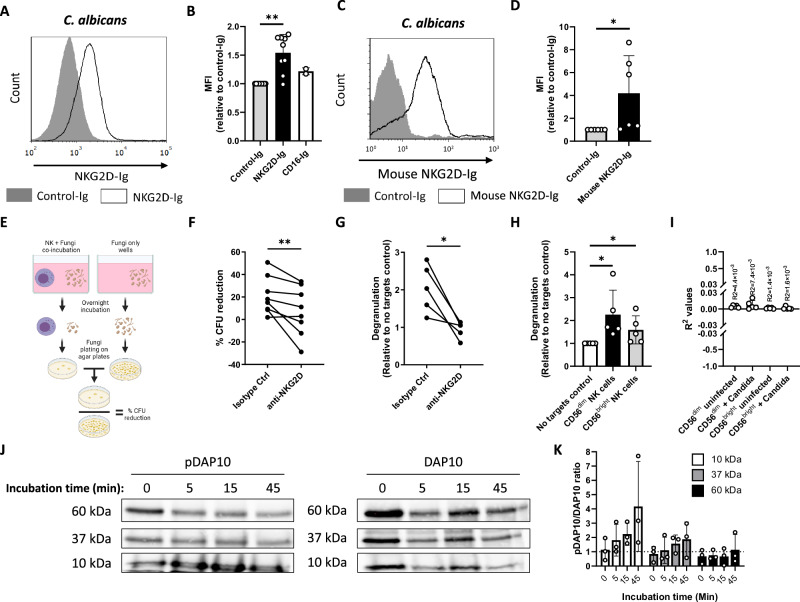
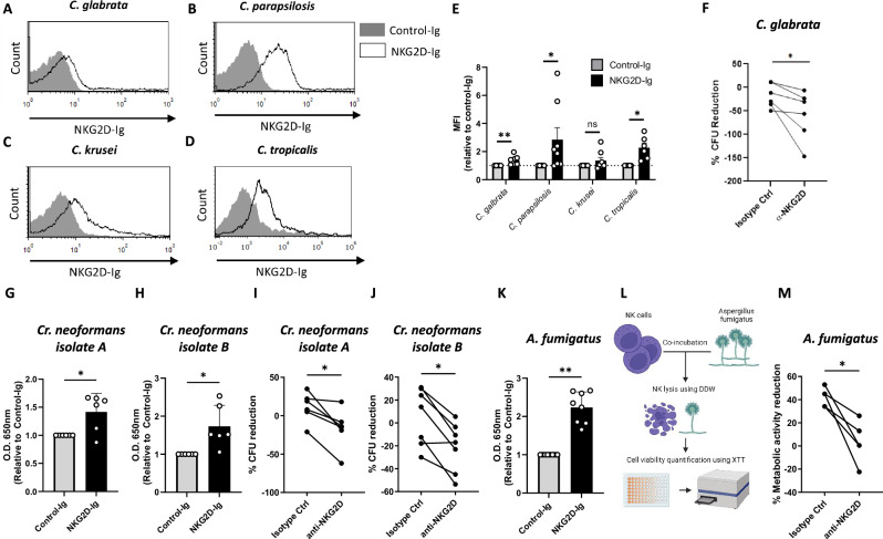
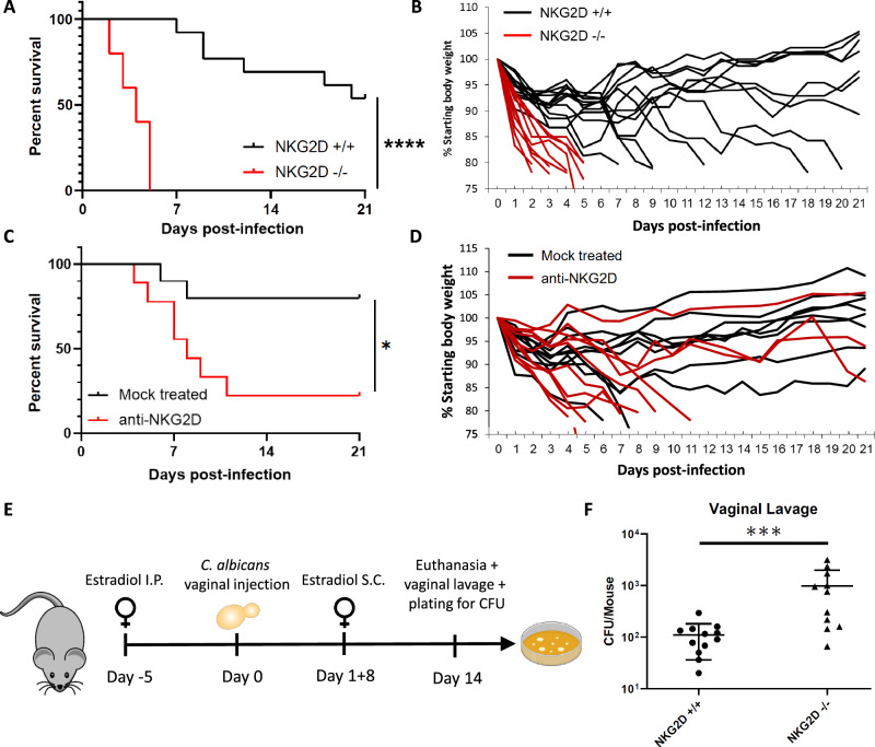
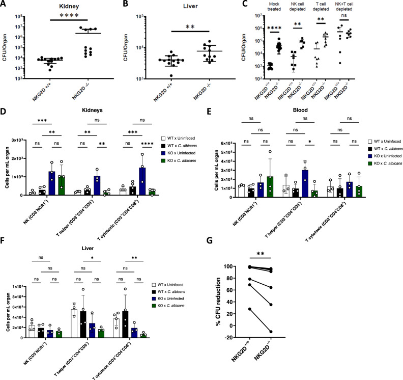
NKG2D is a central activating receptor involved in target recognition and killing by Natural Killer and CD8+ T cells. The known role of NKG2D is to recognize a family of self-induced stress ligands that are upregulated on stressed cells such as cancerous or virally infected cells. Fungal pathogens are a major threat to human health, infecting more than a billion patients yearly and becoming more common and drug resistant. Here we show that NKG2D plays a critical role in the immune response against fungal infections. NKG2D can recognize fungal pathogens from most major families including Candida, Cryptococcus and Aspergillus species, and mice lacking NKG2D are extremely sensitive to fungal infections in models of both invasive and mucosal infections, making NKG2D an anti-fungal pattern recognition receptor.


Conflict of interest statement

The authors declare no competing interests.

Figures

Fig. 1
Fig. 1. NKG2D binds and mediates elimination of C. albicans cells.
A Flow cytometry staining of C. albicans yeasts using human NKG2D-Ig or a Control-Ig. One representative experiment out of 9 is presented. B Quantification of Ig-fusion protein staining followed by flow cytometry of C. albicans yeast. Presented are stainings using Control-Ig, NKG2D-Ig and CD16-Ig. p = 0.0078. C Flow cytometry staining of C. albicans yeasts using mouse NKG2D-Ig or a Control-Ig. One representative experiment out of 6 is presented. D Quantification of the results presented in C. p = 0.03. E A diagram depicting the in vitro anti-fungal NK cytotoxicity model in (F). F Anti-fungal NK cytotoxicity against C. albicans using NK cells blocked with α-NKG2D or isotype control antibodies. Each line represents NK cells isolated from a single independent donor. n = 8, p = 0.0078. G NK cell de-granulation assay against C. albicans in the presence of α-NKG2D antibodies or an isotype control. Shown is the relative change in surface CD107a expression relative to NK cells that were not co-incubated with fungal cells. Each line represents NK cells isolated from a single independent donor. n = 5, p = 0.03. H NK cell de-granulation assay against C. albicans was performed using fresh NK cells stained and gated from the CD56dim and the CD56bright sub-populations. Shown is the relative change in surface CD107a expression relative to NK cells that were not co-incubated with fungal cells. Each dot represents NK cells isolated from a single independent donor. n = 5, p = 0.03 for both sub-populations. I R2 values measuring the correlation between NKG2D expression and CD107 upregulation, derived from the data presented in (H). Each dot represents NK cells isolated from a single independent donor. J Immunoblots of NK cell lysates immunoprecipitated with α-DAP10 and stained with different antibodies against DAP10 or antibodies against phospho-tyrosine (pDAP10). Lysates of cells incubated with C. albicans cells for 0, 5, 15, or 45 min are shown. One representative experiment out of 3 is presented. K Quantification of results in (J). Data for (B, D, H, and K) are presented as mean ± SEM. Significance was tested using the Wilcoxon signed-rank test. In data for (B, D), a two-sided test was performed, and for data for (F, G, H) one-sided test was used. * = p < 0.05, ** = p < 0.01. Figure 1E Created in BioRender. Chaouat, A. (2024) BioRender.com/k73p450.
Fig. 2
Fig. 2. NKG2D mediates anti-fungal activity against a broad variety of fungal pathogens.
AD Flow cytometry staining of C. glabrata (A), C. parapsilosis (B), C. krusei (C), or C. tropicalis (D) yeasts using human NKG2D-Ig or a Control-Ig protein. One representative experiment out of 6–8 is presented. E Quantification of the results in (AD). Each dot represents an independent experiment with fresh fungi. n = 6 for C. tropicalis (p = 0.03) and n = 8 for C. glabrata (p = 0.0078) C. parapsilosis (p = 0.0156) C. krusei (p = 0.148). Data are presented as mean values +/− SEM. F Anti-fungal NK cytotoxicity against C. glabrata using NK cells blocked with α-NKG2D or isotype control antibodies. Each line represents NK cells isolated from a single independent donor. n = 6, p = 0.0156. G, H ELISA assay with plate-fixed Cr. neoformans cells from two independent clinical isolates stained with human NKG2D-Ig or a control-Ig. Each dot represents an independent staining of freshly thawed Cr. Neoformans. n = 6, p = 0.0469 (G), p = 0.0156 (H). Data are presented as mean values +/− SD. I, J Anti-fungal NK cytotoxicity against two independent clinical isolates of Cr. neoformans using NK cells blocked using α-NKG2D or isotype control antibodies. Each line represents NK cells isolated from a single independent donor. n = 6, p = 0.0156 (I) and n = 7, p = 0.0156 (J). K ELISA assay with plate-fixed A. fumigatus conidia stained with human NKG2D-Ig or a control-Ig. Each dot represents the staining of an independent a. fumigatus or an individually generated NKG2D-Ig. n = 8, p = 0.0039. Data are presented as mean values +/− SD. L A diagram depicting the in vitro anti-fungal XTT model used in (M). M Anti-fungal activity assay using the cell viability reporter dye XTT. NK cells were incubated with A. fumigatus hyphae in the presence of NKG2D-blocking antibodies or isotype control, followed by their clearance using hypotonic double de-ionized water. Survival rates of the fungal hyphae were examined using XTT and are presented relative to fungal cultures that were not exposed to NK cells. Significance was tested using the Wilcoxon matched-pairs signed rank test. For data in (E), the two-tailed test was performed, and for (FK, M) one-tailed test was used. ns = not significant. * = p < 0.05, ** = p < 0.01. Figure 2L was Created in BioRender. Chaouat, (A). (2024) BioRender.com/j55q074.
Fig. 3
Fig. 3. NKG2D is critical for the in vivo control of Candida infections.
A Survival of C57BL/6 mice (black line) or NKG2D KO mice (red line) following intravenous injection of C. albicans cells. Each line represents 10–13 mice from 2 independent experiments. p < 0.0001. B Change in body weight as a measure of disease severity of C57BL/6 mice (black lines) or NKG2D KO mice (red lines) following intravenous injection of C. albicans cells. Each line represents a single mouse. Data is shared and derived from the same experiments as (A). C Survival of C57BL/6 mice mock-treated (black line) or treated with NKG2D blocking antibodies (red line) following intravenous injection of C. albicans cells. Each line represents 9 or 10 mice from 2 independent experiments. p = 0.0221. D Change in body weight as a measure of disease severity of C57BL/6 mice mock-treated (black line) or treated with NKG2D blocking antibodies (red line) following intravenous injection of C. albicans cells. Each line represents a single mouse. Data is shared and derived from the same experiments as (C). E A diagram depicting the in vivo candida vulvovaginitis model used in (F). F C. albicans burden in vaginal lavage of C57BL/6 or NKG2D KO mice 2 weeks post-inoculation. n = 11 (wt) or n = 12 (NKG2D KO) mice examined over 2 independent experiments. p = 0.0007. Data are presented as mean values +/− SD. For the survival assays significance was tested using the Mantel–Cox log-rank test. For fungal burden, significance was tested using the two-tailed Mann-Whitney test. * = p < 0.05, *** = p < 0.001, **** = p < 0.0001. Figure 3E was Created in BioRender. Chaouat, A. (2024) BioRender.com/w89b021.
Fig. 4
Fig. 4. NK and T cells use NKG2D to eliminate Candida cells in vivo.
A, B. Fungal burden in the kidneys (A) or livers (B) of C57BL/6 or NKG2D KO mice 48 hours after I.V. infection with C. albicans yeasts. The organs were harvested, processed, and seeded on Sabouraud agar plates. n = 11 (NKG2D KO) or n = 13 (wt) mice examined over 2 independent experiments. p < 0.0001(A), p = 0.0076 (B). Data are presented as mean values +/− SD. C Fungal burden in the kidneys of C57BL/6 or NKG2D KO mice 48 h after I.V. infection with C. albicans yeasts. The mice were injected intraperitoneally with depleting antibodies against NK cells, T cells, or both on days − 1 and 1 post-infection. The organs were harvested, processed, and seeded on Sabouraud agar plates. n = 10 (Mock treated wt), n = 11 (Mock treated NKG2D KO), n = 7 (NK depleted wt and T cell-depleted wt and NKG2D KO, NK and T cell Depleted NKG2D KO), n = 6 (NK depleted NKG2D KO, NK and T cell Depleted wt), group examined over 2 independent experiments. Data are presented as mean values +/− SD. DF. Leukocytes were isolated from kidneys (D), blood (E), or livers (F) of uninfected or C. albicans-infected NKG2D WT or KO mice. The cells were stained with antibodies against CD3 and NCR1 or CD3, CD4, and CD8 and divided into NK (CD3-NCR1+), T helper (CD3+CD4+CD8-), and T cytotoxic cells (CD3+CD4-CD8+). Data is presented as mean ± SEM. G Anti-fungal NK cytotoxicity against C. albicans using splenic NK cells isolated from WT or NKG2D KO mice. Each line represents an average of at least 3 repeats from NK cells isolated from a single mouse. p = 0.0078. Significance was tested using the two-tailed Mann-Whitney test (AC), two-way ANOVA with Tukey’s multiple comparisons test (DF), or one-tailed Wilcoxon matched-pairs signed rank test (G). ns = not significant, * = p < 0.05, ** = p < 0.01, *** = p < 0.001, **** = p < 0.0001.

References

    1. Bongomin, F., Gago, S., Oladele, R. O. & Denning, D. W. Global and multi-national prevalence of fungal diseases-estimate precision. J. Fungi3, 57 (2017). - PMC - PubMed
    1. Rokas, A. Evolution of the human pathogenic lifestyle in fungi. Nat. Microbiol.7, 607–619 (2022). - PMC - PubMed
    1. Narunsky-Haziza, L. et al. Pan-cancer analyses reveal cancer-type-specific fungal ecologies and bacteriome interactions. Cell185, 3789–3806 (2022). - PMC - PubMed
    1. Hoenigl, M. et al. COVID-19-associated fungal infections. Nat. Microbiol.7, 1127–1140 (2022). - PMC - PubMed
    1. Kariyawasam, R. M. et al. Defining COVID-19-associated pulmonary aspergillosis: systematic review and meta-analysis. Clin. Microbiol. Infect.28, 920–927 (2022). - PMC - PubMed

Publication types

MeSH terms

Substances

LinkOut - more resources