. 2024 Oct 7;15(1):8676.
doi: 10.1038/s41467-024-52886-2.

Transcript errors generate amyloid-like proteins in huwman cells

Affiliations

Transcript errors generate amyloid-like proteins in huwman cells

Claire S Chung et al. Nat Commun. .

Abstract

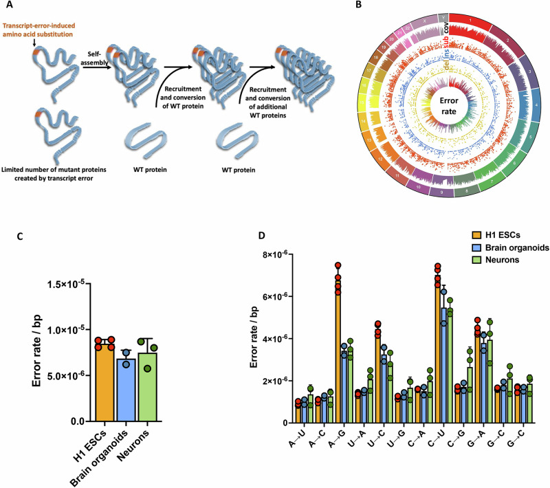
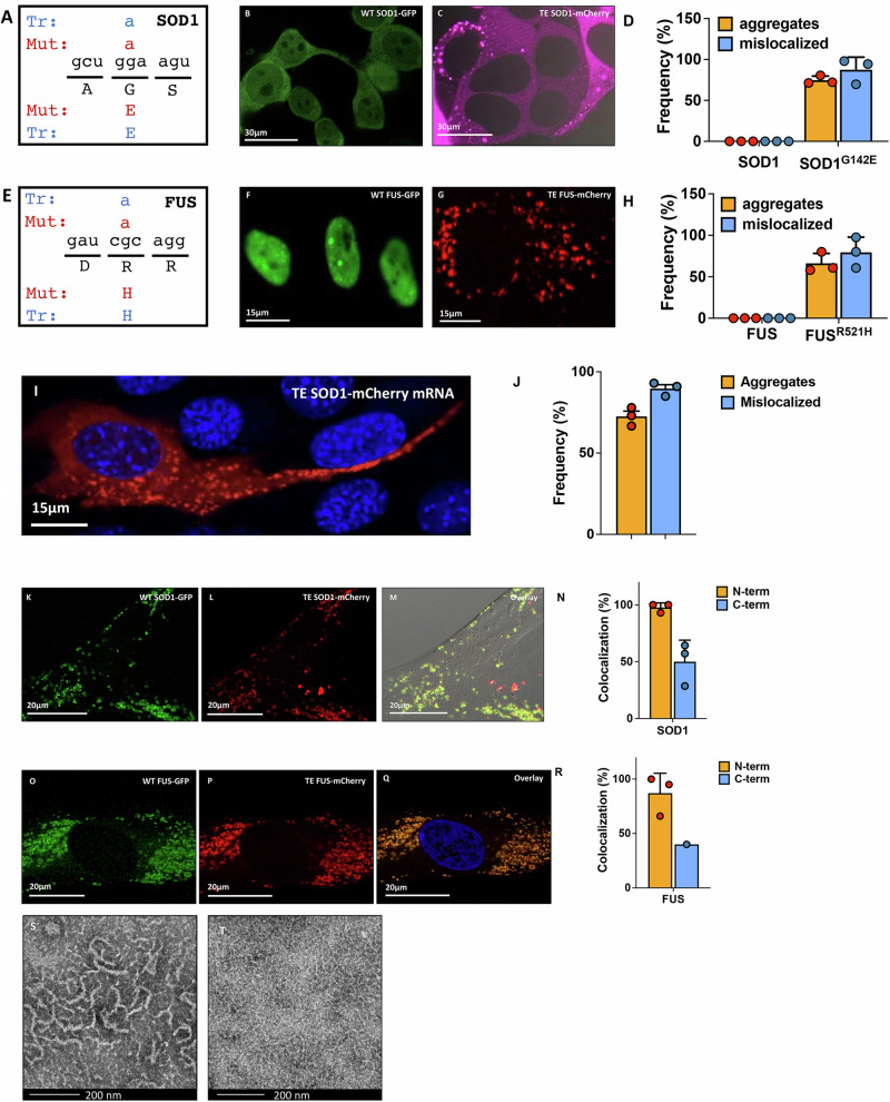
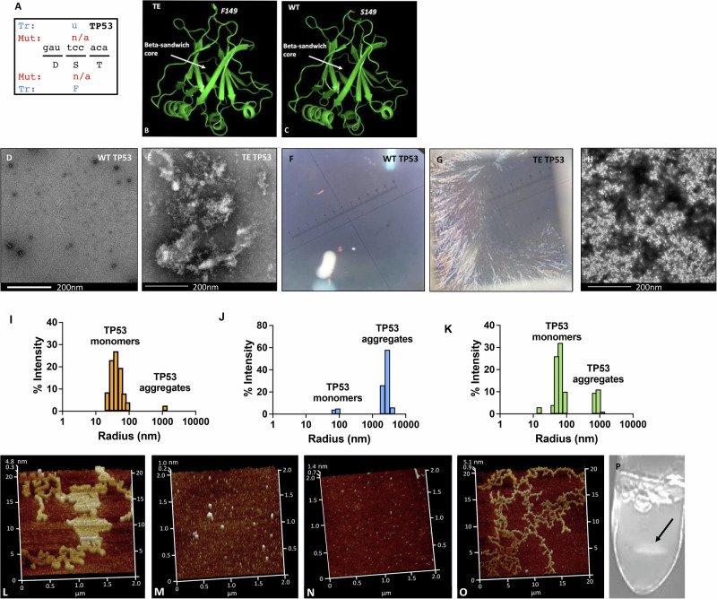
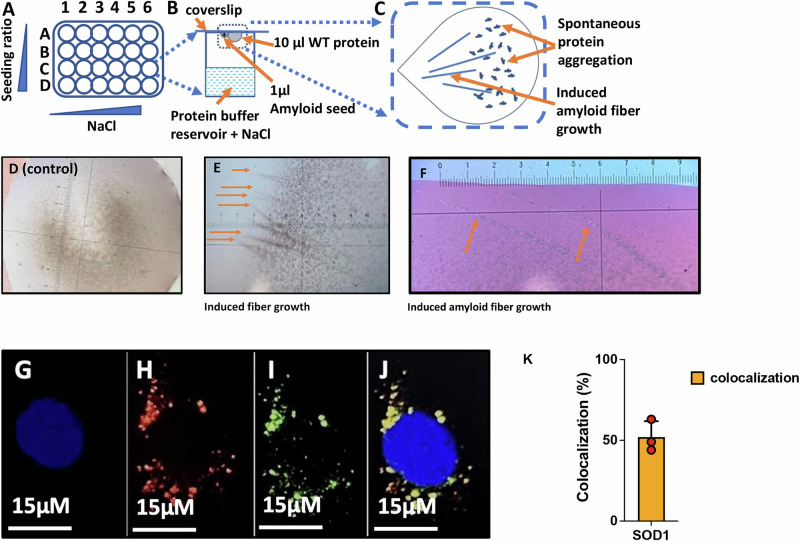
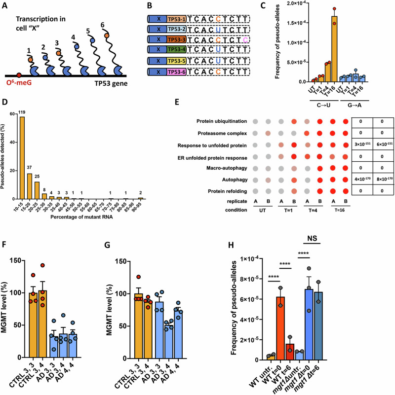
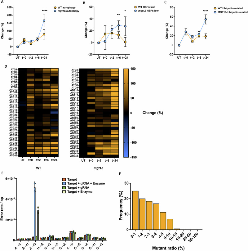
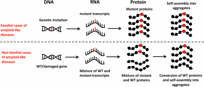
Aging is characterized by the accumulation of proteins that display amyloid-like behavior. However, the molecular mechanisms by which these proteins arise remain unclear. Here, we demonstrate that amyloid-like proteins are produced in a variety of human cell types, including stem cells, brain organoids and fully differentiated neurons by mistakes that occur in messenger RNA molecules. Some of these mistakes generate mutant proteins already known to cause disease, while others generate proteins that have not been observed before. Moreover, we show that these mistakes increase when cells are exposed to DNA damage, a major hallmark of human aging. When taken together, these experiments suggest a mechanistic link between the normal aging process and age-related diseases.


Conflict of interest statement

The authors declare no competing interests.

Figures

Fig. 1
Fig. 1. Graphical representation of hypothesis and summary of transcription error data.
A We hypothesize that transcription errors could give rise to amyloid- and prion-like proteins. This relatively small cache of mutant proteins can then form a seed that recruits WT proteins and converts them to an amyloid-like state to generate the large amyloid fibers and amorphous deposits that characterize protein aggregation diseases. B Transcription errors were identified across the genome of H1 human embryonic stem cells (n = 4, ESCs), brain organoids (n = 2) and human neurons (n = 3). All replicates are independent biological replicates. C,D The error rate and spectrum of H1 ESCs, brain organoids and human neurons are similar, with the exception of A → G errors, which most likely indicate off-target A to I RNA editing (n is identical to B). Error bars indicate standard error of the mean. Source data are provided as a Source Data file.
Fig. 2
Fig. 2. Transcription errors result in proteins with increased amyloid-like behavior.
A A transcription error (Tr) was identified in the SOD1 transcript that mimics a mutation (Mut) implicated in ALS. This error substitutes a guanine for an adenine base, resulting in a glycine (G) to glutamine (E) mutation at residue 142. B WT SOD1 is soluble and present throughout the cell, including the nucleus. C In contrast, SOD1G142E proteins form aggregates and are excluded from the nucleus. D Quantification of WT and mutant SOD1 aggregation and mislocalization. Depicted are the % of cells with aggregates or mislocalized proteins (n = 3 biological replicates). For aggregates, P = 0.0015, for mislocalization, P = 0.0102. E A transcription error was identified in the FUS transcript that mimics a mutation implicated in ALS. This error substituted a guanine for an adenine base, resulting in an arginine (R) to histidine (H) mutation at residue 521. F WT FUS is present in a soluble state in the nucleus, while FUSR521H (G) forms aggregates outside of the nucleus. H Quantification of FUS aggregation and mislocalization (n = 3 biological replicates). Depicted are the % of cells with aggregates or mislocalized proteins. For aggregates, P = 0.0108, for mislocalization, P = 0.0174. I, J Transfection of cells with 500 ng of SOD1G142E mRNA resulted in cells with clearly visible mislocalized protein aggregates (n = 3 biological replicates). KM When cells are transducted with lentiviral particles carrying both WT SOD1 and SOD1G142E simultaneously, WT SOD1 is excluded from the nucleus and recruited into extranuclear aggregates. N Quantification of SOD1 colocalization with either N-terminal or C-terminal tags. OQ WT and FUSR521H co-expression in primary human fibroblasts, demonstrating that mutant and WT FUS co-localize in cytoplasmic aggregates (n = 3 biological replicates). R Quantification of FUS colocalization with either N-terminal or C-terminal tags. (n = 3 biological replicates). S WT SOD1 does not form fibers under TEM, but SOD1G142E does (T). TEM experiments were performed 3 times with similar results. *P < 0.05. **P < 0.01 according to a two-tailed unpaired t-test with Welch’s correction. Data are presented as mean values ± SEM. Source data are provided as a Source Data file.
Fig. 3
Fig. 3. Biophysical examination of WT and mutant TP53.
A A transcription error (Tr) was identified in a TP53 transcript that substitutes a uracil for a cytosine base, resulting in a serine (S) to phenyl-alanine (F) mutation at residue 149. B,C Predicted structure of WT (B) and mutant TP53 (C). D Transmission electron microscopy showed little or no aggregates of WT TP53, while TP53S149F induces large protein aggregates (E). These experiments were performed 3–6 times with similar results. F,G Congo-red birefringence under polarized light indicates that TP53S149F forms amyloid fibrils (G), while WT TP53 does not (F). H After addition of 1% TP53S149F to a solution of WT TP53 (v/v), the WT solution generated countless aggregates. This experiment was performed 3 times with similar results. IK Dynamic light scattering, which can be used to determine the radius of protein particles, indicates that WT TP53 is primarily in a monomeric form (I), while mutant TP53 consists of aggregates greater than 1000 nm (J). After 2% TP53S149F is added to a solution of WT TP53 (v/v), a large amount of TP53 aggregates emerges (K). L TP53S149F aggregates into a variety of structures. M TP53 aggregates were sonicated to create a seed solution of particles that are around 0.1 µm in size, which equates to 800-1000 proteins. (N) WT TP53 solution shows no apparent aggregation; (O) Adding the TP53S149F amyloid seed solution to WT TP53 in a 1:100 ratio induced fibril growth. P Protein aggregates created by mutant TP53 form spontaneously and can be seen by the naked eye (arrow).
Fig. 4
Fig. 4. A hanging drop method and mRNA transfections to assess WT:mutant ratios required for protein aggregation.
A 4 × 6 screening tray was set up with a 1 ml reservoir that contains protein buffer and an increasing concentration of NaCl. B A 10 µl WT TP53 solution (60 µM) was then added to a siliconized coverslip and a 1 µl drop of TP53S149F seed particles was placed immediately adjacent at decreasing concentrations. C If the mutant seed particles have amyloid potential, this event will trigger conversion of WT proteins at the drop-drop interface and lead to localized fiber growth D If no TP53S149F is provided as seeding material, no fiber-like material forms in the WT TP53 solution. E However, if TP53S149F seeding material is provided, fiber-like material grows out of the WT solution. F These fibers show strong birefringence under polarized light, suggestive of amyloid structures. GJ. Primary fibroblasts were transfected with 90% WT and 10% mutant SOD1G142E transcripts display co-localized WT and mutant proteins inside protein aggregates G DAPI. H SOD1G142E-mCherry. I WT SOD1-eGFP. J Overlay of GI. These experiments were performed 3 times with similar results. K Quantitation of GJ (n = 3 biological replicates), data is presented as mean values ± SEM. Source data are provided as a Source Data file.
Fig. 5
Fig. 5. DNA damage and off-target RNA editing affect the fidelity of transcription.
A If DNA damage results in multiple rounds of error prone transcription, then multiple transcripts in a single cell should carry identical errors. B When these transcripts are captured and tagged with UMIs, they can be grouped together, and their sequences can be compared to each other to search for identical errors that occur in multiple transcripts. In contrast, sequencing errors or RNA damage will only be present in one transcript. Blue bar: cell-specific barcode. Multi-colored bar: transcript UMI. Blue base: WT. Orange base: transcription error. Pink base: Sequencing error/RNA damage C C → U pseudo-alleles emerge after MNNG treatment of mouse neural stem cells, while G → A errors (which would indicate conventional mutagenesis is occurring as well) do not (n = 2 biological replicates). *P < 0.05, ****P < 0.0001 according to a Chi-squared test with Yates’ continuity correction. D Ratio of WT:mutant mRNAs identified. Only alleles with more than 10% mutant mRNAs are depicted. E Dot plots of single cell gene expression profiles grouped by GO-terms indicate markers of proteotoxic stress are elevated in treated cells, particularly at 16 h, when the transcript error rate is the highest. Significance was ascertained by ANOVA test. FDR= False Discovery Rate. F MGMT levels are decreased in all females with AD compared to control females with an APOE3,3 genotype. For AD 3,3, P = 0.0022, for AD 3,4, P = 0.0034, for AD 4,4, P = 0.0022. n = 4 biological replicates, a two-tailed unpaired t-test with Welch’s correction. G MGMT levels are not decreased in males with AD, except for those with a APOE3/APOE4 genotype, P = 0.0066. n = 4 biological replicates, a two-tailed unpaired t-test with Welch’s correction. H Loss of MGT1, the yeast homolog of MGMT allows O6-me-G lesions to remain on the genome, resulting in greatly increased numbers of pseudo-alleles over time. n = 2 biological replicates. ****P < 0.0001 according to a Chi-squared test with Yates’ continuity correction. Source data are provided as a Source Data file.
Fig. 6
Fig. 6. DNA damage induces markers of proteotoxic stress.
Consistent with the idea that these errors result in misfolded proteins, these cells displayed markers of proteotoxic stress, including upregulated autophagy genes (A), heat shock proteins (B) and proteins implicated in the ubiquitin-proteasome system (C). Depicted in figure A and C is the average percentage change for all autophagy and ubiquitination-related genes identified by bulk RNA-seq. The genes depicted in B have been separated from several heat shock proteins that displayed unusually large increases in transcript levels (Supplementary Fig. 9, Supplementary Table 4). n = 3 biological replicates, for autophagy P < 0.0001, for heat-shock proteins P = 0.0016 at 6 h and P = 0.0287 at 24 h. For ubiquitin P < 0.0001. *P < 0.05, **P < 0.01, ****P < 0.0001 according to a two-tailed paired t-test with Welch’s correction. D. Heat map of autophagy genes detected in WT and mutant cells. E Error spectrum of human cells after transformation with plasmid that carries an editing target, the gRNA required to edit the target, and the editing enzyme. If the editing enzyme is present, large numbers of A to I editing events (A to G errors) were observed. n = 2 biological replicates. F Percentage of editing events that generate mRNAs with various mutant:WT ratios. Data are presented as mean values ± SEM. Source data are provided as a Source Data file.
Fig. 7
Fig. 7. Model for the contribution of transcription errors to non-familial cases of disease.
Familial cases of protein aggregation diseases are caused by genetic mutations that generate mutant proteins with increased amyloid or amyloid-like behavior. In non-genetic cases, identical mutant proteins (and potentially unique mutant proteins) are generated by non-genetic mutations such as transcription errors. Over time, these proteins convert WT proteins to an amyloid state, leading to a later onset of amyloid and amyloid-like diseases compared to familial cases. Because these amyloid and amyloid-like proteins are generated by mutations that are only present in transcripts though, they have thus far gone undetected in the clinic.

References

    1. Lopez-Otin, C., Blasco, M. A., Partridge, L., Serrano, M. & Kroemer, G. The hallmarks of aging. Cell153, 1194–1217 (2013). - PMC - PubMed
    1. Hipp, M. S., Kasturi, P. & Hartl, F. U. The proteostasis network and its decline in ageing. Nat Rev Mol Cell Biol20, 421–435 (2019). - PubMed
    1. Eisenberg, D. & Jucker, M. The amyloid state of proteins in human diseases. Cell148, 1188–1203 (2012). - PMC - PubMed
    1. Scheckel, C. & Aguzzi, A. Prions, prionoids and protein misfolding disorders. Nature reviews. Genetics19, 405–418 (2018). - PubMed
    1. de Oliveira, G. A. P. et al. The status of p53 oligomeric and aggregation states in cancer. Biomolecules10, 548 (2020). - PMC - PubMed

Publication types