. 2024 Oct 7;14(1):171.
doi: 10.1038/s41408-024-01153-0.

MYC-rearranged mature B-cell lymphomas in children and young adults are molecularly Burkitt Lymphoma

Affiliations

MYC-rearranged mature B-cell lymphomas in children and young adults are molecularly Burkitt Lymphoma

Sara Mato et al. Blood Cancer J. .

Abstract

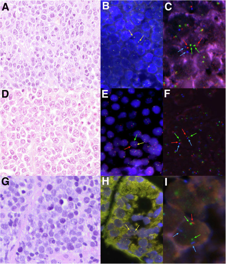
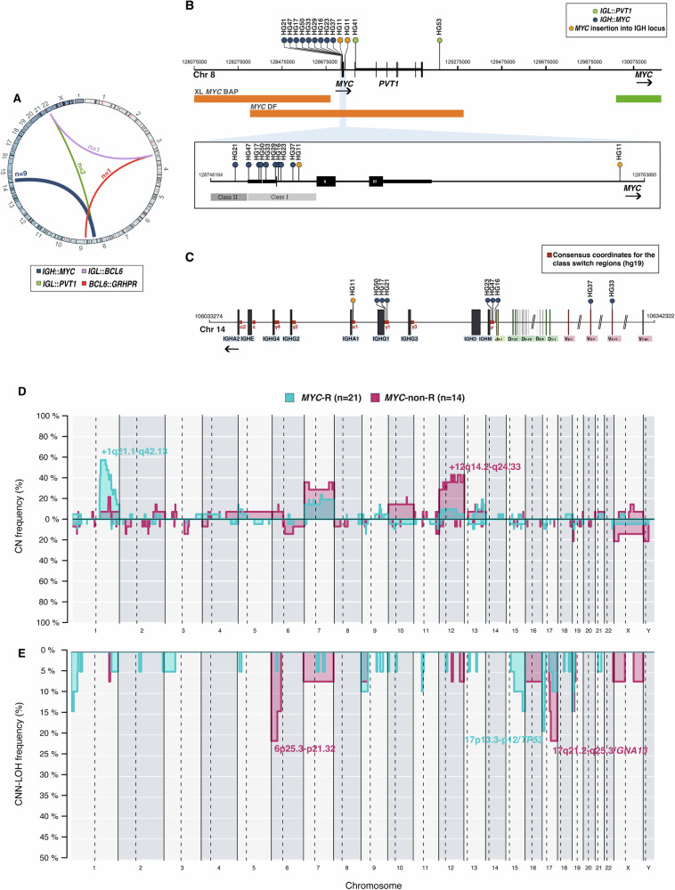
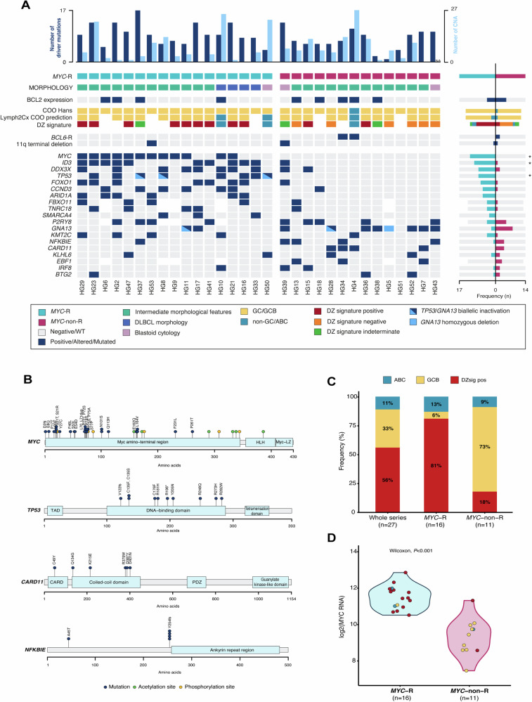
Aggressive B-cell non-Hodgkin lymphomas (NHL) in children, adolescents, and young adults (CAYA) include Burkitt lymphoma (BL), diffuse large B-cell lymphoma (DLBCL), and a subset of high-grade tumors with features intermediate between these entities whose genetic and molecular profiles have not been completely elucidated. In this study, we have characterized 37 aggressive B-NHL in CAYA, 33 with high-grade morphology, and 4 DLBCL with MYC rearrangement (MYC-R), using targeted next-generation sequencing and the aggressive lymphoma gene expression germinal center B-cell-like (GCB), activated B-cell-like (ABC), and dark zone signatures (DZsig). Twenty-two tumors had MYC-R without BCL2 breaks, and two MYC-non-R cases had BCL6 translocations. MYC-R cases, including DLBCL, carried BL-related mutations and copy number alterations. Conversely, MYC-non-R lymphomas had alterations in the B-cell receptor signaling/NF-κB pathway (71%). DZsig was expressed in 12/13 of MYC-R tumors but only in 2/10 of MYC-non-R GCB tumors (P < 0.001). The 3-year event-free survival (EFS) of the whole cohort was 79.6%. TP53 and KMT2C mutations conferred inferior outcome (3-year EFS P < 0.05). Overall, MYC-R lymphomas in CAYA have a molecular profile similar to BL regardless of their high-grade or DLBCL morphology, whereas MYC-non-R has more heterogeneous genetic alterations closer to that of DLBCL.


Conflict of interest statement

The authors declare no competing interests.

Figures

Fig. 1
Fig. 1. Morphological, immunohistochemical, and genetic features of three high-grade cases.
A Case HG28 showed a diffuse proliferation of medium-sized lymphocytes with slight pleomorphism, irregular nuclei, and apoptotic bodies consistent with features intermediate between BL and DLBCL (H&E, original magnification at 400×). B MYC break-apart FISH probe showed two colocalizations in each cell consistent with the absence of gene rearrangement. C FISH analysis for 11q-aberration showed 2 green (11q23.3 minimal region gain, MRG), 2 orange (11q24.3 minimal region loss, MRL), and 2 aqua (CEN11 D11Z1) signals, compatible with a non-altered 11q chromosomal region. D Case HG33 depicted a DLBCL morphology showing a predominance of large centroblastic lymphocytes (H&E, original magnification at 400×). E MYC break-apart FISH probe showed one red signal, one green signal, and one colocalization, consistent with the rearrangement of the gene. F FISH analysis for 11q-aberration showed 2 green (11q23.3 MRG), 2 orange (11q24.3 MRL), and 2 aqua (CEN11 D11Z1) signals constellation, compatible with a non-altered 11q chromosomal region. G Case HG39 consisted of monotonous medium-sized lymphocytes with blastoid appearance (H&E, original magnification at 400×). H MYC break-apart FISH probe showed two colocalizations consistent with a non-rearranged pattern. I A non-prototypic terminal deletion of 11q24.3 region was identified in case HG39. The FISH constellation showed 2 green (11q23.3 MRG), 1 orange (22q24.3 MRL), and 2 aqua (CEN11 D11Z1) signals.
Fig. 2
Fig. 2. SV architecture of 35 aggressive B-cell lymphomas with overlapping features between BL and DLBCL.
A SV discovered by targeted NGS sequencing (n = 26). B Location of the breakpoints identified in the MYC gene. C Location of breakpoints in the IGH locus according to current consensus coordinates [37]. D, E Comparative plot of CN and CNN-LOH alterations among the 2 genetic groups based on MYC rearrangement status. Light blue identifies MYC-R cases (n = 21), whereas dark pink identifies MYC-non-R tumors (n = 14).
Fig. 3
Fig. 3. Molecular CN and mutational information on 31 B-cell lymphomas with overlapping features between BL and DLBCL.
A Oncoprint showing gene expression, mutations, and CN characteristics by case. Each column corresponds to a case, the light blue bars in the top histogram depict the number of CNA while the dark blue bars represent the number of driver mutations. Each row of the bottom plot represents a gene. Only genes with driver mutations in more than 2 cases are represented. Asterisks identify significant differences according to adjusted (FDR) Fisher’s exact test (P-adjusted < 0.05). B Diagram of the relative positions of driver mutations is shown for MYC, TP53, CARD11, and NFKBIE genes, x-axes indicate amino acid position. MYC domains (HLH, helix-loop-helix; MYC-LZ, leucine zipper; TAD, transactivation domain). TP53 domains (DBD: DNA-binding domain); TAD: transcription activation domain; CARD11 (CARMA1) domains (CARD, caspase activation, and recruitment domain; PDZ, PDZ containing domain). C Proportion of DZsig positive tumors in the whole analyzed series (n = 27) and according to MYC-R status. D MYC RNA expression (NM_002467.3) according to DZsig and COO categorization and MYC-R status.

References

    1. Swerdlow SH, Campo E, Harris NL, Jaffe ES, Pileri SA, Stein H, et al., editors. WHO classification of tumours of haematopoietic and lymphoid tissues. Revised 4th ed. Lyon: IARC; 2017.
    1. Alaggio R, Amador C, Anagnostopoulos I, Attygalle AD, Araujo IBO, Berti E, et al. The 5th edition of the World Health Organization classification of haematolymphoid tumours: lymphoid neoplasms. Leukemia. 2022;36:1720–48. - PMC - PubMed
    1. Campo E, Jaffe ES, Cook JR, Quintanilla-Martinez L, Swerdlow SH, Anderson KC, et al. The international consensus classification of mature lymphoid neoplasms: a report from the Clinical Advisory Committee. Blood. 2022;140:1229–53. - PMC - PubMed
    1. Olszewski AJ, Kurt H, Evens AM. Defining and treating high-grade B-cell lymphoma, NOS. Blood. 2022;140:943–54. - PubMed
    1. Ok CY, Medeiros LJ. High-grade B-cell lymphoma: a term re-purposed in the revised WHO classification. Pathology. 2020;52:68–77. - PubMed

Publication types

Substances